

 Find Duplicates
 Check Document
 Submission(new)
 Experts Office
 Editorial Office
2023 Vol. 46, No. 1
Display Method:
2023, 46(1): 1-6.
doi: 10.12122/j.issn.1674-4500.2023.01.01
Abstract:
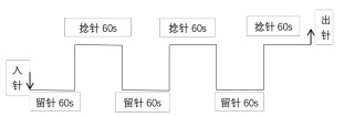
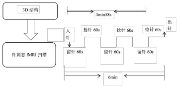
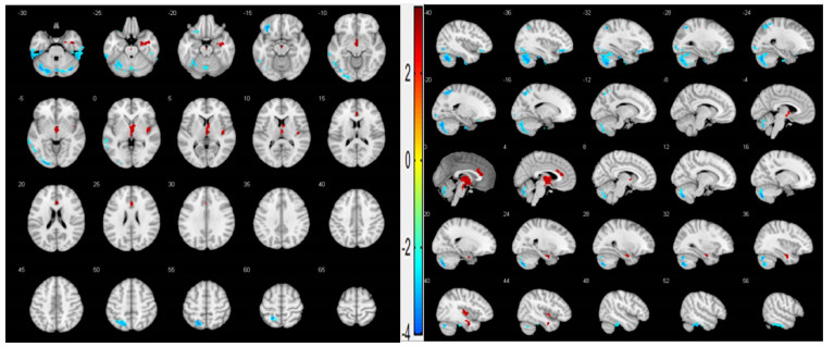
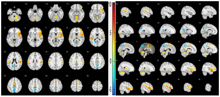
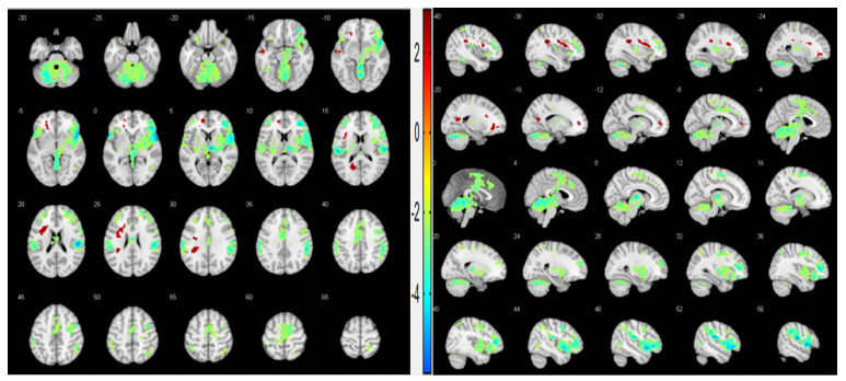
Objective To observe the changes of brain functional activities of the source and connecting needling (Taixi-Feiyang acupoint) of patients with ischemic vascular dementia by event-related cerebral functional magnetic resonance imaging. Methods A 6-minute task state experiment was performed on 12 patients with ischemic vascular dementia using a 3.0T magnetic resonance scanner. The 3D structural image scan for 4 minutes was collected first, and then the needle-prick data of the source connecting points (Taixi-Feiyang acupoint) was collected for 6 minutes. The needle-prick data of the single points of the Feiyang acupoint was collected again for 6 minutes at an interval of 2 weeks. The obtained image data was processed and analyzed by a general linear model.The brain function partitions with altered neuronal activities in patients with needle-prick ischemic vascular dementia were obtained. Results Brain activation areas of needling the source connecting points " Taixi and Feiyang" in patients with ischemic vascular dementia were right hippocampal gyrus, right horn gyrus, left thalamus, right thalamus, right insula, left anterior cingulate and central paraloids. The negatively activated brain areas were left cerebellar foot area 1, left lower half moon lobule, cerebellar worm eighth area, left cerebellar posterior lobe, left fusiform gyrus, left inferior temporal gyrus, right inferior temporal gyrus, left orbital subfrontal gyrus, left occipital regress, left temporal recirculation, left occipital recurrent, left glossal gyrus, left sub-parietal lobules, and left parietal lobes. Conclusion The immediate effect of needling the source connecting points "Taixi-Feiyang acupoints" in patients with ischemic acupuncture vascular dementia can directly affect and regulate the neuronal activities of the brain function partitions related to cognitive activities. The treatment of source and connecting needling has a wider range of brain activation areas.
2023, 46(1): 7-11.
doi: 10.12122/j.issn.1674-4500.2023.01.02
Abstract:
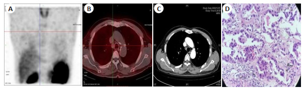
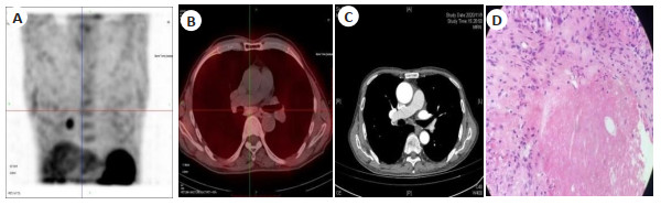
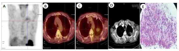
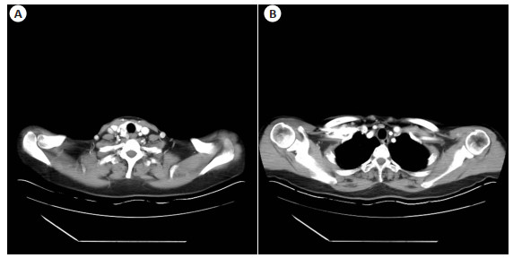
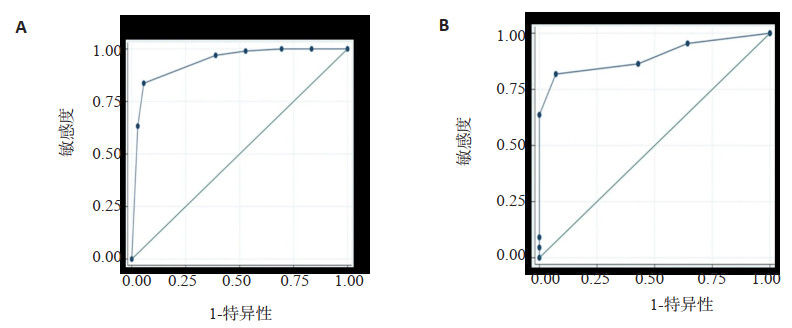
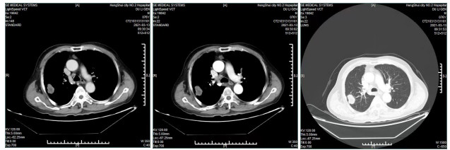
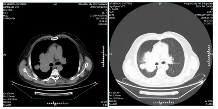
Objective To investigate the diagnostic value of 99mTc-3PRGD2 SPECT/CT imaging combined with enhanced CT for the evaluation of lymph node metastasis in chest masses. Methods Forty-one patients with suspected lung lesions were prospectively recruited and underwent 99mTc-3PRGD2 SPECT/CT imaging and enhanced CT imaging with tumor resection and lymph node dissection. The detection of lymph node metastasis in lung tumors was recorded. The pathological results were used as the "gold standard" to analyze the diagnostic ability of 99mTc-3PRGD2 imaging, lung-enhanced CT and the combination of both to determine whether lymph node metastasis of lung malignant tumors were detected. The sensitivity, specificity, accuracy, positive predictive value and negative predictive value were measured. Results The sensitivity, specificity, accuracy, positive predictive value, and negative predictive value of 99mTc-3PRGD2 imaging, enhanced CT, and the combination of the two for the diagnosis of lymph node metastasis in lung tumors were 54.6% (6/11), 68.4% (13/19), 63.3% (19/30), 50.0% (6/12), 72.2% (13/18); 72.3% (8/11), 47.4% (9/19), 56.7% (17/30), 44.4% (8/18), 75.0% (9/12); 81.8% (9/11), 89.5% (17/19), 86.7% (26/30), 81.8% (9/11), 89.5% (17/19). The diagnostic power of 99mTc-3PRGD2 imaging relative to enhanced CT ability was relatively high in specificity and low in sensitivity compared to enhanced CT. The diagnostic efficacy of both methods was higher than that of enhanced CT (χ2=6.914, P=0.009) and 99mTc-3PRGD2 imaging (χ2=7.751, P=0.005) alone for the diagnosis of malignant lymph node metastasis in the chest. The concordance of diagnostic and pathological findings using Kappa test comparing 99mTc-3PRGD2 SPECT/CT imaging, enhanced CT and both combined were 0.225, 0.177, 0.713, respectively, which were not statistically different (P>0.05). Conclusion Chest-enhanced CT combined with 99mTc-3PRGD2 SPECT/CT imaging has high accuracy in diagnosing lymph node metastasis of lung tumors and helps patients' surgical decision.
2023, 46(1): 12-20.
doi: 10.12122/j.issn.1674-4500.2023.01.03
Abstract:
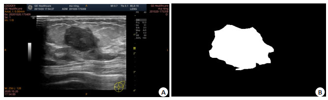
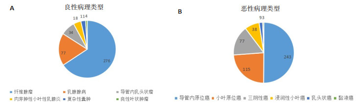
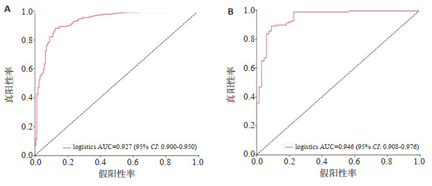
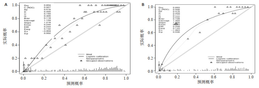
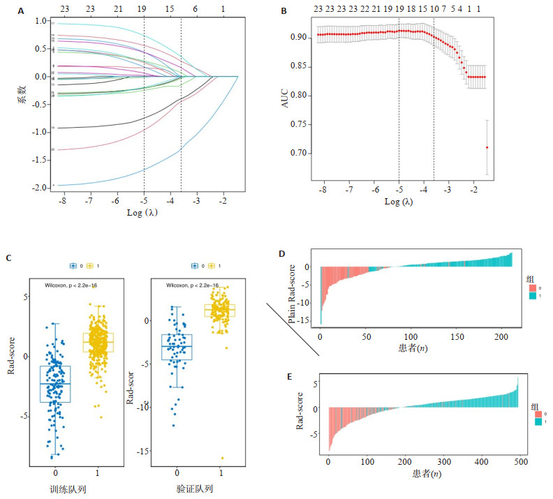
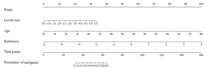
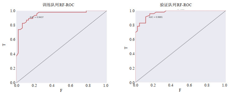
Objective To investigate the value of ultrasound-based radiomics in discriminating benign and malignant BI-RADS category 4a irregular breast nodules, and to assess its value in reducing unnecessary biopsies by establishing a nomogram through combine radiomics, ultrasound features and clinical independent risk factor characteristics. Methods A total of 905 cases of BI- RADS 4a irregular breast nodules screened by conventional ultrasonography were retrospectively collected and randomly divided into a training set (n=634) and a validation set (n=271) with a ratio of 7:3. Pyradiomics was used to extract 851 features.The radiomics model was constructed by Logistics regression model using surgical pathology as the gold standard. The diagnostic efficacy of the radiomics model and the nomogram model on the diagnosis of irregular breast nodules with ultrasound BI-RADS 4a pattern was evaluated by ROC curve. Results Among 905 irregular breast nodules, 485 were malignant nodules and 420 were benign nodules, with the age of 22-83(50.05±11.13) years old. The age, Rad-score value, and mass diameter of the two groups were not significantly different (P>0.05). The AUC value of the training cohort radiomics model was 0.927 (95% CI: 0.900-0.950), and the AUC value of the validation cohort radiomics model was 0.946 (95% CI: 0.908-0.976). The sensitivity, accuracy, specificity, F1 value, and precision for the training cohort of this model were 0.879, 0.879, 0.877, 0.909 and 0.940, respectively. The sensitivity, accuracy, specificity, F1 value, and precision for the validation cohort of this model were 0.890, 0.896, 0.909, 0.921 and 0.956, respectively. The calibration curves showed good calibration between the training and validation cohorts of this mode; The AUC value was 0.943 (95% CI: 0.912-0.960) for the training cohort normogram model and 0.968 (95% CI: 0.924-0.970) for the validation cohort normogram model. Conclusion Ultrasound-based radiomics and the nomogram model have important value in improving the diagnostic efficacy of benign and malignant BIRADS 4a morphologically irregular breast nodules, have better predictive efficacy for BI-RADS 4a irregular breast nodules, and can reduce unnecessary biopsies.
2023, 46(1): 21-27.
doi: 10.12122/j.issn.1674-4500.2023.01.04
Abstract:
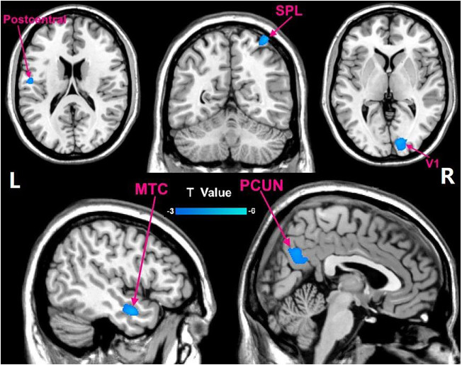
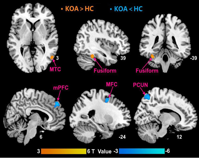
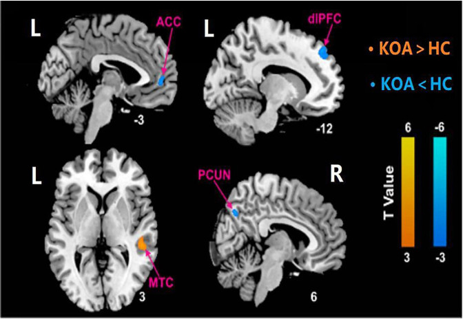
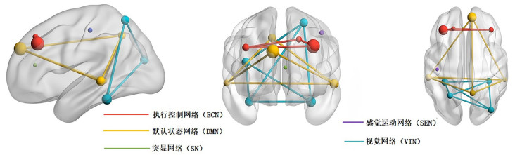
Objective To explore the change of brain gray matter volume, neuron activity and functional connectivity strength based on seed points in patients with knee osteoarthritis (KOA) at rest through combined voxel based morphology and rsfMRI technology, and comprehensively analyze KOA-related abnormal brain networks. Methods 3D high-resolution T1WI images and rs- fMRI images of 30 patients with KOA (KOA group) and 30 healthy controls (HC group) were collected prospectively. Voxel based morphology, amplitude of low-frequency fluctuation, and functional connectivity were used to analyze the differences in gray matter structural and functional data between the two groups. Results Compared with the HC group, the amplitude of low-frequency fluctuation values of bilateral fusiform gyrus and right middle temporal gyrus in KOA group increased, while the amplitude of low-frequency fluctuation values of right precuneus, right medial prefrontal cortex and left middle frontal gyrus decreased (voxel level P<0.005, cluster level P<0.05). The gray matter volume of right precuneus, right inferior parietal lobule, right primary visual cortex, left middle temporal gyrus, left postcentral gyrus decreased (voxel level P<0.002, cluster level P<0.05). With the right precuneus as the seed point, the functional connectivity with right middle temporal gyrus was enhanced, and the functional connectivity with left anterior cingulate gyrus, left dorsolateral prefrontal cortex was reduced (voxel level P<0.005, cluster level P<0.05). Conclusion The changes of BOLD signal and gray matter microstructure were found in both perceptual cortex system and combined cortex system of KOA patients, There are overlap, mainly involving in the visual network, sensorimotor network, default mode network, executive control network within and between partial brain regions of default mode network, executive control network and salience network. This may suggest that KOA patients are not only in the pathological state of chronic pain, but also accompanied by abnormalities in functional activities such as information integration, attentional control, emotional response, and emotional interpretation.
2023, 46(1): 28-32.
doi: 10.12122/j.issn.1674-4500.2023.01.05
Abstract:
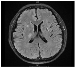
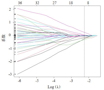
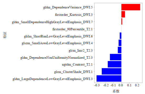
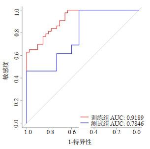
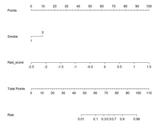
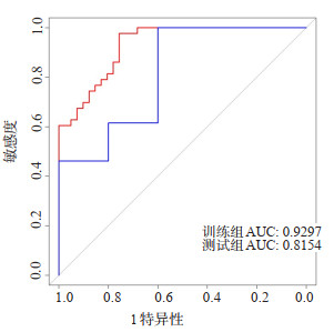
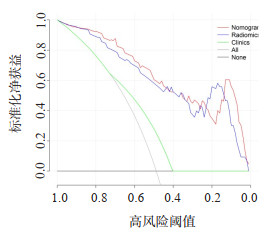
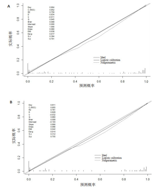
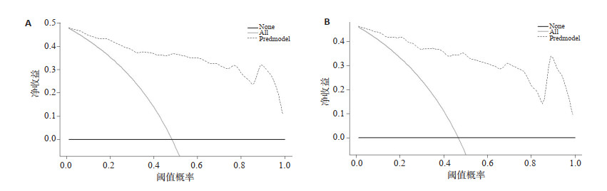
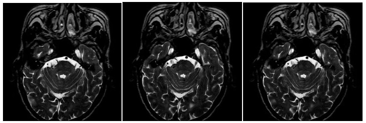
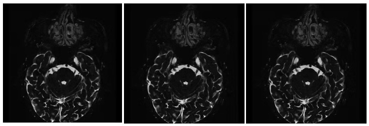
Objective To explore the clinical value of the diffusion weighted imaging (DWI) and T2 Flair radiomics model in prognosis of patients with acute cerebral infarction in the basal ganglia region after non-thrombolytic therapy. Methods Seventy-eight patients with acute cerebral infarction in the basal ganglia region which were given the brain MRI scan from the Department of Neurology of Binzhou Medical University Hospital in 2021 were included. They were diagnosed as acute cerebral infarction by the clinical symptoms and auxiliary examinations. According to the changes of modified Rankin scale scores at admission and discharge, the patients who did not accept thrombolytic therapy for one week were divided into good prognosis group and poor prognosis group. Firstly, 3D Slicer was used to delineate the infarct region of high signal intensity in DWI images and T2 Flair images. The Radiomics module was used to collect the radiomics features from the delineated images. Combined with modified Rankin scale scores of patients on admission and discharge, the data were analyzed by univariate analysis and then carried on multivariate analysis respectively by multivariate logistic regression algorithm. The predictive efficacy of various factors was analyzed by ROC curve to assess the prognostic value of the study subjects. Results By establishing the combined radiomics model of 3.0T MRI-DWI sequence and T2 Flair sequence, the area under the ROC curve (AUC) of the prediction model in the training group was 0.9189 respectively, and the prediction model in the validation group was 0.7846. Smoking and rad-scores were found to be independent risk factors for poor prognosis after nonthrombolytic therapy in patients with acute cerebral infarction. In clinical model, the AUC of the training group and the test group were 0.6497 and 0.5468, respectively. The nomogram model was constructed based on these models, and the AUC of the training group and the test group were 0.9297 and 0.8154, respectively. The differences between nomogram model and clinical model, nomogram model and radiography model were statistically significant (P<0.05). Conclusion The radiomics model combined with DWI and T2 Flair sequence can evaluate the prognosis of non- thrombolytic therapy in patients with acute cerebral infarction in the basal ganglia region. Nomogram model has better prediction efficiency than clinical prediction model and omics model.
2023, 46(1): 33-36.
doi: 10.12122/j.issn.1674-4500.2023.01.06
Abstract:
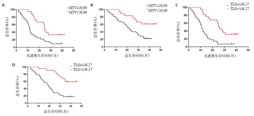
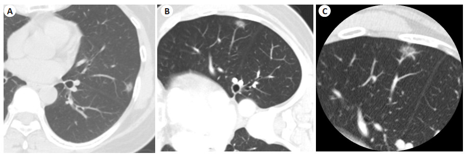
Objective To explore the evaluation value of PET/CT metabolism parameters for prognosis in middle aged and elderly patients with non-small cell lung cancer (NSCLC). Methods Fifty middle aged and elderly patients with NSCLC treated in the hospital were enrolled from March 2018 to August 2019. All patients underwent PET/CT examinations and follow-up. The overall survival (OS) and progression-free survival (PFS) were recorded. The relationship between PET/CT metabolism parameters and prognosis was analyzed by Kaplan-Meier method, Log-rank test and Cox regression analysis. Results Overall median OS and median PFS were 28.25 months and 15.25 months of the 50 patients, respectively. The 1-year, 2- year and 3-year OS and PFS were 80.00%, 56.00%, 36.00% and 60.00%, 34.00%, 18.00%, respectively. The diagnostic coincidence rates of PET/CT for T, N and M staging were 92.00%, 94.00% and 98.00%, respectively. Univariate analysis showed that TNM staging, maximum lesion diameter, surgery, SUVmax, MTV and TLG were influencing factors of PFS and OS (P<0.05). Multivariate analysis showed that MTV and TLG were independent risk factors of PFS, while TNM staging, MTV and TLG were independent risk factors of OS. Conclusion MTV and TLG are independent risk factors of PFS and OS in patients with NSCLC. PET/CT metabolism parameters have certain evaluation value for disease progression.
2023, 46(1): 37-42.
doi: 10.12122/j.issn.1674-4500.2023.01.07
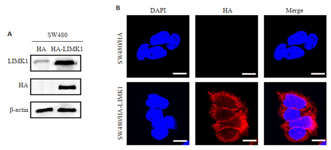
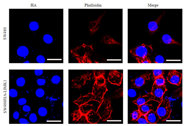
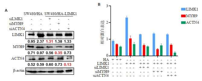
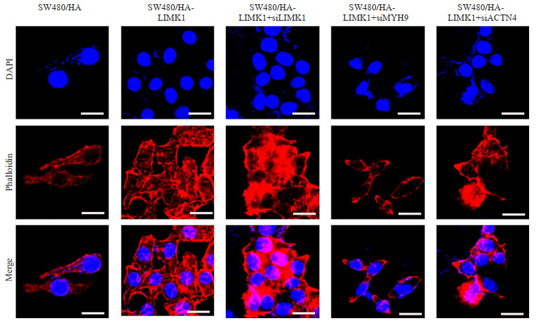
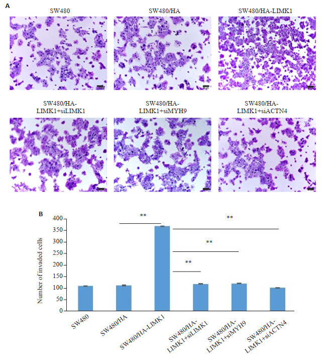
Abstract:
Objective To elucidate the specific mechanism of colorectal metastasis induced by LIMK1 based on molecular visualization imaging technique of cytoskeleton. Methods The effects of overexpressed LIMK1 on the cytoskeletal of SW480 cells were detected by molecular visualization imaging technique, and the effects of silencing LIMK1, ACTN4 and MYH9 on the cytoskeleton were detected by Phalloidin molecular visualization imaging technique. Transwell migration assay was used to detect the effects of silenced ACTN4 and MYH9 on LIMK1-mediated cell migration. Results In SW480 cell line, the staining of cytoskeleton protein F-actin was significantly enhanced after overexpression of LIMK1, but this increase could be restored after silencing the expression of ACTIN4 or MYH9 with siRNA. In addition, silencing these two proteins in SW480 cell lines inhibited the phenotype of LIMK1-mediated malignant migration(P<0.05). Conclusion LIMK1 may regulate the cytoskeleton and promote the migration and invasion of colorectal cancer cells by interacting with ACTIN4 or MYH9.
2023, 46(1): 43-47.
doi: 10.12122/j.issn.1674-4500.2023.01.08
Abstract:
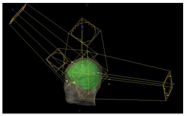
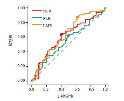
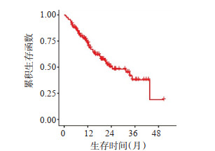
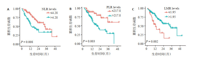
Objective To investigate the relationship between peripheral blood neutrophil to lymphocyte ratio (NLR), platelet to lymphocyte ratio (PLR) and lymphocyte to monocyte ratio (LMR) and prognosis of patients with brain metastases. Methods A total of 118 patients with brain metastases treated with whole brain radiotherapy were retrospectively analyzed. The relationship of NLR, PLR, LMR and clinical data with the prognosis patients were analyzed. Receiver operating characteristic curve was used to calculate the optimal cut-off values of NLR, PLR and LMR. The overall survival analysis was performed by Kaplan- Meier method. Significant differences were analyzed by Log-rank test. Multivariate analysis for survival was performed by Cox proportional regression model. Results The median overall survival time of 188 patients with brain metastases was 12.5 months. The 1-year, 2-year survival rates were 72.8% and 50.1%, respectively. The optimal cut-off values of NLR, PLR and LMR were determined by ROC curve to be 4.28, 217.8 and 1.95, respectively. The tumor stage, targeted therapy, NLR, PLR, and LMR were all associated with the prognosis of patients with brain metastases (P=0.001, 0.032, 0.008, 0.003, 0.002). The PLR were an independent prognostic factors for the patients with brain metastases (P=0.003). Conclusion The peripheral blood PLR before whole brain radiotherapy may be an independent prognostic factor in the patients with brain metastases.
2023, 46(1): 48-52.
doi: 10.12122/j.issn.1674-4500.2023.01.09
Abstract:
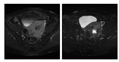
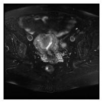
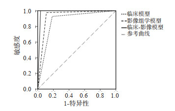
Objective To investigate the value of microsatellite instability (MSI) assessment model of endometrial carcinoma (EC) established by clinical factors combined with imaging omics. Methods Sixty- eight patients with EC diagnosed by preoperative MR imaging and postoperative pathological examination from June 2018 to January 2022 were retrospectively analyzed, with imaging and clinicopathologic data collected. Patients were divided into unstable group (n=27) and stable group (n=41) according to microsatellite stability. Logistic regression analysis was used to screen the clinical factors. ITK-SNAP software was used to delineate the region of interest of the lesion and extract the image omics characteristics. The minimum absolute contraction and selection operator algorithm were used for feature dimension reduction. ROC curve was plotted to assess the predictive power of the image-omics model, the clinical model, and the combined clinical-imaging model, and Delong test was used to compare the predictive power of the three models. Results Logistic regression analysis showed that mismatch repair protein MutL homolog 1, post-meiotic segregation protein 2 expression, tumor differentiation and depth of myometrial invasion were clinical risk factors of EC MSI. Six image-omics features were screened for construction of imageomics models (P < 0.05). Through ROC curve analysis, the combined clinic-image model had good prediction and evaluation performance in EC MSI, and the AUCs of the clinical model, the image-omics model, and the combined clinic-image model were 0.871, 0.932 and 0.981, respectively. The results of Delong test showed that the difference between the clinical imaging combined model and the imaging omics model and the clinical model was statistically significant (Z=1.933, 2.735, P=0.046, 0.006). Conclusion The assessment model established by clinical factors combined with MR imaging omics characteristics has a good predictive value for EC MSI.
2023, 46(1): 53-57.
doi: 10.12122/j.issn.1674-4500.2023.01.10
Abstract:
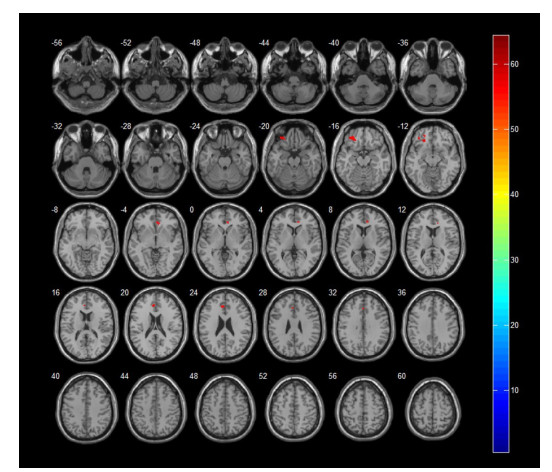
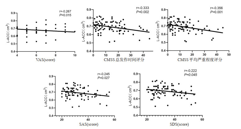
Objective To investigate voxel- based morphometry in primary dysmenorrhea patients with altered brain gray matter volume and the correlation between it and clinical scale scores. Methods Forty-one patients with primary dysmenorrhea and 41 healthy controls who met the inclusion criteria were collected to undergo high-resolution T1 structural imaging scans during days 1-3 of menstruation They completed the dysmenorrhea severity scale, the COX dysmenorrhea symptom scale, the anxiety self-assessment scale and the depression self-assessment scale. The SPM8 software was used to compare gray matter volumes at the whole brain level. We observed the brain areas with significant changes in gray matter volumes in both groups. Results The gray matter volume of the patients with primary dysmenorrhea was reduced compared with that of the healthy control group. The differences mainly included the left orbital middle frontal gyrus, left anterior cingulate gyrus and right anterior cingulate gyrus (P < 0.001), but no brain regions with a significant increase in gray matter volume were found. There were significant differences in dysmenorrhea severity scale, the COX dysmenorrhea symptom scale, the anxiety self-assessment scale and the depression self-assessment scale scores between the dysmenorrhea group and the healthy group (P < 0.001). Moreover, the left anterior cingulate gyrus was negatively correlated with the scores of dysmenorrhea severity scale, the COX dysmenorrhea symptom scale, the anxiety self-assessment scale and the depression selfassessment scale. Conclusion Based on voxel- based morphometry analysis, we can accurately assess the gray matter microstructural changes in the brain of patients with primary dysmenorrhea, and these brain regions with reduced gray matter volume are mainly involved in brain regions related to pain transmission, processing and emotion regulation. The changes in gray matter volume may play an important role in the occurrence and persistence of primary dysmenorrhea.
2023, 46(1): 58-62.
doi: 10.12122/j.issn.1674-4500.2023.01.11
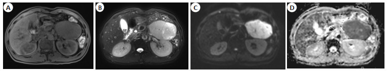
Abstract:
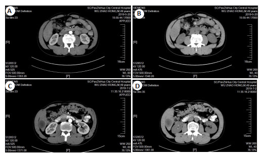
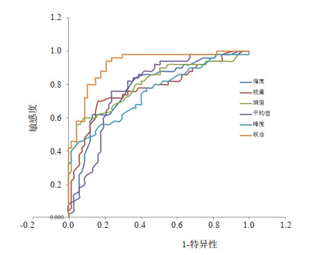
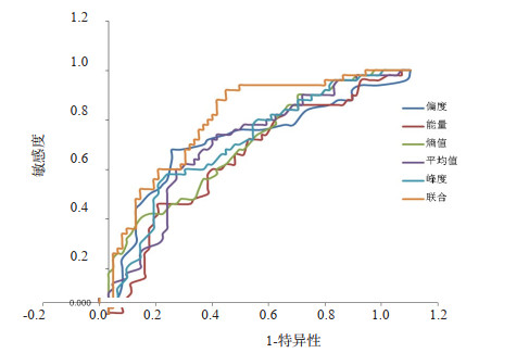
Objective To explore the changes in the contrast-enhanced CT texture parameters such as skewness, energy and entropy before and after drug-eluting beads transarterial chemoembolization (DEB-TACE) for hepatitis B virus-related hepatocellular carcinoma (HBV-related HCC), and discuss their clinical significance. Methods A total of 117 patients with HBV-related HCC in our hospital from June 2018 to June 2021 were enrolled. All patients received DEB-TACE treatment. Contrast-enhanced CT scans were performed before and 6 weeks after treatment, and the textural parameters were recorded. The value of contrast-enhanced CT texture parameters in evaluating the efficacy of DEB-TACE treatment was analyzed. Results Compared with data before treatment, the skewness, entropy, mean and kurtosis of enhanced CT texture parameters in HCC patients were decreased, and energy was increased in patients after DEB-TACE treatment, with statistical difference (P < 0.05). Of 117 patients, 86 patients had good prognosis and 31 had poor prognosis after treatment. The skewness, entropy, mean and kurtosis were decreased in patients with different prognosis status, and the energy was increased in patients after DEB-TACE treatment (P < 0.05). The skewness, entropy, mean and kurtosis of good prognosis group were lower, and energy was higher than those of poor prognosis group before and after treatment (P < 0.05). The textural parameters before DEB-TACE treatment were all highly effective in predicting the prognosis of HCC patients (AUC>0.75), and the predictive value of combined detection of all parameters was the highest (AUC=0.920, 95%CI: 0.870-0.970). The textural parameters after DEBTACE treatment were also all highly effective in predicting the prognosis of HCC patients (AUC>0.70). The predictive value of combined detection of all parameters was the highest (AUC=0.810, 95% CI: 0.731-0.888). Conclusion Contrast-enhanced CT texture parameters are valuable in assessing the prognosis of HCC patients underwent DEB-TACE treatment, which can provide a reference for clinical HCC treatment.
2023, 46(1): 63-69.
doi: 10.12122/j.issn.1674-4500.2023.01.12
Abstract:
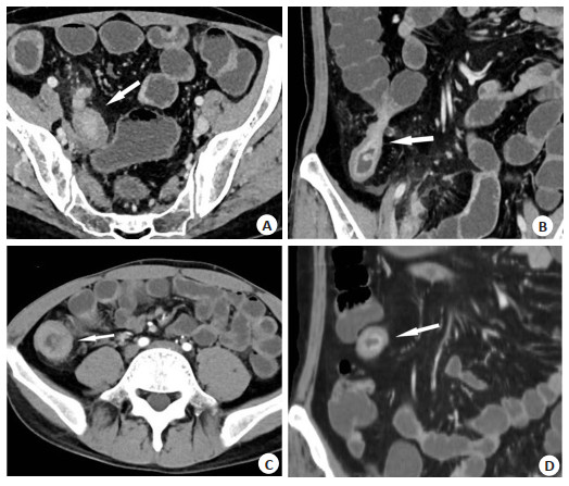
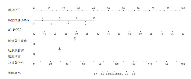
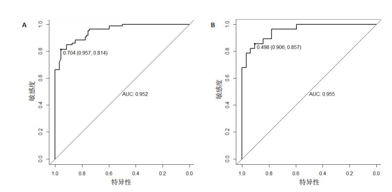
Objective To explore the CT enterography characteristics of moderate to severe Crohn's disease (CD) and structure a nomogram to predict moderate to severe CD. Methods A total of 180 patients with CD admitted to the Affiliated Hospital of Yangzhou University from January 2019 to June 2022 were randomly divided into modeling group (n=120) and validation group (n=60). The activity of CD was assessed on the basis of the Simplified Endoscopic Score for Crohn's Disease and subsequently divided into two groups: remission to mild (n=94) and moderate to severe (n=86). The CT enterography signs of the diseased intestinal segment of the two groups were compared and analyzed. The model was constructed using R package of Rstudio4.1.2 software. ROC curve, calibration curve and clinical decision-making curve were drawn. Results Binomial Logistic regression analysis showed mural thickness (OR=1.746, 95% CI: 1.085-2.811), ΔV-P (difference in CT value of intestinal wall in venous phase and plain scan) (OR=1.148, 95% CI: 1.062-1.241), mural stratification and hyperenhancement (OR=14.183, 95%CI: 3.737-53.824) and increased mesenteric fat density (OR=5.332, 95% CI: 1.278-22.246) were independent parameters for the diagnosis of moderate to severe CD. The areas under the ROC curve of the model in the modeling group and the validation group was 0.952 (95% CI: 0.925-0.979, P < 0.05) and 0.955 (95% CI: 0.911-0.997, P < 0.05). The calibration curve of the model fitted well with the ideal curve, and the clinical decision-making curve has indicated large net benefit within a certain threshold range. Conclusion Mural thickness, ΔV-P, Mural stratification and hyperenhancement and Increased mesenteric fat density are independent parameters for determining moderate to severe stage of the disease and the correspondingly constructed nomogram can predict disease activity in moderate-to-severe CD.
2023, 46(1): 70-73.
doi: 10.12122/j.issn.1674-4500.2023.01.13
Abstract:
Objective To explore the effect of MRI labyrinth hydroneuroimaging in the diagnosis of trigeminal neuralgia and neurovascular compression. Methods Retrospective analysis was performed on clinical data of 168 patients with trigeminal neuralgia in the hospital. All patients were confirmed by surgical treatment and performed MRI labyrinth hydroneuroimaging. The imaging examination results of neurovascular compression were analyzed and compared with the surgical results. The morphological parameters of the trigeminal cistern segment (maximum length and area of the trigeminal cisternal segment, trigeminal nerve-pontine angle, cerebellopontine angle cistern section) were compared among the patients. Results Among the 168 patients with trigeminal neuralgia, 80 cases were compressed on the left side by MRI imaging detection, which was 90.91% consistent with the clinical distribution of left trigeminal neuralgia, and 74 cases were compressed on the right side which was 92.50% consistent with the clinical distribution of right trigeminal neuralgia. No compression was observed in 14 cases. There was no significant difference in the compression position between the affected and healthy sides (P> 0.05). The main compression artery was the middle superior cerebellar artery, accounting for 70.83% (119/168). The degree of compression on the affected side was higher (P < 0.05). There was no significant difference in the maximum length of trigeminal nerve between the two sides (P>0.05), and the cross-sectional area of trigeminal nerve, trigeminal nerve-pontine angle and cross-sectional area of cerebellopontine angle cistern were smaller in the affected side (P < 0.05). Conclusion MRI labyrinth hydroneuroimaging can effectively diagnose trigeminal neuralgia, show the specific conditions around the nerve, judge the morphological changes of the trigeminal cisternal segment. It can provide a basis for the diagnosis of neurovascular compression in clinical trigeminal neuralgia.
2023, 46(1): 74-77.
doi: 10.12122/j.issn.1674-4500.2023.01.14
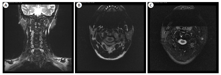
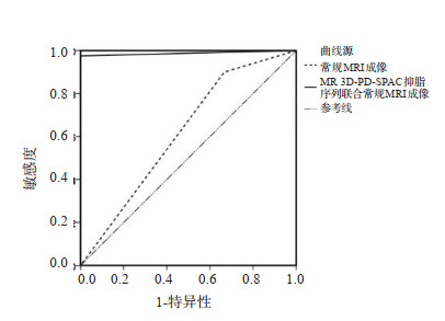
Abstract:
Objective To analyze the diagnostic value of magnetic resonance three- dimension sampling perfection with application optimized contrasts using different flip angle evolution (MR 3D-PD-SPACE) fat saturation sequence combined with conventional MRI for atlantoaxial subluxation. Methods A total of 43 patients with suspected atlantoaxial subluxation admitted to our hospital from January 2017 to January 2020 were selected. All patients received MR 3D-PD-SPACE fat saturation sequence combined with conventional MRI examinations. Taking spiral CT angiography in surface displaytechnique as gold standard, the positive rates of the two examination methods were compared. ROC curve was drawn to analyze the value of MR 3D-PD-SPACE fat saturation sequence combined with conventional MRI in the diagnosis of atlantoaxial subluxation. Results The gold standard identified 40 patients with atlantoaxial subluxation in 43 suspected cases with a positive rate of 93.02%. MRI diagnosis of atlantoaxial subluxation screened 36 cases, with a positive rate of 83.72%, while MR 3D-PD-SPACE fat saturation sequence combined with conventional MRI screened 39 cases, with a positive rate of 90.69%. The sensitivity and specificity of MR 3D-PD-SPACE fat saturation sequence combined with conventional MRI for the diagnosis of atlantoaxial subluxation were 97.5% and 100.0%, while the sensitivity and specificity of conventional MRI for the diagnosis of atlantoaxial subluxation were 90.00% and 33.3%, The diagnostic value of MR 3D-PD-SPACE fat saturation sequence combined with conventional MRI was significantly better than that of MRI diagnosis (P < 0.05). Conclusion MR 3DPD-SPACE fat saturation sequence combined with conventional MRI is of great clinical value in the diagnosis of atlantoaxial subluxation.
2023, 46(1): 78-82.
doi: 10.12122/j.issn.1674-4500.2023.01.15
Abstract:
Objective To explore the MRI imaging analysis of gastrointestinal stromal tumors and the differential diagnosis of benign and malignant tumors by apparent dispersion coefficient (ADC) images. Methods This study was a prospective study, 120 patients with gastrointestinal stromal tumor who were treated in our hospital from October 2019 to October 2021 were selected. Disease severity: 11 patients with very low risk, 22 patients with low risk, 41 patients with medium risk and 46 patients with high risk. According to the analysis of pathological results, 35 patients with benign tumor and 85 patients with malignant tumor were enrolled. MRI examination was performed on all patients after enrollment, and the differences of imaging characteristics and ADC values between benign group, malignant group and patients with different risk were compared. Results The differences of ADC, variation, skewness, maximum, minimum, total distance, median, 10% value, 90% value and evenness between benign group and malignant group were statistically significant (P < 0.05). There were statistically significant differences among ADC, variance, skewness, maximum, minimum, total distance, median, 10% value, 90% value and evenness of patients with very low risk, low risk, medium risk and high risk group (P < 0.05). The differences of maximum diameter, tumor morphology and boundary between benign group and malignant group were significant(P < 0.05). The differences in maximum diameter, tumor morphology and boundary among patients with different risk levels were statistically significant (P < 0.05). Conclusion There are significant differences in the maximum diameter, tumor morphology and boundary in MRI imaging of benign and malignant gastrointestinal stromal tumors with different severity, and their ADC images have significant differential diagnosis significance for benign and malignant tumors.
2023, 46(1): 83-87.
doi: 10.12122/j.issn.1674-4500.2023.01.16
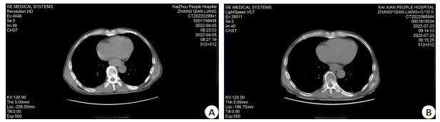
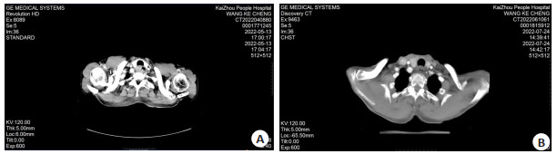
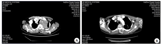
Abstract:
Objective To investigate the efficacy and CT manifestations of intensity-modulated radiotherapy (IMRT) combined with chemotherapy in the treatment of esophageal cancer. Methods A total of 134 patients with esophageal cancer admitted to Chongqing Kaizhou District People's Hospital from May 2020 to May 2021 were selected. They were randomly divided into concurrent chemoradiotherapy group (n=68) and combined with intensity-modulated radiotherapy group (n=68). The concurrent chemoradiotherapy group was given three-dimensional conformal radiotherapy combined with chemotherapy, while the combined with intensity-modulated radiotherapy group was given IMRT combined with chemotherapy. The clinical efficacy and CT manifestations, immunoglobulin [immunoglobulin A (IgA), IgM and IgG], vascular endothelial growth factor (VEGF) and its receptor 1 (VEGFR-1), serum tumor markers (CEA, SCC-AG, CA125) and adverse reactions were compared between the two groups. Results The total effective rate of combined with intensity-modulated radiotherapy group was significantly higher than that of concurrent chemoradiotherapy group (P < 0.05). CT could be used to evaluate the clinical efficacy of patients. After treatment, compared with the concurrent chemoradiotherapy group, the levels of IgA, IgM and IgG in the combined with intensity-modulated radiotherapy group were significantly higher (P < 0.05), the levels of VEGF and VEGFR-1 were significantly lower (P < 0.05), and the levels of CEA, SCC-AG and CA125 were significantly lower (P < 0.05). There was no difference in the total incidence of adverse reactions between the two groups (P>0.05). Conclusion IMRT combined with chemotherapy has good efficacy in the treatment of esophageal cancer patients and less effect on the level of immunoglobulin. It can also down-regulate the levels of VEGF and VEGFR-1, reduce the level of serum tumor markers, and has good safety.
2023, 46(1): 88-92.
doi: 10.12122/j.issn.1674-4500.2023.01.17
Abstract:
Objective To analyze the diagnostic efficiency of cervical contrast-enhanced CT on cervical lymph node metastasis in thyroid cancer. Methods The clinical data of 76 patients with papillary thyroid carcinoma were retrospectively analyzed. Taking the pathological diagnosis result as the gold standard, the diagnostic value of contrast-enhanced CT examination on cervical lymph node metastasis in thyroid cancer was analyzed. Results Pathological examination confirmed 153 cervical lymph node metastases and 533 non-metastases, with 60.13% central lymph node metastasis. Taking pathological diagnosis as the gold standard, the accuracy rate of contrast-enhanced CT was 83.61% in diagnosing central cervical lymph node metastasis and was 89.26% in the diagnosis of lateral cervical lymph node metastasis. Contrast-enhanced CT showed that the average short diameter of cervical metastatic lymph nodes in patients was significantly higher than that in non-metastatic lymph nodes (8.91±2.62 mm vs 8.02±2.51 mm). There were significant differences between metastatic and non-metastatic lymph nodes in terms of short diameter >10 mm, obvious enhancement, obvious heterogeneous enhancement, cystic degeneration, calcification in the lesion and surrounding tissue invasion (P < 0.05). Conclusion The diagnostic efficiency of contrast-enhanced CT for central cervical lymph node metastasis in thyroid cancer is lower than that in the lateral cervical lymph node metastasis. It is recommended to combine ultrasound or MRI for comprehensive evaluation.
2023, 46(1): 93-97.
doi: 10.12122/j.issn.1674-4500.2023.01.18
Abstract:
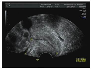
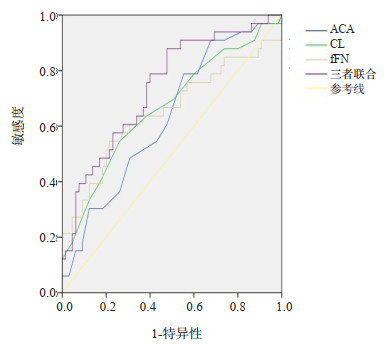
Objective To analyze the clinical value of the transvaginal ultrasound measurement of the anterior uterocervical angle (ACA) and cervical length (CL) combined with fetal fibronectin (fFN) for predicting spontaneous preterm birth (SPB). Methods Ninety-eight pregnant women with threatened preterm birth were selected as research objects from June 2019 to June 2022. They were divided into SPB group (n=33) and full-term group (n=65) according to whether SPB occurred. Univariate analysis and multivariate logistic regression analysis were used to analyze the risk factors of SPB in pregnant women with threatened premature labor. The predictive efficiency of ACA, CL and fFN to SPB were evaluated by drawing ROC curve. Results Univariate analysis showed that there were statistically significant differences in pre-pregnancy BMI, premature rupture of membranes, intrauterine infection, excessive amniotic fluid, gestational diabetes and ACA, CL and fFN levels between the two groups (P < 0.05). Multivariate logistic regression analysis showed that premature rupture of membranes, ACA and fFN were risk factors for SPB in pregnant women with threatened premature labor (OR>1, P < 0.05). BMI and CL before pregnancy were protective factors for SPB in pregnant women with threatened premature labor (OR < 1, P < 0.05). ROC analysis showed that when ACA ≥116.500°, the area under curve (AUC) of predicting SPB in pregnant women with threatened preterm birth was 0.630, the sensitivity was 48.5%, and the specificity was 69.2%. When CL ≤25.000 mm, the AUC was 0.667, the sensitivity was 54.5%, and the specificity was 73.8%. When fFN ≥96.155 μg/L, the AUC was 0.652, the sensitivity was 60.6%, and the specificity was 69.2%. The AUC of combined prediction was 0.740, the sensitivity was 72.7%, and the specificity was 75.4%. Conclusion The ACA, CL and fFN measured by transvaginal ultrasound have a certain predictive value for predicting SPB in pregnant women with threatened premature labor, and the combined predictive efficacy of the three is better.
2023, 46(1): 98-102.
doi: 10.12122/j.issn.1674-4500.2023.01.19
Abstract:
Objective To investigate the effects of Jiangu capsule combined with alendronate sodium on bone mineral density, serum tyrosine YWHAH and inflammatory indexes in elderly patients with osteoporosis. Methods Ninety-eight elderly patients with osteoporosis were selected, and they were divided into control group (n=49) and observation group (n=49) by random number table method. The control group was treated with alendronate sodium, and the observation group was given Jiangu capsule on the basis of the control group. After 6 months, bone mineral density, TCM syndrome score, pain intensity and serum levels of YWHAH, osteocalcin, alkaline phosphatase, TNF-α and IL-6 were compared between the two groups. Results After treatment, the bone mineral density of lumbar vertebra and femoral neck in two groups were higher than that before treatment (P < 0.05). After treatment, the TCM syndrome score of the observation group was lower than that before treatment. After treatment, the resting and activity VAS scores of the observation group was lower than that before treatment (P < 0.05). After treatment, the level of serum YWHAH in the observation group was lower than that before treatment (P < 0.05). After treatment, the serum osteocalcin level in the observation group was higher than that before treatment(P < 0.05), and the serum alkaline phosphatase level in the observation group was lower than that before treatment(P < 0.05). After treatment, TNF-α and IL-6 in the observation group was lower than that before treatment (P < 0.05). Conclusion On the basis of alendronate sodium, combined with Jiangu capsule can effectively improve bone mineral density and relieve pain in elderly patients with osteoporosis. The mechanism may be related to down-regulating serum YWHAH level, improving bone metabolism indexes and alleviating inflammatory response.
2023, 46(1): 103-107.
doi: 10.12122/j.issn.1674-4500.2023.01.20
Abstract:
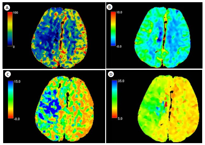
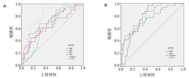
Objective To explore the changes in CT perfusion imaging parameters in patients with acute ischemic stroke (AIS) after thrombolytic therapy and their early predictive value on prognosis. Methods The clinical data of 90 patients with AIS in the hospital were enrolled from March 2019 to March 2022 for retrospective analysis. All patients were followed up for 3 months with modified Rankin scale (mRS) score and grouped according to the results. Patients with MRS≤2 points were assigned to good prognosis group (n=31), and patients with score >2 points were classified into poor prognosis group (n=59). The penumbra CT perfusion imaging parameters including cerebral blood flow (CBF), cerebral blood volume (CBV), mean transit time (MTT) and time to peak (TTP) were compared between the two groups after thrombolytic therapy. The healthy side was used as the control to calculate the relative values of rCBF, rCBV, rMTT and rTTP, and the predictive value of each parameter on the prognosis of AIS patients was analyzed. Results After thrombolytic therapy, the CBF, CBV, rCBF and rCBV in penumbra in poor prognosis group were significantly lower than those in good prognosis group (P < 0.05).The MTT and TTP were significantly longer than those in good prognosis group (P < 0.05), but there were no statistical differences in rMTT and rTTP between the two groups (P>0.05). The AUCs of CBF, CBV, MTT and TTP in predicting the prognosis of AIS patients were 0.701, 0.662 and 0.698 respectively. The AUC, sensitivity and specificity of the combined prediction of the four parameters were 0.817, 93.55% and 64.41%. The AUCs of rCBF and rCBV in predicting the prognosis of AIS patients were 0.724 and 0.798 respectively. The AUC, sensitivity and specificity of the combined prediction of the two parameters were 0.812, 96.77% and 57.63%. Conclusion CT perfusion imaging can quantitatively analyze the blood perfusion level in the penumbra of AIS patients, and the detection of CBF, CBV, MTT and TTP parameters after thrombolytic therapy and their relative values in brain tissue in the mirror region of the healthy side can provide a reference for early assessment of prognosis of patients.
2023, 46(1): 108-114.
doi: 10.12122/j.issn.1674-4500.2023.01.21
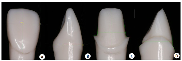
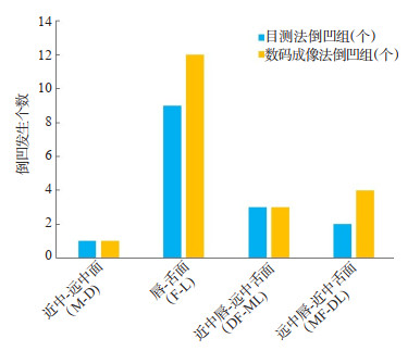
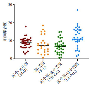
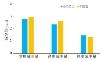
Abstract:
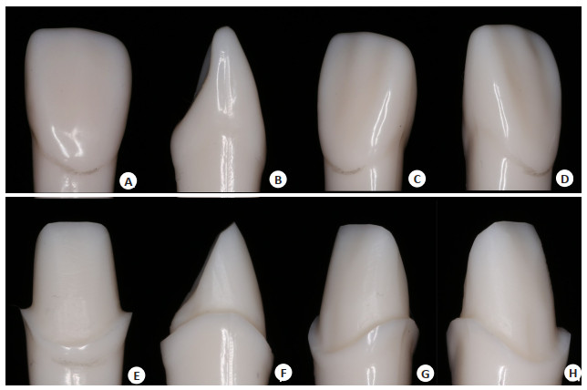
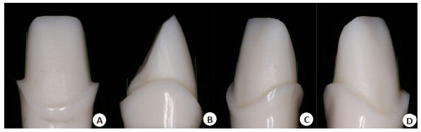
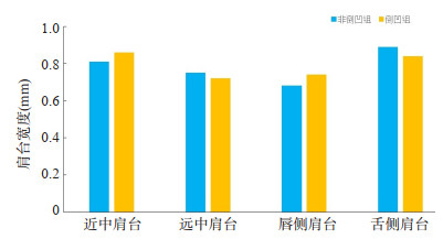
Objective To explore the multi-plane total occlusal convergence measurement and undercut analysis of all‐ceramic crown preparation. The dental preparation operation is targeted to guide residents, so as to improve the operation skills of fixed restoration. Methods Thirteen residents participated in standardized training in Beijing Shijitan Hospital Affiliated to Capital Medical University from 2019 to 2022. According to the requirement of all-ceramic crown, 2-3 right maxillary central incisors were prepared on the standard tooth model of head mold. Finally, 37 preparers conforming to the process were collected. Four different planes of the preparation were determined by visual and digital imaging methods (mesial-distal surface, labial-lingual surface, mesial labial-distal lingual surface, distal labial-mesial lingual surface) to measure total occlusal convergence and undercut. According to the results of the digital imaging method, the preparation was divided into undercut group and non-undercut group. Whether the existence of undercut is related to the height, width, thickness reduction and shoulder width of the preparation were analyzed. Results The analysis of undercut observation showed that there were differences between the visual method and the digital imaging method. The data of total occlusal convergence by digital imaging showed that the undercut ratio of the four different plane preparations was statistically different (χ2=16.188, P < 0.05). Bonferroni correction was used for comparison. The labial-lingual surface undercut ratio was higher than that of other planes (P < 0.05). There was no statistical difference between the undercut group and the non-undercut group in the height and width reduction of the mesial-distal surface, the thickness reduction of the labial-lingual surface, and the shoulder width of the mesial, distal, labial and lingual sides (P>0.05). Conclusion There is undercut phenomenon in the axial plane of the preparation. Compared with the visual method, the digital imaging method could more accurately judge the degree of undercut and total occlusal convergence.
2023, 46(1): 115-119.
doi: 10.12122/j.issn.1674-4500.2023.01.22
Abstract:
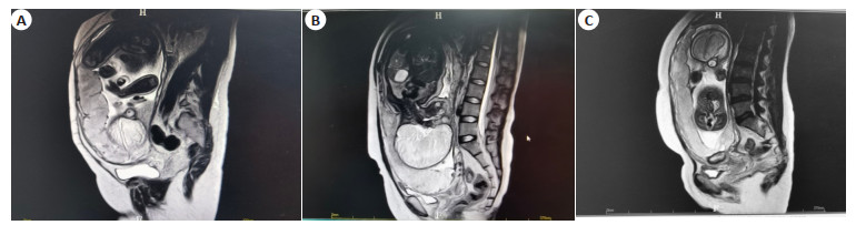
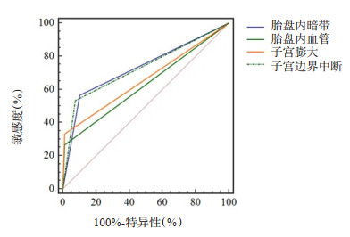
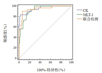
Objective To analyze the value of serum creatine kinase (CK), soluble vascular endothelial growth factor receptor-1 (sFlt-1) combined with MRI examination in evaluating placenta accreta. Methods A total of 108 patients with suspected placenta accreta who were admitted to our hospital from April 2015 to April 2021 were selected. According to the results of postpartum pathological diagnosis, the patients were divided into placenta accreta group (n=30) and non-placenta accreta group (n=78). Prenatal serum CK and sFlt-1 levels of all pregnant women were detected, and MRI was performed. The serum CK and sFlt-1, and the incidence of abnormal signs on MRI (intraplacental dark zone, intraplacental blood vessels, uterine enlargement, and uterine border interruption) were compared between the two groups. Pearson correlation was used to analyze the correlation between serum CK and sFlt-1 levels and placenta accreta. Spearman correlation was used to analyze the correlation between placental dark zone, intraplacental blood vessels, uterine enlargement, uterine border interruption and placenta accreta. The clinical value of placenta accreta was evaluated by ROC curve of serum CK and sFlt-1 levels combined with MRI examination. Results The serum CK in the placenta accreta group was higher than that in the non-placenta accreta group, and the sFlt-1 was lower than that in the non-placenta accreta group (P < 0.05). The uterine enlargement and the incidence of uterine border interruption were higher than those in the non-placenta accreta group (P < 0.05). Correlation showed that serum CK, intraplacental dark zone, intraplacental blood vessels, uterine enlargement, and uterine border disruption were positively correlated with placenta accreta (r=0.503, 0.511, 0.416, 0.422, 0.436, P < 0.05). Serum sFlt-1 level was negatively correlated with placenta accreta (r=-0.486, P < 0.05). The results of ROC curve analysis showed that the AUC of serum CK and sFlt-1 levels combined with MRI in evaluating placenta accreta was 0.945, the sensitivity was 90.0%, and the specificity was 93.6%, which was significantly better than the evaluation of each index alone. Conclusion Serum CK and sFlt-1 levels and MRI signs such as dark zone in placenta, intraplacental blood vessels, uterine enlargement and uterine border interruption are closely related to the occurrence of placenta accreta. Serum CK and sFlt-1 levels combined with MRI have significant clinical value in placenta accreta.
2023, 46(1): 120-123.
doi: 10.12122/j.issn.1674-4500.2023.01.23
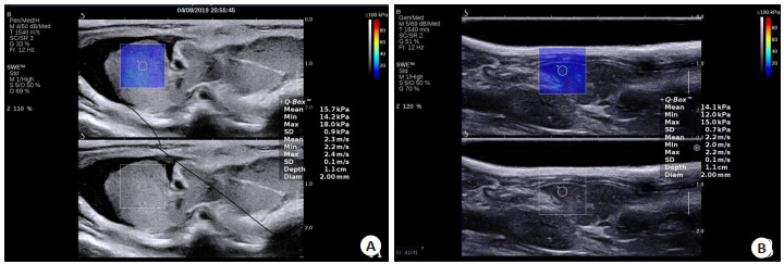
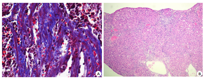
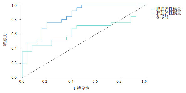
Abstract:
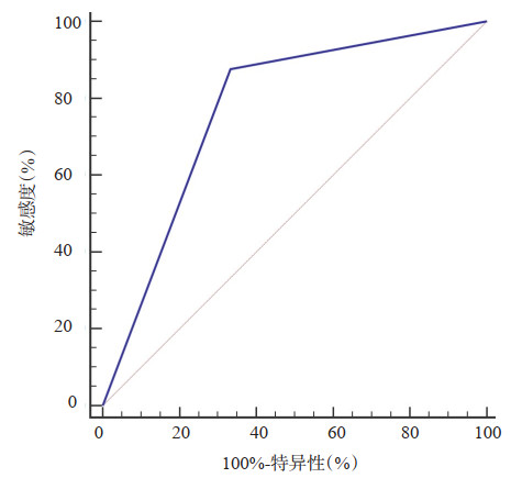
Objective To analyze the value of spleen stiffness (SS) and liver stiffness (LS) in the evaluation of clinically significant portal hypertension by two-dimensional shear wave elastic imaging (2D-SWE). Methods Fifty SD rats were randomly divided into portal hypertension group (PH group, n=25) and normal control group (NC group, n=25). The animal model of PH group was established by CCl4 induction method, and the NC group was only injected with corn oil. SS and LS of rats were detected by 2D-SWE, portal vein pressure (PVP) was measured by direct portal vein puncture and the pathological changes of spleen and liver were observed at 12 weeks. Furthermore, the correlation between SS, LS and PVP was analyzed, and the area under ROC curve was obtained by using PVP as the gold standard. Results SS and LS were positively correlated with PVP, but the correlation between SS and PVP (r=0.797, P < 0.001) was stronger than LS (r=0.505, P < 0.001). With PVP≥10 mmHg as the gold standard, the area under ROC curve of SS and LS detected by 2D-SWE were 0.875 and 0.673 respectively, which indicated that SS was better than LS (P < 0.05). Conclusion The diagnostic efficacy of 2D-SWE measurement of SS in evaluating clinically significant portal hypertension is better than LS.
2023, 46(1): 124-128.
doi: 10.12122/j.issn.1674-4500.2023.01.24
Abstract:
Objective To explore the relationship between CT signs and Ki-67, p53 in peripheral lung cancer (PLC). Methods A total of 108 patients with PLC treated in the hospital were enrolled between January 2016 and February 2022. CT signs were examined by Philips 256-slice spiral CT. The expression levels of Ki-67 and p53 in PLC tissues were detected. The relationship between pathological tissue types, differentiation degree, CT signs and Ki-67, p53 was analyzed. Results In PLC tissues, positive expression rates of Ki- 67 and p53 were 69.44% (75/108) and 51.85% (56/108), respectively. The main CT findings included pleural indentation, tumor diameter ≥3.0 cm, lobulation sign, spiculate protuberance sign, spicule sign, enhancement value ≥20 Hu, hilar and mediastinal lymph node enlargement, while CT findings (air bronchus sign, cavity sign, glass sign, vacuole sign, calcification sign) were relatively rare. The positive expression rate of Ki-67 in adenocarcinoma was lower than that in squamous cell carcinoma (P < 0.05), but there was no significant difference in the positive expression rate of p53 between adenocarcinoma and squamous cell carcinoma (P > 0.05). The lower the differentiation degree of PLC, the higher the positive expression rates of Ki-67 and p53 (P < 0.05). The positive expression rate of Ki-67 was higher in patients with tumor diameter ≥ 3.0 cm, lobulation sign, spiculate protuberance sign, spicule sign, enhancement value ≥20 Hu, hilar and mediastinal lymph node enlargement, while which was lower in patients with glass sign and vacuole sign (P < 0.05). The positive expression rate of p53 was higher in patients with tumor diameter ≥3.0 cm, lobulation sign, spiculate protuberance sign, spicule sign, enhancement value ≥20 Hu, hilar and mediastinal lymph node enlargement (P < 0.05). Conclusion The expressions of Ki-67 and p53 are closely related to CT signs in PLC patients.

2023, 46(1): 129-134.
doi: 10.12122/j.issn.1674-4500.2023.01.25
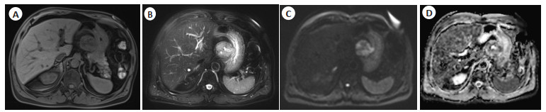
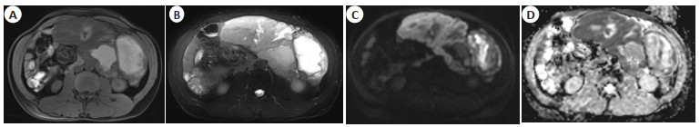
Abstract:
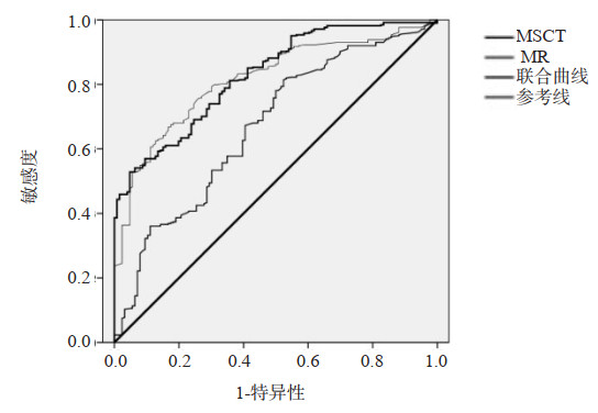
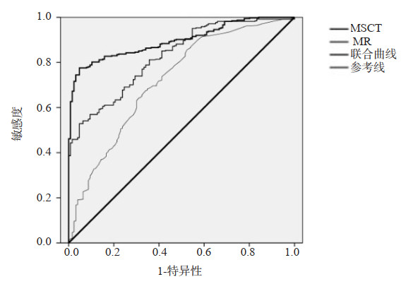
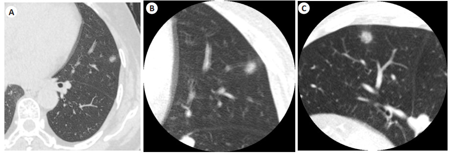
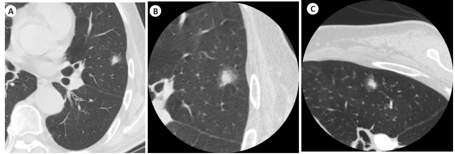
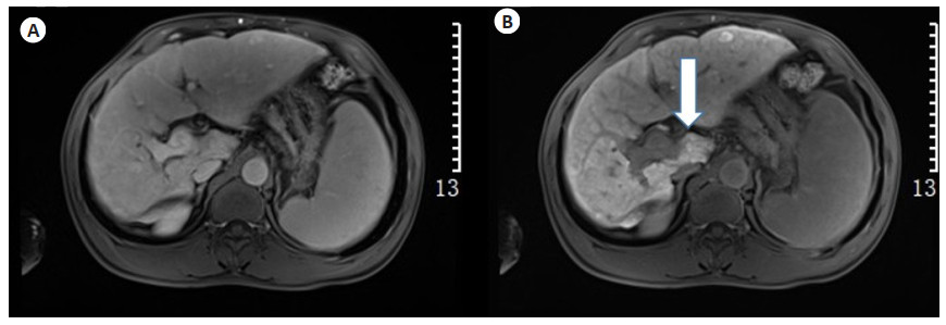
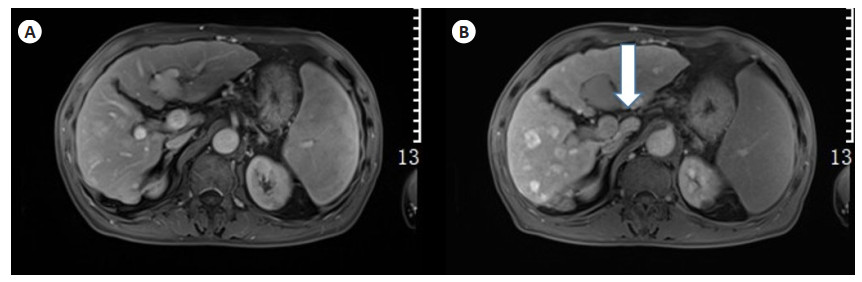
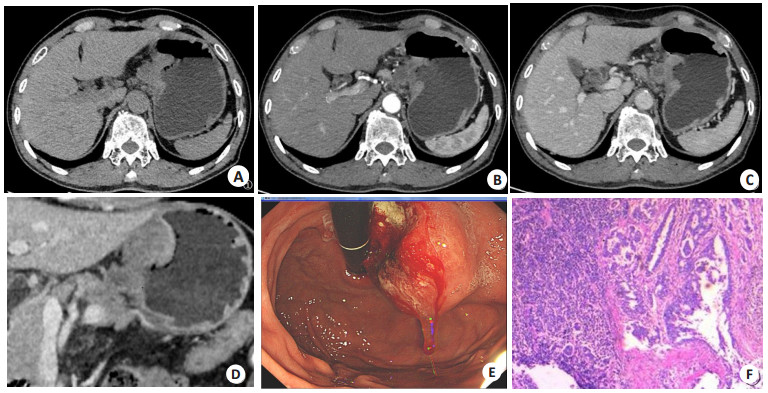
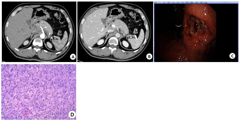
Objective To investigate the difference between MSCT and MRI in the pathological grading and assessment of tumor blood supply in pancreatic neuroendocrine tumors. Methods In this prospective study, 70 patients with pancreatic neuroendocrine tumors diagnosed and treated in our hospital from January 2019 to June 2022 were selected as the research objects, including 25 patients with G1 grade, 23 patients with G2 grade, and 22 patients with G3 grade, as well as 29 patients with blood-rich pancreatic neuroendocrine tumors. Another 70 healthy volunteers who underwent physical examination at the same time were selected as the control group. The differences of MSCT and MR indexes between the two groups, patients with different tumor grades, and patients with different tumor blood supply were compared. The diagnostic significance of MSCT and MRI in the pathological grade and assessment of tumor blood supply in pancreatic neuroendocrine tumors were analyzed. Results The CT absolute enhancement values of arterial phase and portal phase in the observation group were significantly lower than those in the control group (P < 0.05). The high signal in arterial phase and portal phase in the observation group were significantly lower than those in the control group (P < 0.05). There were significant differences in CT absolute enhancement value, margin, shape, cystic degeneration and necrosis, main pancreatic duct dilatation and distant metastasis in arterial phase and portal phase of patients with different grades and blood supplies by MSCT (P < 0.05). There were significant differences in high signal in arterial phase and portal phase, margin, shape, cystic necrosis, main pancreatic duct dilatation and distant metastasis in patients with different grades and blood supplies by MRI (P < 0.05). The specificity of combination diagnosis of MSCT and MRI was significantly higher than that of single detection (P < 0.05). Conclusion MSCT and MRI have positive significance in pathological grading and assessment of tumor blood supply in pancreatic neuroendocrine tumors.
2023, 46(1): 135-138.
doi: 10.12122/j.issn.1674-4500.2023.01.26
Abstract:
Objective To explore the application of two-dimensional speckle tracking technique in assessing the fetal cardiac function in patients with thyroid dysfunction during pregnancy. Methods Sixty-three pregnant women with hyperthyroidism and another 55 healthy pregnant women underwent prenatal examination in our hospital from August 2021 to June 2022 were enrolled as study group and control group, respectively. Two-dimensional speckle tracking technology was used to detect fetal cardiac function. Comparison was conducted on parameters including right ventricular end-diastolic area (RVDA), left ventricular end-diastolic area (LVDA), right ventricular end-diastolic circumference (RVDC), left ventricular end-diastolic circumference (LVDC), right ventricular end-systolic area (RVSA), left ventricular end-systolic area (LVSA), right ventricular end-systolic circumference (RVSC), left ventricular end-systolic circumference (LVSC), tricuspid valve velocity (TV), mitral valve velocity (MV), peak E-wave velocity, peak A-wave velocity, E/A ratio, left ventricular shortening fraction 1 (LVSF1), left ventricular shortening fraction 2 (LVSF2), right ventricular systolic fraction 1 (RVSF1), right ventricular systolic fraction 2 (RVSF1). The fetal cardiac structures were observed, such as ventricular septal defect, tetralogy of Fallot, pulmonary stenosis, endocardial cushion defects, coarctation of aorta and tricuspid atresia. Results The differences of age, gestational weeks, BMI, systolic blood pressure, diastolic blood pressure and other clinical data between two groups were not significant (P > 0.05). LVSA, RVSA, RVSC and LVSC at end-systolic stage in study group were significantly higher than those in control group (P < 0.05), while LVDA, RVDA, RVDC and LVDC at end-diastolic stage were significantly higher than those in control group (P < 0.05). The fetal diastolic cardiac function indexes MVA, MVE, MV-E/A, TVA, TVE and TV-E/A in study group were significantly higher than those in control group (P < 0.05), and the end-systolic cardiac function indexes LVSF1, RVSF1, LVSF2 and RVSF2 were also higher than those in control group (P < 0.05). There were 9 cases (14.29%) of fetal cardiac structural abnormalities in study group, and the total incidence rate was significantly higher than that in control group (P < 0.05). Conclusion Fetal cardiac function is abnormal in patients with thyroid dysfunction during pregnancy, and cardiac structure may also be abnormal. Two- dimensional speckle tracking technology can accurately monitor fetal cardiac function and structure in the early stage.
2023, 46(1): 139-143.
doi: 10.12122/j.issn.1674-4500.2023.01.27
Abstract:
Objective To compare the CT imaging effects of personalized and conventional scanning schemes on pulmonary nodules≤1 cm, and explore the best scanning scheme. Methods A total of 142 patients were included from January 2020 to June 2022 according to inclusion and exclusion criteria. According to the random control table, they were randomly divided into personalized group and conventional group by balanced control method. Patients in the personalized group were subjected to rigorous hyperinspiratory training, develop personalized position and personalized scanning direction. The obtained images were compared with the conventional scan group for image quality. Results The image quality of CT plain scan, pulmonary artery phase and pulmonary vein phase in the personalized group was better than that in the conventional scan group, the difference was statistically significant (χ2=8.204, P=0.017, χ2=6.625, P=0.036, χ2=7.139, P=0.028). The image quality of CT plain scan, pulmonary artery phase and pulmonary vein phase was compared within the two groups. In the conventional group, there was a statistically significant difference in image quality between CT plain scan and pulmonary venous stage (χ2=9.188, P=0.010), but there was no statistical significance between the rest phases in the conventional group, also between all the phases in the personalized group(P > 0.05). Conclusion The personalized scanning scheme can provide stable imaging quality, and improve the image quality of pulmonary nodules≤1cm CT imaging.
2023, 46(1): 144-148.
doi: 10.12122/j.issn.1674-4500.2023.01.28
Abstract:
Objective To analyze the predictive effect of functional liver imaging score (FLIS) on liver function in gadolinium serate enhanced MRI. Methods A total of 134 patients admitted to our hospital from October 2019 to February 2022 who were diagnosed with cirrhosis or chronic liver disease (CLD) by gadolinium serate enhanced MRI were enrolled. Three parameters of FLIS hepatobiliary phase images were evaluated: hepatic parenchyma enhancement, bile excretion and portal vein signal intensity, which were divided into CLD (n=11), Child-pugh (CP) GRADE A (n=87), CP B (n=22) and CP C (n=14). Spearman rank correlation method was used to evaluate the correlation between CP score and FLIS and its components. The critical value of FLIS was obtained by ROC curve analysis to distinguish different CP categories. The relationship between patient characteristics, serum markers, FLIS, and liver decompensation were assessed using Cox proportional risk model. Results FLIS and three parameters were strongly correlated with CP score (r=-0.68, -0.60, -0.82, -0.80, P < 0.001). ROC curve analysis showed that FLIS≥5 was the optimal threshold for predicting CP CLASS A or CLD (sensitivity was 83.7%, specificity was 94.4% and the area under the curve was 0.93). FLIS < 5 was independently associated with the occurrence of first liver decompensation in patients with CP A (HR was 50.0, 95% CI: 6.2-400.4). Conclusion FLIS is strongly correlated with liver function, which can be used to grade CP. FLIS can help predict the development of first decompensation.
2023, 46(1): 149-153.
doi: 10.12122/j.issn.1674-4500.2023.01.29
Abstract:
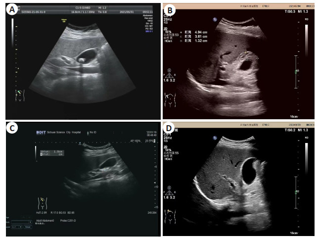
Objective To analyze the diagnostic efficacy of preoperative color Doppler flow image (CDFI) and the risk factors of postoperative recurrence in patients with cholecystolithiasis with minimally invasive cholelithotomy. Methods A total of 90 patients with cholecystolithiasis who were proposed to undergo minimally invasive gallbladder-preserving lithotomy treated in our hospital from January 2017 to January 2020 were selected. All patients underwent CDFI examination before surgery, and the manifestations of CDFI in patients with cholecystolithiasis were observed. Using surgical results as the gold standard, Kappa test was used to evaluate the consistency of the diagnostic results, ROC curve was used to evaluate the efficacy of CDFI in diagnosing gallbladder stones, and AUC was calculated. Follow-up for 2 years after operation, the recurrence of cholecystolithiasis was counted, and univariate and multivariate Logistic regression was used to analyze the high-risk factors for postoperative recurrence. Results Before operation, the gallbladder stones were irregular, round or quasi-round low signal shadows. After gallbladder hepatoprotective lithotripsy, the size was about 4.9 cm ×1.8 cm, the wall was thickened, with the thickness of 1.3 cm, and the inner wall saw dot strong echo attachment, rear accompanied by the comet tail, good sound in the cavity, gallbladder did not see obvious effusion. Among the 90 patients, 81 cases were positive and 9 cases were negative by gold standard diagnosis, 74 cases were positive and 16 cases were negative by CDFI diagnosis, the diagnostic results were in good agreement with surgery (Kappa value=0.524). The ROC curve showed that the AUC of CDFI in diagnosing gallstones was 0.840, with a sensitivity of 90.12% and a specificity of 77.78%. After 2 years of postoperative follow-up, there were 12 cases had relapsed. Univariate and multivariate Logistic regression analysis showed that the family history of cholelithiasis, the thickness of the gallbladder wall, and the number of stones were all high risk factors for postoperative recurrence in patients with cholecystolithiasis (P < 0.05). Conclusion CDFI has a good effect on the preoperative diagnosis of cholecystolithiasis patients with minimally invasive gallbladder-preserving lithotomy. Family history of cholelithiasis, thickness of gallbladder wall, and number of stones are high risk factors for postoperative recurrence in patients with cholecystolithiasis.
2023, 46(1): 154-158.
doi: 10.12122/j.issn.1674-4500.2023.01.30
Abstract:
Objective To explore the diagnostic value of multi-slice spiral CT (MSCT) with hypotonic water-filling method on TNM staging of gastric cancer. Methods A total of 116 patients with gastric cancer who came to the hospital for treatment were selected from July 2017 to July 2020. They were divided into control group and observation group by the random number table method, with 58 cases in each group. The control group underwent routine MSCT examination while the observation group was given MSCT with hypotonic water-filling method. The pathological examination result was used as the standard to analyze the accuracy rates of the two groups in the diagnosis of TNM staging of gastric cancer. Results There were no statistical differences in TNM staging results of pathological examination between the two groups (P > 0.05). The diagnostic accuracy rates of T1-T4 stages of gastric cancer and overall diagnostic accuracy rate were 80.00%, 70.59%, 70.59%, 50.00% and 70.69% in control group, 100%, 84.62%, 90.48%, 80.00% and 87.93% in observation group. The diagnostic accuracy rates of N0- N2 stages of gastric cancer and overall diagnostic accuracy rate were 84.00%, 73.91%, 73.91%, 60.00% and 75.86% in control group, which in observation group were 90.00%, 90.00%, 87.50% and 89.66%, respectively. The diagnostic accuracy rate of M staging was 82.93% in control group and 87.93% in observation group. The diagnostic accuracy rate of TN staging in observation group was higher than that in control group (P < 0.05), but there was no statistical significance in the diagnostic accuracy rate of M staging between the two groups (P > 0.05). Conclusion The accuracy of MSCT with hypotonic water-filling method in diagnosing TN staging of gastric cancer is higher than that of routine MSCT examination. The two methods are similar in diagnosing M staging of gastric cancer.
2023, 46(1): 159-163.
doi: 10.12122/j.issn.1674-4500.2023.01.31
Abstract:
Objective To explore the application value of 256-slice spiral CT combined with serum carcinoembryonic antigen (CEA), cytokeratin 19 fragment (CYFRA21-1) and precursor gastrin-releasing peptide (ProGRP) in the diagnosis of primary lung cancer. Methods Clinical data of 181 patients with primary lung cancer who were admitted from January 2020 to April 2022 were retrospectively analyzed. All the patients received 256-slice MSCT examination and detection of serum tumor markers CEA, CYFRA21-1 and ProGRP. The imaging findings and serological examination results were analyzed.The pathological diagnosis results were used as the gold standard to determine the diagnostic accuracy rate of each indicator on primary lung cancer. Results Histopathological examination showed that among the 181 patients with primary lung cancer. There were 91 cases of adenocarcinoma, 61 cases of squamous cell carcinoma and 29 cases of small cell carcinoma. CT imaging features were shown that adenocarcinoma was mainly with peripheral type, more ground glass shadow, vessel convergence sign, lobulation, spiculated changes, uneven enhancement and pleural indentation. The squamous cell carcinoma and small cell carcinoma were with mainly central type. Squamous cell carcinoma was mainly characterized by air bronchogram, vacuole sign, lobulation and even enhancement while small cell carcinoma was mainly characterized by air bronchogram, vacuole sign and even enhancement. The proportion of lobulation sign of squamous cell carcinoma was higher than small cell carcinoma (P < 0.05). The proportion of even enhancement of small cell carcinoma was higher than squamous cell carcinoma (P < 0.05). All types of lung cancer were accompanied by a small amount of calcification. The levels of CEA and CYFRA21-1 in patients with adenocarcinoma or squamous cell carcinoma were higher than those in patients with small cell carcinoma while the ProGRP level was significantly lower than that in patients with small cell carcinoma. The CEA level in patients with adenocarcinoma was significantly higher than that in patients with squamous cell carcinoma. The positive detection rate of MSCT or CEA in patients with adenocarcinoma was higher than CYFRA21-1 and ProGRP, the positive detection rate of MSCT or CYFRA21-1 in patients with squamous cell carcinoma was higher than CEA and ProGRP. The positive detection rate of MSCT or ProGRP in patients with small cell carcinoma was higher than CEA and CYFRA21-1 (P < 0.05). The positive detection rates of adenocarcinoma, squamous cell carcinoma and small cell carcinoma were all enhanced in the combined detection of the four indicators. Conclusion 256-slice MSCT in the diagnosis of primary lung cancer can clearly display the CT imaging features of lesions. Serum tumor markers CEA, CYFRA21-1 and ProGRP can better diagnose primary lung cancer with different pathological types, and the combined diagnosis of the four can improve the positive detection rate.
2023, 46(1): 164-169.
doi: 10.12122/j.issn.1674-4500.2023.01.32
Abstract:
Radiomics can quantitatively mine and analyze the deep-level information of medical images. At present, MRI-based radiomics has been widely used in tumor research. Still, most studies have ignored the peritumoral radiomics features, which may contain information on the tumor microenvironment. Therefore, more and more studies have begun incorporating MRI peritumoral radiomics features into tumor research. Some progress has been made in tumor differential diagnosis, molecular typing, metastasis prediction, efficacy evaluation, recurrence, and prognosis prediction. This article reviews the application progress of MRI-based peritumoral radiomics in tumor research.
Radiomics can quantitatively mine and analyze the deep-level information of medical images. At present, MRI-based radiomics has been widely used in tumor research. Still, most studies have ignored the peritumoral radiomics features, which may contain information on the tumor microenvironment. Therefore, more and more studies have begun incorporating MRI peritumoral radiomics features into tumor research. Some progress has been made in tumor differential diagnosis, molecular typing, metastasis prediction, efficacy evaluation, recurrence, and prognosis prediction. This article reviews the application progress of MRI-based peritumoral radiomics in tumor research.
2023, 46(1): 170-174.
doi: 10.12122/j.issn.1674-4500.2023.01.33
Abstract:
The existence of significant gender differences in major depressive disorder has been extensively studied. Brain network analysis based on magnetic resonance imaging has been commonly used in clinical medical research on gender differences in major depressive disorder to investigate the neurological mechanisms of the disease and provide a better basis for clinical pathology and diagnosis. The author summarizes several of the most commonly used brain network research methods and discusses the progress and application prospects of MRI-based research on gender differences in major depressive disorder.
The existence of significant gender differences in major depressive disorder has been extensively studied. Brain network analysis based on magnetic resonance imaging has been commonly used in clinical medical research on gender differences in major depressive disorder to investigate the neurological mechanisms of the disease and provide a better basis for clinical pathology and diagnosis. The author summarizes several of the most commonly used brain network research methods and discusses the progress and application prospects of MRI-based research on gender differences in major depressive disorder.
2023, 46(1): 175-180.
doi: 10.12122/j.issn.1674-4500.2023.01.34
Abstract:
Alzheimer's disease (AD) is a neurodegenerative disease that occurs in the elderly and is the most common cause of dementia. The insidious onset and lack of specificity of clinical presentation make early diagnosis difficult. Currently, the biomarker-based AT(N) diagnostic framework has led to a significant improvement in the accuracy of AD diagnosis. With the extensive promotion of cross- fertilization of disciplines, molecular imaging shows great potential and is the future development trend of medical imaging. PET imaging can localize and quantify the pathological process of AD in vivo at the cellular and molecular levels, and is an important imaging tool for the diagnosis of AD. The development of multiple tracers targeting AD glucose metabolism and pathological mechanisms can help to better study the nature of AD pathogenesis. This review will summarize the research advances in PET imaging targeting amyloid, Tau protein, glucose, neuroinflammation, synaptic density, and neurotransmitters in the diagnosis of AD.
Alzheimer's disease (AD) is a neurodegenerative disease that occurs in the elderly and is the most common cause of dementia. The insidious onset and lack of specificity of clinical presentation make early diagnosis difficult. Currently, the biomarker-based AT(N) diagnostic framework has led to a significant improvement in the accuracy of AD diagnosis. With the extensive promotion of cross- fertilization of disciplines, molecular imaging shows great potential and is the future development trend of medical imaging. PET imaging can localize and quantify the pathological process of AD in vivo at the cellular and molecular levels, and is an important imaging tool for the diagnosis of AD. The development of multiple tracers targeting AD glucose metabolism and pathological mechanisms can help to better study the nature of AD pathogenesis. This review will summarize the research advances in PET imaging targeting amyloid, Tau protein, glucose, neuroinflammation, synaptic density, and neurotransmitters in the diagnosis of AD.
2023, 46(1): 181-187.
doi: 10.12122/j.issn.1674-4500.2023.01.35
Abstract:
Stroke and other thromboembolic events in the brain are usually due to carotid atherosclerosis, in which atherosclerotic plaques present with inflammation are more fragile and increase the chance of clinical symptoms. The development and progression of atherosclerotic plaques involves a variety of pathophysiological processes, including inflammation, apoptosis, necrosis, and calcification. PET can not only detect but also quantify the pathophysiological process of atherogenesis. On the basis of PET examination, 18F-FDG has an irreplaceable role in demonstrating the inflammatory process in carotid atherosclerosis, and it is a well-established tool for assessing atherosclerotic inflammation. At the same time, the process of vascular calcification plays an important role in the early stage of atherosclerotic plaque formation, and 18F-NaF is deposited on hydroxyapatite by chemisorption, based on which the presence of hydroxyapatite in atherosclerotic plaques can be deduced to further refine the formation process of calcification during inflammation. In this paper, based on the different imaging principles of 18F-FDG and 18F-NaF on PET/CT and PET/MRI, the latest progress of the two imaging agents on carotid plaques is described.
Stroke and other thromboembolic events in the brain are usually due to carotid atherosclerosis, in which atherosclerotic plaques present with inflammation are more fragile and increase the chance of clinical symptoms. The development and progression of atherosclerotic plaques involves a variety of pathophysiological processes, including inflammation, apoptosis, necrosis, and calcification. PET can not only detect but also quantify the pathophysiological process of atherogenesis. On the basis of PET examination, 18F-FDG has an irreplaceable role in demonstrating the inflammatory process in carotid atherosclerosis, and it is a well-established tool for assessing atherosclerotic inflammation. At the same time, the process of vascular calcification plays an important role in the early stage of atherosclerotic plaque formation, and 18F-NaF is deposited on hydroxyapatite by chemisorption, based on which the presence of hydroxyapatite in atherosclerotic plaques can be deduced to further refine the formation process of calcification during inflammation. In this paper, based on the different imaging principles of 18F-FDG and 18F-NaF on PET/CT and PET/MRI, the latest progress of the two imaging agents on carotid plaques is described.