


Total occlusal convergence measurement and undercut analysis of all-ceramic crown preparation of right maxillary central incisors
-
摘要:
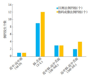
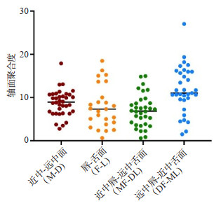
目的 对全瓷冠牙体预备后的预备体进行多平面聚合度测量及倒凹分析,针对性指导住院医师牙体预备操作,从而提高固定修复操作技能。 方法 由2019~2022年在首都医科大学附属北京世纪坛医院参加规范化培训的13位住院医师按照全瓷冠的要求在仿头模标准牙颌模型上对2~3颗右上中切牙进行牙体预备,最终收集符合流程的37颗预备体。首先,分别以目测法及数码成像法判断预备体4个不同平面(近中-远中面、唇-舌面、近中唇-远中舌面和远中唇-近中舌面)轴面聚合度及倒凹;再根据数码成像法的倒凹判断结果,将预备体分为倒凹组和非倒凹组;最后分析倒凹的存在是否与预备体高度、宽度、厚度减少量和肩台宽度有相关性。 结果 倒凹观察分析发现目测法与数码成像法判断有差异;数码成像法对轴面聚合度测量数据显示4个不同平面预备体的倒凹率的差异有统计学意义(χ2=16.188,P < 0.05),采用Bonferroni校正法进行比较,唇-舌面的倒凹率高于其他平面,差异有统计学意义(P < 0.05);倒凹组与非倒凹组在近中-远中面的高度及宽度减少量、唇-舌面厚度减少量,近中、远中、唇侧、舌侧肩台宽度之间的差异无统计学意义(P>0.05)。 结论 本研究对住院医师预备体4个不同平面的轴面聚合度进行测量,发现了预备体轴面存在倒凹的现象,使用数码成像法相比目测法可以更准确的判断倒凹和聚合度。此法可以针对性帮助住院医师提高轴面牙体预备过程的操作水平,提升固定修复操作技能。 Abstract:Objective To explore the multi-plane total occlusal convergence measurement and undercut analysis of all‐ceramic crown preparation. The dental preparation operation is targeted to guide residents, so as to improve the operation skills of fixed restoration. Methods Thirteen residents participated in standardized training in Beijing Shijitan Hospital Affiliated to Capital Medical University from 2019 to 2022. According to the requirement of all-ceramic crown, 2-3 right maxillary central incisors were prepared on the standard tooth model of head mold. Finally, 37 preparers conforming to the process were collected. Four different planes of the preparation were determined by visual and digital imaging methods (mesial-distal surface, labial-lingual surface, mesial labial-distal lingual surface, distal labial-mesial lingual surface) to measure total occlusal convergence and undercut. According to the results of the digital imaging method, the preparation was divided into undercut group and non-undercut group. Whether the existence of undercut is related to the height, width, thickness reduction and shoulder width of the preparation were analyzed. Results The analysis of undercut observation showed that there were differences between the visual method and the digital imaging method. The data of total occlusal convergence by digital imaging showed that the undercut ratio of the four different plane preparations was statistically different (χ2=16.188, P < 0.05). Bonferroni correction was used for comparison. The labial-lingual surface undercut ratio was higher than that of other planes (P < 0.05). There was no statistical difference between the undercut group and the non-undercut group in the height and width reduction of the mesial-distal surface, the thickness reduction of the labial-lingual surface, and the shoulder width of the mesial, distal, labial and lingual sides (P>0.05). Conclusion There is undercut phenomenon in the axial plane of the preparation. Compared with the visual method, the digital imaging method could more accurately judge the degree of undercut and total occlusal convergence. -
Key words:
- all‐ceramic crown /
- tooth preparation /
- total occlusal convergence
-

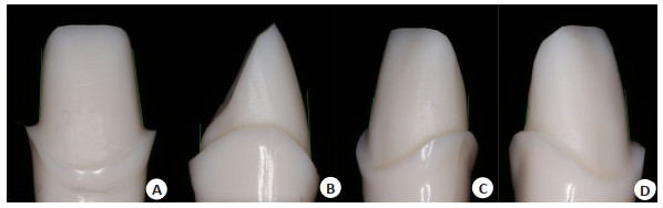
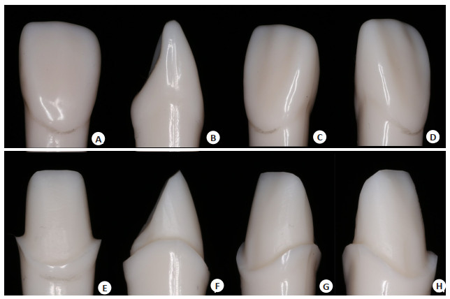
图 1 A-D单直根恒牙(A~D)及预备体(E~H)的4个不同平面
A~D: 单直根恒牙的4个不同平面[A: 近中-远中面(M-D); B: 唇-舌面(F-L); C: 近中唇-远中舌面(MF-DL); D: 远中唇- 近中舌面(DF-ML)]; E~H: 预备体的4个不同平面[E: 近中-远中面(M-D); F: 唇-舌面(F-L); G: 近中唇-远中舌面(MF-DL); H: 远中唇-近中舌面(DF-ML)].
Figure 1. The four different planes of permanent tooth model (A-D) and preparations model (E-H).

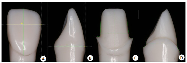
图 2 预备体4个不同平面轴面聚合度测量线
A: 近中-远中面(M-D); B: 唇-舌面(F-L); C: 近中唇-远中舌面(MF-DL); D: 远中唇-近中舌面(DF-ML).
Figure 2. Preparations model's measurement line of total occlusal convergence of four different planes

图 3 各平面图像的测量
A: 近中-远中面(M-D)单直根恒牙高度及宽度测量线; B: 唇-舌面(F-L)单直根恒牙厚度测量线; C: 近中-远中面(M-D)预备体唇面高度、宽度、近中肩台宽度、远中肩台宽度测量线; D: 唇-舌面(F-L)预备体厚度、唇侧肩台宽度、舌侧肩台宽度测量线.
Figure 3. Measurement of each plane image.

图 4 目测法及数码成像法判断倒凹发生个数的比较
Figure 4. Comparison between visual method and digital imaging method to determine the number of undercut cases.

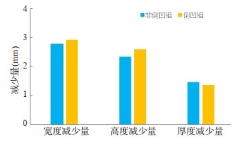
图 6 倒凹组与非倒凹组的宽度减少量、高度减少量、厚度减少量
Figure 6. Width reduction, height reduction and thickness reduction in undercut and nonundercut groups.

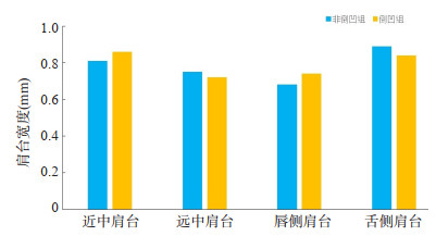
图 7 倒凹组与非倒凹组的近中肩台宽度、远中肩台宽度、唇侧肩台宽度、舌侧肩台宽度
Figure 7. The shoulder width of the mesial, distal, labial and lingual sides in undercut and non-undercut group.
表 1 目测法判断倒凹发生情况
Table 1. The occurrence of undercut were determined by visual method [n(%)]
测量平面 非倒凹组 倒凹组 近中-远中面(M-D) 36(97.3) 1(2.7) 唇-舌面(F-L) 28(75.7) 9(24.3) 近中唇-远中舌面(DF-ML) 34(89.2) 3(10.8) 远中唇-近中舌面(MF-DL) 35(94.6) 2(5.4)  下载: 导出CSV
表 2 数码成像判断倒凹发生情况
Table 2. The occurrence of undercut were determined by digital imaging methods [n(%)]
测量平面 非倒凹组 倒凹组(%) χ2 P 近中-远中面(M-D) 36(97.3%) 1(2.7) 16.188 0.001 唇-舌面(F-L) 25(67.6) 12(32.4) 近中唇-远中舌面(MF-DL) 34(89.2) 3(10.8) 远中唇-近中舌面(DF-ML) 33(91.9) 4(8.1)
下载: 导出CSV
表 3 宽度减少量、高度减少量、厚度减少量、近中肩台宽度、远中肩台宽度、唇侧肩台宽度、舌侧肩台宽度测量值
Table 3. The width and height reduction, the thickness reduction, and the shoulder width of the mesial, distal, labial and lingual sides measurement value (mm)
指标 最小值 最大值 测量值(Mean±SD) 宽度减少量 1.51 3.85 高度减少量 1.07 3.36 2.82±0.40 厚度减少量 0.96 2.53 1.43±0.29 近中肩台宽度 0.55 1.12 0.83±0.13 远中肩台宽度 0.40 1.04 0.74±0.14 唇侧肩台宽度 0.29 1.42 0.70±0.21 舌侧肩台宽度 0.53 1.32 0.88±0.19
下载: 导出CSV
表 4 倒凹组与非倒凹组的宽度减少量、高度减少量、厚度减少量比较
Table 4. Comparison of width reduction, height reduction and thickness reduction in undercut and non-undercut groups (mm, Mean±SD)
减少量 非倒凹组 倒凹组 t P 宽度减少量 2.78±0.40 2.92±0.40 -1.005 0.322 高度减少量 2.35±0.53 2.60±0.33 -1.513 0.139 厚度减少量 1.46±0.18 1.36±0.45 0.709 0.491
下载: 导出CSV
表 5 倒凹组与非倒凹组的近中肩台宽度、远中肩台宽度、唇侧肩台宽度、舌侧肩台宽度
Table 5. The shoulder width of the mesial, distal, labial and lingual sides in undercut and non-undercut group (mm, Mean±SD)
肩台位置 非倒凹组 倒凹组 t P 近中肩台 0.81±0.12 0.86±0.14 -1.207 0.235 远中肩台 0.75±0.13 0.72±0.17 0.572 0.571 唇侧肩台 0.68±0.18 0.74±0.28 -0.746 0.460 舌侧肩台 0.89±0.21 0.84±0.11 0.700 0.489
下载: 导出CSV
表 6 Logistic回归分析
Table 6. Logistic regression analysis
指标 β P OR 95% CI 宽度减少量 2.819 0.213 16.766 (0.198~1422.354) 高度减少量 0.743 0.504 2.102 (0.238~18.573) 厚度减少量 -3.993 0.087 0.018 (0.000~1.796) 近中肩台宽度 5.952 0.188 384.551 (0.054~2719104.663) 远中肩台宽度 -6.720 0.153 0.001 (0.000~12.067) 唇侧肩台宽度 1.982 0.545 7.260 (0.012~4437.184) 舌侧肩台宽度 -0.122 0.968 0.885 (0.002~371.339) 常量 -6.392 0.169 0.002
下载: 导出CSV
-
[1] 中华口腔医学会口腔美学专业委员会, 中华口腔医学会口腔材料专业委员会. 全瓷美学修复材料临床应用专家共识[J]. 中华口腔医学杂志, 2019, 54(12): 825-8. [2] Zhang Y, Kelly JR. Dental ceramics for restoration and metal veneering[J]. Dent Clin North Am, 2017, 61(4): 797-819. doi: 10.1016/j.cden.2017.06.005 [3] 卢嘉仪, 赵君仪, 高静, 等. 1312件单冠预备体数字化模型的关键预备质量指标的分析研究[J]. 华西口腔医学杂志, 2022, 40(1): 52-60. https://www.cnki.com.cn/Article/CJFDTOTAL-HXKQ202201008.htm [4] Strain KJ, Mackie J, Bonsor SJ, et al. Crown taper angles achieved by dental students: a systematic review[J]. J Dent Educ, 2018, 82 (11): 1203-12. doi: 10.21815/JDE.018.125 [5] 蒋文翔, 沈燕青, 赵鹃, 等. 上中切牙全瓷冠牙体预备的数字化评价[J]. 浙江医学教育, 2019, 18(4): 7-9, 22. https://www.cnki.com.cn/Article/CJFDTOTAL-ZYJT201904003.htm [6] Kim YK, Kim JH, Jeong Y, et al. Comparison of digital and conventional assessment methods for a single tooth preparation and educational satisfaction[J]. Eur J Dent Educ, 2022: 2022Apr5. [7] Yoon SS Jr, et al. Measurement of total occlusal convergence of 3 different tooth preparations in 4 different planes by dental students [J]. J Prosthet Dent, 2014, 112(2): 285-92. doi: 10.1016/j.prosdent.2014.01.021 [8] Jørgensen KD. The relationship between retention and convergence angle in cemented veneer crowns[J]. Acta Odontol Scand, 1955, 13 (1): 35-40. doi: 10.3109/00016355509028171 [9] Walton JN. A survey of crown and fixed partial denture failures: length of service and reasons for replacement[J]. J Prosthet Dent, 1986, 56(4): 416-21. doi: 10.1016/0022-3913(86)90379-3 [10] 毛勇, 高媛, 王忠义, 等. 计算机辅助设计与辅助制作全瓷冠的八年临床随访[J]. 中华口腔医学杂志, 2008, 43(12): 752-3. https://www.cnki.com.cn/Article/CJFDTOTAL-KQZZ201203006.htm [11] Wake R, Buck R, DuVall N, et al. Effect of molar preparation axial height on retention of adhesively-luted CAD-CAM ceramic crowns [J]. J Adhes Dent, 2019, 21(6): 545-50. [12] Yahya MM, Abdulrahman MB, Arwa OH, et al. Evaluation of undergraduate dental students in typodont preparation for allceramic crowns[J]. J Int Med Dent, 2018, 5(2): 72-9 https://www.researchgate.net/publication/328036110_Evaluation_of_undergraduate_dental_students_in_typodont_preparation_for_all-ceramic_crowns [13] 刘明月, 谭建国. 一步一步做好牙体缺损修复体类型的选择[J]. 中华口腔医学杂志, 2021, 56(7): 720-5. [14] Tiu J, Al-Amleh B, Waddell JN, et al. Clinical tooth preparations and associated measuring methods: a systematic review[J]. J Prosthet Dent, 2015, 113(3): 175-84. https://www.sciencedirect.com/science/article/pii/S0022391314004338 [15] Leempoel PJB. The convergence angle of tooth preparations for complete crowns[J]. J Prosthet Dent, 1987, 58(4): 414-6. https://www.sciencedirect.com/science/article/pii/0022391387902654 [16] Mack PJ. A theoretical and clinical investigation into the taper achieved on crown and inlay preparations[J]. J Oral Rehabil, 1980, 7(3): 255-65. [17] Tiu J, et al. Clinical tooth preparations and associated measuring methods: a systematic review[J]. J Prosthet Dent, 2015, 113(3): 175-84. https://www.sciencedirect.com/science/article/pii/S0022391314004338 [18] 周永胜. 口腔修复学[M]. 3版. 北京: 北京大学医学出版社, 2020. [19] Dorriz H, Nokar S, Naini RB, et al. The convergence angle of fullcoverage crown preparations made by dental students[J]. J Dent, 2008, 5: 37-41. https://www.researchgate.net/publication/268872147_The_Convergence_Angle_of_Full-coverage_Crown_Preparations_Made_by_Dental_Students [20] Rafeek RN, Marchan SM, Seymour KG, et al. Abutment taper of full cast crown preparations by dental students in the UWI School of Dentistry[J]. Eur J Prosthodont Restor Dent, 2006, 14(2): 63-6. https://pubmed.ncbi.nlm.nih.gov/16808106/ [21] 刘思璇, 吕培军, 孙玉春, 等. 一种牙冠预备体三维数字化模型的就位倒凹检测方法[J]. 科学技术与工程, 2011, 11(14): 3165-7, 3171. https://www.cnki.com.cn/Article/CJFDTOTAL-KXJS201114012.htm [22] Seet RH, Soo PR, Leong KJM, et al. Crown preparations by undergraduate dental students: a comparison of conventional versus digital assessment via an intraoral scanner[J]. J Dent Educ, 2020, 84(11): 1303-13. [23] Sato T, Al Mutawa N, Okada D, et al. A clinical study on abutment taper and height of full cast crown preparations[J]. J Med Dent Sci, 1998, 45(3): 205-10. https://pubmed.ncbi.nlm.nih.gov/11186212/ [24] Al-Omari WM, Al-Wahadni AM. Convergence angle, occlusal reduction, and finish line depth of full-crown preparations made by dental students[J]. Quintessence Int, 2004, 35(4): 287-93. https://pubmed.ncbi.nlm.nih.gov/15119714/ -

 点击查看大图
计量
- 文章访问数: 225
- HTML全文浏览量: 180
- PDF下载量: 12
- 被引次数: 0