


Multimodal magnetic resonance imaging study of brain structure and function changes in patients with knee osteoarthritis
-
摘要:
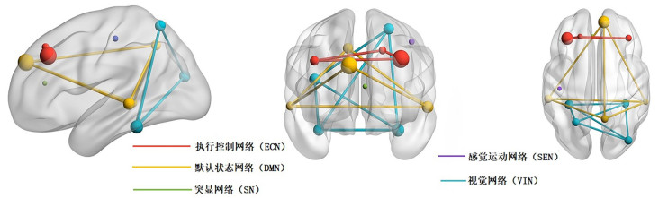
目的 联合基于体素的形态学测量(VBM)技术和静息态功能磁共振成像(rs-fMRI)技术探究膝骨性关节炎(KOA)患者静息状态下脑灰质体积、脑神经元活动强度及基于种子点的功能连接强度改变,综合分析KOA相关异常脑网络。 方法 前瞻性收集30例KOA患者(KOA组)及30例健康人(HC组)的3D高分辨率T1WI像和rs-fMRI图像,采用VBM、低频振幅、功能连接3种方法分析两组间脑灰质结构和功能数据差异。 结果 与HC组相比,KOA组双侧梭状回、右侧颞中回低频振幅值增高,右侧楔前叶、右侧内侧前额叶皮质、左侧额中回低频振幅值减低(体素水平P<0.005,团块水平P<0.05);右侧楔前叶、右侧顶下小叶、右侧初级视觉皮层、左侧颞中回、左侧中央后回灰质体积减小(体素水平P<0.002,团块水平P<0.05);以右侧楔前叶为种子点,与右侧颞中回的功能连接增强,与左侧前扣带回、左侧背外侧前额叶皮质的功能连接降低(体素水平P<0.005,团块水平P<0.05)。 结论 KOA患者感知皮层系统及联合皮层系统阵营均存在血氧水平依赖信号及灰质微结构的改变且有重叠,主要涉及视觉网络、感觉运动网络、默认状态网络、执行控制网络内及默认状态网络、执行控制网络、突显网络的部分脑区间,这可能提示KOA患者不仅处于慢性疼痛的病理状态,还伴有信息整合、注意力控制、情绪反应、情感解读等功能活动的异常。 -
关键词:
- 膝骨性关节炎 /
- 基于体素的形态学测量 /
- 静息态功能磁共振成像 /
- 低频振幅 /
- 功能连接
Abstract:Objective To explore the change of brain gray matter volume, neuron activity and functional connectivity strength based on seed points in patients with knee osteoarthritis (KOA) at rest through combined voxel based morphology and rsfMRI technology, and comprehensively analyze KOA-related abnormal brain networks. Methods 3D high-resolution T1WI images and rs- fMRI images of 30 patients with KOA (KOA group) and 30 healthy controls (HC group) were collected prospectively. Voxel based morphology, amplitude of low-frequency fluctuation, and functional connectivity were used to analyze the differences in gray matter structural and functional data between the two groups. Results Compared with the HC group, the amplitude of low-frequency fluctuation values of bilateral fusiform gyrus and right middle temporal gyrus in KOA group increased, while the amplitude of low-frequency fluctuation values of right precuneus, right medial prefrontal cortex and left middle frontal gyrus decreased (voxel level P<0.005, cluster level P<0.05). The gray matter volume of right precuneus, right inferior parietal lobule, right primary visual cortex, left middle temporal gyrus, left postcentral gyrus decreased (voxel level P<0.002, cluster level P<0.05). With the right precuneus as the seed point, the functional connectivity with right middle temporal gyrus was enhanced, and the functional connectivity with left anterior cingulate gyrus, left dorsolateral prefrontal cortex was reduced (voxel level P<0.005, cluster level P<0.05). Conclusion The changes of BOLD signal and gray matter microstructure were found in both perceptual cortex system and combined cortex system of KOA patients, There are overlap, mainly involving in the visual network, sensorimotor network, default mode network, executive control network within and between partial brain regions of default mode network, executive control network and salience network. This may suggest that KOA patients are not only in the pathological state of chronic pain, but also accompanied by abnormalities in functional activities such as information integration, attentional control, emotional response, and emotional interpretation. -

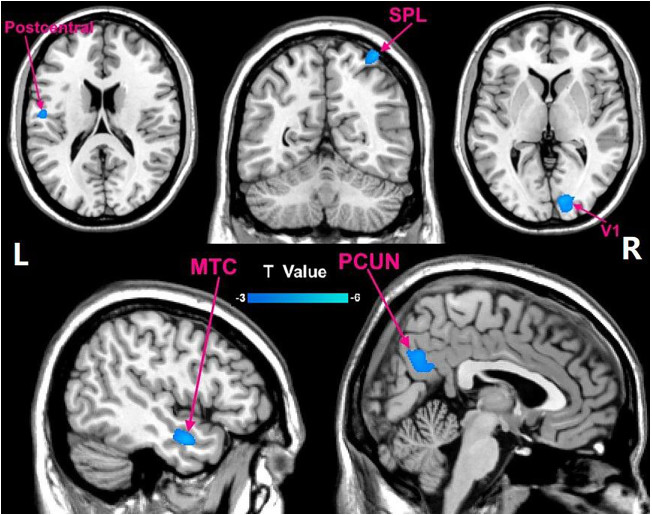
图 1 KOA组与HC组VBM值差异脑图(冷色代表KOA组灰质体积低于HC组的脑区)
SPL: 顶上小叶; V1: 初级视觉皮层; MTC: 颞中回; PCUN: 楔前叶.
Figure 1. Brain map of the difference in VBM value between KOA group and HC group (Cool colors represent brain regions where the gray matter volume of KOA group is lower than that of HC group).

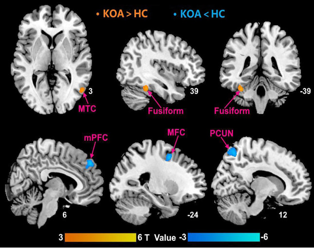
图 2 KOA组与HC组ALFF值差异脑图(暖色代表KOA组神经元活性高于HC组的脑区,冷色代表KOA组神经元活性低于HC组的脑区)
MFC: 额中回; mPFC: 内侧前额叶皮层.
Figure 2. Brain map of the difference in ALFF value between KOA group and HC group (Warm colors represent brain regions with higher neuronal activity in KOA group than HC group, cool colors represent brain regions with lower neuronal activity in KOA group than HC group).

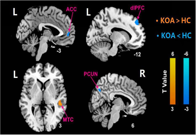
图 3 KOA组与HC组功能连接差异脑图(暖色代表KOA患者FC值高于健康人的脑区,冷色代表KOA患者FC低于健康人的脑区)
ACC: 前扣带回; dlPFC: 背外侧前额叶皮层.
Figure 3. Brain map of the difference in FC value between KOA group and HC group (Warm colors represent brain regions with higher FC value in KOA group than HC group, cool colors represent brain regions with lower FC value in KOA group than HC group).
表 1 两组受试者一般资料比较
Table 1. Comparison of baseline data between two groups of subjects (n=30, Mean±SD)
组别 性别(n) 年龄(岁) 受教育时间(年) 身高(m) 体质量(kg) 男 女 KOA组 6 24 57.40±9.95 10.20±2.73 1.65±0.05 63.67±8.22 HC组 5 25 58.70±10.04 10.10±2.76 1.64±0.05 65.27±9.45 t/χ2 0.111 0.254 0.141 0.206 0.489 P 0.739 0.616 0.888 0.838 0.487 KOA: 膝骨性关节炎; HC: 健康对照.  下载: 导出CSV
表 2 KOA组与HC组VBM值显著差异脑区信息
Table 2. Brain region information of significant difference in VBM value between KOA group and HC group
AAL脑区 大脑半球 MNI坐标 BA分区 团块大小
(体素数)t X Y Z 楔前叶 R 3 -63 33 7 602 -3.49 颞中回 L -48 -3 -21 21 230 -3.96 顶上小叶 R 36 -57 69 7 139 -3.87 中央后回 L -54 -9 -18 20 62 -3.27 初级视觉皮层 R 15 -87 3 17 666 -3.92 L: 左; R: 右; AAL: 解剖学自动标记; BA: 布罗德曼分区; MNI: 蒙特利尔神经学研究所.
下载: 导出CSV
表 3 KOA组与HC组ALFF值显著差异脑区信息
Table 3. Brain region information of significant difference in ALFF value between KOA group and HC group
AAL脑区 大脑半球 MNI坐标 BA分区 团块大小
(体素数)t X Y Z 梭状回 L -33 -39 -27 37 59 4.1 梭状回 R 39 -42 -24 37 68 3.91 颞中回 R 54 -66 3 37 44 3.58 楔前叶 R 12 -63 57 7 99 -4.3 额中回 L -24 3 51 - 25 -4.66 内侧前额叶皮质 R 6 48 39 9 54 -4.6
下载: 导出CSV
表 4 KOA组与HC组FC值显著差异脑区信息
Table 4. Brain region information of significant difference in FC value between KOA group and HC group
AAL脑区 大脑半球 MNI坐标 BA分区 团块大小
(体素数)t X Y Z 颞中回 R 51 -33 3 21 64 3.67 楔前叶 R 6 -69 39 7 28 -3.59 背外侧前额叶皮质 L -12 45 48 9 52 -3.47 前扣带回 L -3 51 0 10 41 -3.13
下载: 导出CSV
-
[1] Murray CJL, et al. Disability-adjusted life years (DALYs) for 291 diseases and injuries in 21 regions, 1990-2010: a systematic analysis for the Global Burden of Disease Study 2010[J]. Lancet, 2012, 380(9859): 2197-223. doi: 10.1016/S0140-6736(12)61689-4 [2] 中华医学会骨科学分会关节外科学组. 骨关节炎诊疗指南(2018年版)[J]. 中华骨科杂志, 2018(12): 705-15. doi: 10.3760/cma.j.issn.0253-2352.2018.12.001 [3] Duchaine B, Yovel G. A revised neural framework for face processing[J]. Annu Rev Vis Sci, 2015, 1: 393-416. doi: 10.1146/annurev-vision-082114-035518 [4] Matsuyoshi D, Morita T, Kochiyama T, et al. Dissociable cortical pathways for qualitative and quantitative mechanisms in the face inversion effect[J]. J Neurosci, 2015, 35(10): 4268-79. doi: 10.1523/JNEUROSCI.3960-14.2015 [5] 王钰萍, 张宾, 黄佳滨, 等. 神经性贪食患者额叶-纹状体神经环路静息态fMRI功能连接研究[J]. 中华行为医学与脑科学杂志, 2018, 27(4): 316-21. doi: 10.3760/cma.j.issn.1674-6554.2018.04.006 [6] 曲冰, 王瀚, 赵晨雨, 等. 针刺法治疗慢性膝骨关节炎的临床疗效评价及中枢机制研究[J]. 新疆医科大学学报, 2021, 44(5): 600-4. https://www.cnki.com.cn/Article/CJFDTOTAL-XJYY202105017.htm [7] 卿伦学. 推拿治疗慢性膝骨关节炎疼痛的fMRI和股直肌超声造影研究[D]. 北京: 北京中医药大学, 2019: 41. [8] Guo H, Wang YQ, Qiu LH, et al. Structural and functional abnormalities in knee osteoarthritis pain revealed with multimodal magnetic resonance imaging[J]. Front Hum Neurosci, 2021, 15: 783355. doi: 10.3389/fnhum.2021.783355 [9] 郭雅, 李瑞阳, 卢富强, 等. 多模态功能磁共振成像在前庭性偏头痛的研究进展[J]. 磁共振成像, 2021, 12(4): 86-8. https://www.cnki.com.cn/Article/CJFDTOTAL-CGZC202104022.htm [10] Kang BX, Ma J, Shen J, et al. Altered brain activity in end-stage knee osteoarthritis revealed by resting-state functional magnetic resonance imaging[J]. Brain Behav, 2022, 12(1): e2479. [11] Barroso J, Vigotsky AD, Branco P, et al. Brain gray matter abnormalities in osteoarthritis pain: a cross-sectional evaluation[J]. Pain, 2020, 161(9): 2167-78. doi: 10.1097/j.pain.0000000000001904 [12] 李伟雄. 基于视觉皮层下通路的拓扑知觉与情绪认知的研究[D]. 昆明: 云南大学, 2020: 1-2. [13] Baliki MN, Mansour AR, Baria AT, et al. Functional reorganization of the default mode network across chronic pain conditions[J]. PLoS One, 2014, 9(9): e106133. doi: 10.1371/journal.pone.0106133 [14] Liao X, Mao CP, Wang Y, et al. Brain gray matter alterations in Chinese patients with chronic knee osteoarthritis pain based on voxel-based morphometry[J]. Medicine, 2018, 97(12): e0145. doi: 10.1097/MD.0000000000010145 [15] Cottam WJ, Iwabuchi SJ, Drabek MM, et al. Altered connectivity of the right anterior insula drives the pain connectome changes in chronic knee osteoarthritis[J]. Pain, 2018, 159(5): 929-38. doi: 10.1097/j.pain.0000000000001209 [16] 汤臣建. 针刺对膝骨性关节炎患者大脑默认网络功能活动的影响研究[D]. 成都: 成都中医药大学, 2019: 65. [17] Liu J, et al. Different exercise modalities relieve pain syndrome in patients with knee osteoarthritis and modulate the dorsolateral prefrontal cortex: a multiple mode MRI study[J]. Brain Behav Immun, 2019, 82: 253-63. doi: 10.1016/j.bbi.2019.08.193 [18] 刘书奇, 金军标. MRI在骨性膝关节炎膝关节置换术后脑功能评估中的应用分析[J]. 浙江创伤外科, 2019, 24(5): 1079-81. https://www.cnki.com.cn/Article/CJFDTOTAL-ZJCW201905107.htm [19] Kai U, Kazuyoshi N, Yukio M, et al. Altered resting-state connectivity with pain-related expectation regions in female patients with severe knee osteoarthritis[J]. J Pain Res, 2020, 2020: 3227-34. -

 点击查看大图
计量
- 文章访问数: 224
- HTML全文浏览量: 116
- PDF下载量: 12
- 被引次数: 0