


Detection of liver and spleen stiffness in clinically significant portal hypertensive rats by two-dimensional shear wave elastography
-
摘要:
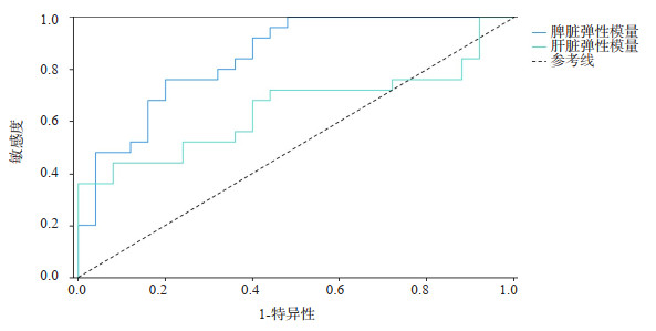
目的 分析二维剪切波弹性成像(2D-SWE)检测脾、肝脏硬度评估临床显著性门静脉高压的价值。 方法 将50只雄性SD大鼠随机分为门脉高压组(PH组,n=25)和正常组(n=25)。PH组采用CCl4诱导法,正常组仅注射玉米油,于12周采用2D-SWE检测大鼠脾、肝脏硬度,运用门静脉主干直接穿刺法测量门静脉压力(PVP),观察脾、肝脏病理改变。分析脾硬度(SS)、肝脏硬度(LS)与PVP相关性,以PVP为金标准求得ROC曲线下面积。 结果 SS、LS与PVP均呈正相关关系,SS与PVP的相关性(r=0.797,P < 0.001)强于LS与PVP(r=0.505,P < 0.001)。以PVP≥10 mmHg作为金标准,2D-SWE检测SS、LS的ROC曲线下面积分别为0.875和0.673,SS优于LS(P < 0.05)。 结论 2D-SWE测量SS评估临床显著性门静脉高压诊断效能优于LS。 -
关键词:
- 临床显著性门静脉高压 /
- 二维剪切波弹性成像 /
- 脾硬度 /
- 肝脏硬度
Abstract:Objective To analyze the value of spleen stiffness (SS) and liver stiffness (LS) in the evaluation of clinically significant portal hypertension by two-dimensional shear wave elastic imaging (2D-SWE). Methods Fifty SD rats were randomly divided into portal hypertension group (PH group, n=25) and normal control group (NC group, n=25). The animal model of PH group was established by CCl4 induction method, and the NC group was only injected with corn oil. SS and LS of rats were detected by 2D-SWE, portal vein pressure (PVP) was measured by direct portal vein puncture and the pathological changes of spleen and liver were observed at 12 weeks. Furthermore, the correlation between SS, LS and PVP was analyzed, and the area under ROC curve was obtained by using PVP as the gold standard. Results SS and LS were positively correlated with PVP, but the correlation between SS and PVP (r=0.797, P < 0.001) was stronger than LS (r=0.505, P < 0.001). With PVP≥10 mmHg as the gold standard, the area under ROC curve of SS and LS detected by 2D-SWE were 0.875 and 0.673 respectively, which indicated that SS was better than LS (P < 0.05). Conclusion The diagnostic efficacy of 2D-SWE measurement of SS in evaluating clinically significant portal hypertension is better than LS. -

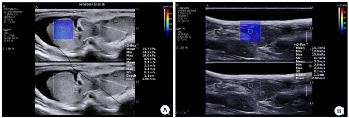
图 1 2D-SWE测量弹性模量
A:2D-SWE测量大鼠肝脏弹性模量; B:2D-SWE测量大鼠脾弹性模量.
Figure 1. 2D-SWE measurement of the elastic modulus.

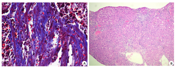
图 2 PH组鼠脾和肝脏病理表现
A:PH组脾纤维化大部分分级为Ⅱ级(Masson染色, ×100); B:PH组肝脏纤维化大部分分期为F4期(HE染色, ×100).
Figure 2. Pathological manifestations of rat liver and spleen in PH groups

图 3 2D-SWE检测鼠肝脾硬度诊断CSPH的ROC曲线
Figure 3. The ROC curve of CSPH was determined by 2D-SWE.
表 1 脾病理分级结果
Table 1. Pathological classification of spleen (n)
组别 0 Ⅰ Ⅱ Ⅲ Ⅳ 总计 PH组脾 0 6 14 4 1 25 正常组脾 19 6 0 0 0 25 总计 19 12 14 4 1 50  下载: 导出CSV
表 2 肝脏病理分期结果
Table 2. Pathological staging of liver (n)
组别 F0 F1 F2 F3 F4 总计 PH组肝脏 0 6 2 5 12 25 正常组肝脏 21 4 0 0 0 25 总计 21 10 2 5 12 50
下载: 导出CSV
表 3 鼠肝脾弹性模量
Table 3. Rat liver and spleen elastic modulus(n=25, kPa, Mean±SD)
组别 SS LS PH组 14.90±4.45 9.22±4.01 正常组 8.73±2.32 6.27±1.23 SS: 脾硬度; LS: 肝脏硬度
下载: 导出CSV
-
[1] Zhu YL, Ding H, Fu TT, et al. Portal hypertension in hepatitis B-related cirrhosis: diagnostic accuracy of liver and spleen stiffness by 2-D shear-wave elastography[J]. Hepatol Res, 2019, 49(5): 540-9. doi: 10.1111/hepr.13306 [2] de Franchis R, Faculty BV. Expanding consensus in portal hypertension: report of the Baveno Ⅵ Consensus Workshop: Stratifying risk and individualizing care for portal hypertension[J]. J Hepatol, 2015, 63(3): 743-52. doi: 10.1016/j.jhep.2015.05.022 [3] 李小梅, 雒博晗, 王钲钰, 等. BAVENO Ⅶ门静脉高压共识更新: 门静脉高压的个体化治疗[J]. 中华肝脏病杂志, 2022, 30(1): 21-9. https://www.cnki.com.cn/Article/CJFDTOTAL-LCGD202206010.htm [4] Colecchia A, et al. Spleen stiffness measurement can predict clinical complications in compensated HCV-related cirrhosis: a prospective study[J]. J Hepatol, 2014, 60(6): 1158-64. doi: 10.1016/j.jhep.2014.02.024 [5] Colecchia A, et al. Measurement of spleen stiffness to evaluate portal hypertension and the presence of esophageal varices in patients with HCV-related cirrhosis[J]. Gastroenterology, 2012, 143 (3): 646-54. doi: 10.1053/j.gastro.2012.05.035 [6] 王磊, 宋清坤, 岳振东, 等. 门静脉高压患者PPG与HVPG相关性研究[J]. 中华肝脏病杂志, 2022(7): 722-7. [7] Königshofer P, et al. Animal models of portal hypertension[J]. Biochim Biophys Acta BBA Mol Basis Dis, 2019, 1865(5): 1019-30. doi: 10.1016/j.bbadis.2018.07.018 [8] 周琴, 郭海欣, 李靖云, 等. 剪切波弹性成像评估大鼠非酒精性脂肪肝病肝纤维化程度[J]. 临床超声医学杂志, 2020, 22(3): 161-4. https://www.cnki.com.cn/Article/CJFDTOTAL-LCCY202003001.htm [9] Kutlu R, Karaman I, Akbulut A, et al. Quantitative Doppler evaluation of the splenoportal venous system in various stages of cirrhosis: differences between right and left portal veins[J]. J Clin Ultrasound, 2002, 30(9): 537-43. doi: 10.1002/jcu.10114 [10] 吴厚慧, 陈家林. 脾纤维化程度对免疫功能的影响分析[[ J]. 中华肝胆外科杂志, 2005, 11(5): 346-7. https://www.cnki.com.cn/Article/CJFDTOTAL-ZHGD200505029.htm [11] Karagiannakis DS, Voulgaris T, Siakavellas SI, et al. Evaluation of portal hypertension in the cirrhotic patient: hepatic vein pressure gradient and beyond[J]. Scand J Gastroenterol, 2018, 53(10/11): 1153-64. [12] Sun XH, Ni HB, Xue J, et al. Bibliometric-analysis visualization and review of non-invasive methods for monitoring and managing the portal hypertension[J]. Front Med (Lausanne), 2022, 9: 960316. [13] Fofiu R, et al. Spleen and liver stiffness for predicting high-risk varices in patients with compensated liver cirrhosis[J]. Ultrasound Med Biol, 2021, 47(1): 76-83. doi: 10.1016/j.ultrasmedbio.2020.09.004 [14] Stefanescu H, Rusu C, Lupsor-Platon M, et al. Liver stiffness assessed by ultrasound shear wave elastography from general electric accurately predicts clinically significant portal hypertension in patients with advanced chronic liver disease[J]. Ultraschall Med, 2020, 41(5): 526-33. [15] Attia D, Schoenemeier B, Rodt T, et al. Evaluation of liver and spleen stiffness with acoustic radiation force impulse quantification elastography for diagnosing clinically significant portal hypertension[J]. Ultraschall Med, 2015, 36(6): 603-10. doi: 10.1055/s-0041-107971 [16] Procopet B, et al. Real-time shear-wave elastography: applicability, reliability and accuracy for clinically significant portal hypertension [J]. J Hepatol, 2015, 62(5): 1068-75. [17] Jansen C, Bogs C, Verlinden W, et al. Shear-wave elastography of the liver and spleen identifies clinically significant portal hypertension: a prospective multicentre study[J]. Liver Int, 2017, 37(3): 396-405. doi: 10.1111/liv.13243 [18] Elkrief L, Rautou PE, Ronot M, et al. Prospective comparison of spleen and liver stiffness by using shear-wave and transient elastography for detection of portal hypertension in cirrhosis[J]. Radiology, 2015, 275(2): 589-98. doi: 10.1148/radiol.14141210 [19] Takuma Y, Nouso K, Morimoto Y, et al. Portal hypertension in patients with liver cirrhosis: diagnostic accuracy of spleen stiffness [J]. Radiology, 2016, 279(2): 609-19. [20] 张智林, 周惠惠, 张君, 等. 二维剪切波弹性成像技术鉴别特发性门静脉高压与肝硬化门静脉高压的价值[J]. 中国超声医学杂志, 2021, 37(9): 1017-21. https://www.cnki.com.cn/Article/CJFDTOTAL-ZGCY202109019.htm [21] 王鹏, 唐少珊, 任卫东. 实时剪切波弹性成像测量脾脏硬度评价肝硬化门静脉高压[J]. 中国医学影像技术, 2018, 34(5): 697-700. https://www.cnki.com.cn/Article/CJFDTOTAL-ZYXX201805020.htm [22] Wagner M, Hectors S, Bane O, et al. Noninvasive prediction of portal pressure with MR elastography and DCE-MRI of the liver and spleen: preliminary results[J]. J Magn Reson Imaging, 2018, 48 (4): 1091-103. doi: 10.1002/jmri.26026 [23] Song JZ, et al. Performance of spleen stiffness measurement in prediction of clinical significant portal hypertension: a metaanalysis[J]. Clin Res Hepatol Gastroenterol, 2018, 42(3): 216-26. https://www.sciencedirect.com/science/article/pii/S2210740117302589 [24] Kim TY, Jeong WK, Sohn JH, et al. Evaluation of portal hypertension by real-time shear wave elastography in cirrhotic patients[J]. Liver Int, 2015, 35(11): 2416-24. [25] Goldschmidt I, Brauch C, Poynard T, et al. Spleen stiffness measurement by transient elastography to diagnose portal hypertension in children[J]. J Pediatr Gastroenterol Nutr, 2014, 59 (2): 197-203. -

 点击查看大图
计量
- 文章访问数: 111
- HTML全文浏览量: 143
- PDF下载量: 7
- 被引次数: 0