


Diagnostic value of MRI labyrinth hydroneuroimaging on neurovascular compression in trigeminal neuralgia
-
摘要:
目的 探究MRI迷路水神经成像诊断三叉神经痛和神经血管压迫的效果。 方法 回顾性分析我院168例三叉神经痛患者的临床资料,均行手术治疗证实,且进行MRI迷路水神经成像检查,分析患者神经血管压迫的影像学检查结果,与手术结果对比,并比较患者三叉神经池段形态学参数(三叉神经脑池段最大长度及面积、三叉神经-桥脑夹角、桥小脑角池截面)。 结果 168例三叉神经痛患者中MRI成像检测处左侧受压80例,与临床左侧三叉神经痛分布区的相符率为90.91%,右侧受压74例,与临床右侧三叉神经痛分布区的相符率为92.50%,未见压迫14例;患侧和健侧压迫位置的差异无统计学意义(P>0.05);主要压迫动脉为中小脑上动脉,占70.83%(119/168);患侧压迫程度较高(P < 0.05);两侧三叉神经最大长度的差异无统计学意义(P>0.05),患侧三叉神经横截面积、三叉神经桥脑夹角、桥小脑角池截面积较低(P < 0.05)。 结论 MRI迷路水神经成像能够有效诊断三叉神经痛,显示神经周围具体情况,判断三叉神经脑池段形态学改变,为临床三叉神经痛神经血管压迫的诊断提供依据。 Abstract:Objective To explore the effect of MRI labyrinth hydroneuroimaging in the diagnosis of trigeminal neuralgia and neurovascular compression. Methods Retrospective analysis was performed on clinical data of 168 patients with trigeminal neuralgia in the hospital. All patients were confirmed by surgical treatment and performed MRI labyrinth hydroneuroimaging. The imaging examination results of neurovascular compression were analyzed and compared with the surgical results. The morphological parameters of the trigeminal cistern segment (maximum length and area of the trigeminal cisternal segment, trigeminal nerve-pontine angle, cerebellopontine angle cistern section) were compared among the patients. Results Among the 168 patients with trigeminal neuralgia, 80 cases were compressed on the left side by MRI imaging detection, which was 90.91% consistent with the clinical distribution of left trigeminal neuralgia, and 74 cases were compressed on the right side which was 92.50% consistent with the clinical distribution of right trigeminal neuralgia. No compression was observed in 14 cases. There was no significant difference in the compression position between the affected and healthy sides (P> 0.05). The main compression artery was the middle superior cerebellar artery, accounting for 70.83% (119/168). The degree of compression on the affected side was higher (P < 0.05). There was no significant difference in the maximum length of trigeminal nerve between the two sides (P>0.05), and the cross-sectional area of trigeminal nerve, trigeminal nerve-pontine angle and cross-sectional area of cerebellopontine angle cistern were smaller in the affected side (P < 0.05). Conclusion MRI labyrinth hydroneuroimaging can effectively diagnose trigeminal neuralgia, show the specific conditions around the nerve, judge the morphological changes of the trigeminal cisternal segment. It can provide a basis for the diagnosis of neurovascular compression in clinical trigeminal neuralgia. -

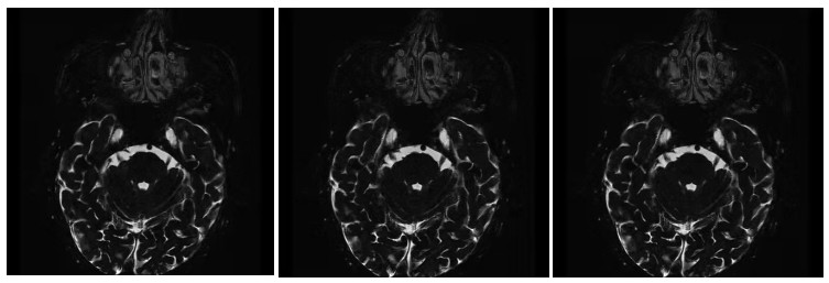
图 1 术前右侧三叉神经区域压迫占位的MRI迷路水神经成像图片
Figure 1. Preoperative MRI images of labyrinstral hydrologic nerve in right trigeminal nerve area compression and occupying.

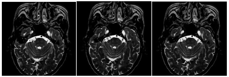
图 2 术后右侧三叉神经无压迫占位的MRI迷路水神经成像图片
Figure 2. Postoperative MRI images of labyrinstral nerve without compression and mass of right trigeminal nerve.
表 1 MRI迷路水神经成像检查结果与临床症状部位的相符率
Table 1. Coincidence rate between MRI hydrolabyrinthian neuroimaging results and clinical symptom sites
检杳结果 MRI成像(n) 临床症状(n) 相符率(%) 阳性 仅右侧 80 88 90.91 仅左侧 74 80 92.50 阴性 14 0 - 合计 168 168  下载: 导出CSV
表 2 三叉神经血管压迫责任血管分布情况
Table 2. Distribution of responsible vessels for trigeminal vascular compression
位置 倒数(%) 主要压迫血管(n=168) 小脑上动脉 119(70.83) 小脑前下动脉 2(12.50) 岩动脉 7(44.64) 椎动脉 5(2.98) 患侧压迫点(n=210) 小脑上动脉 112(53.33) 岩动脉 70(33.33) 小脑前下动脉 20(9.52) 椎动脉 8(3.81
下载: 导出CSV
表 3 两侧压迫位置比较
Table 3. Comparison of compression positions on both sides [n(%)]
检测侧 Ⅰ型 Ⅱ型 Ⅲ型 患侧 5(2.98) 120(71.43) 43(25.60) 健侧 68(40.48) 100(59.52) 0 χ2 9.941 P < 0.001
下载: 导出CSV
表 4 两侧压迫程度比较
Table 4. Comparison of compression degree on both sides [n=168, n(%)]
检测侧 近端压迫 远端压迫 患侧(n=210) 124(59.05) 86(40.95) 健侧(n=11) 6(54.55) 5(45.4) χ2 0.088 P 0.767
下载: 导出CSV
表 5 三叉神经池段形态学参数比较
Table 5. Comparison of morphological parameters of trigeminal cisterna segment (n=168, Mean±SD)
分侧 三叉神经最大长度(mm) 三叉神经横截面积(mm2) 三叉神经桥脑夹角(°) 桥小脑角池截面积(mm2) 患侧 9.85士2.49 27.60士7.92 41.87士7.89 206.07士8.31 健侧 9.92±2.62 33.84士9.52 47.47士9.82 228.78士35.62 t 0.251 6.531 5.762 8.048 P 0.802 < 0.001 < 0.001 < 0.001
下载: 导出CSV
-
[1] Di Stefano G, Yuan JH, Cruccu G, et al. Familial trigeminal neuralgia-a systematic clinical study with a genomic screen of the neuronal electrogenisome[J]. Cephalalgia, 2020, 40(8): 767-77. doi: 10.1177/0333102419897623 [2] Bendtsen L, et al. Advances in diagnosis, classification, pathophysiology, and management of trigeminal neuralgia[J]. Lancet Neurol, 2020, 19(9): 784-96. doi: 10.1016/S1474-4422(20)30233-7 [3] Kc E, Islam J, Park YS. Trigeminal ganglion itself can be a viable target to manage trigeminal neuralgia[J]. J Headache Pain, 2022, 23 (1): 150. doi: 10.1186/s10194-022-01512-x [4] 郭楠, 汪秀玲. 原发性三叉神经痛的MRI研究进展[J]. 放射学实践, 2021, 36(7): 942-6. https://www.cnki.com.cn/Article/CJFDTOTAL-FSXS202107035.htm [5] Danyluk H, Ishaque A, Ta D, et al. MRI texture analysis reveals brain abnormalities in medically refractory trigeminal neuralgia[J]. Front Neurol, 2021, 12: 626504. doi: 10.3389/fneur.2021.626504 [6] 黄雨, 柴学, 肖朝勇, 等. 三维双激发平衡式稳态自由进动序列和三维时间飞跃MRA序列在原发性三叉神经痛中的诊断价值[J]. 临床神经病学杂志, 2020, 33(5): 362-6. https://www.cnki.com.cn/Article/CJFDTOTAL-LCSJ202005015.htm [7] 李胜凯, 李文, 于昭, 等. 磁共振三维双回波稳态水激发3D DESSWE序列在颅神经颅外段成像中的应用[J]. 实用医学杂志, 2020, 36 (19): 2709-13. https://www.cnki.com.cn/Article/CJFDTOTAL-SYYZ202019020.htm [8] Araya EI, Claudino RF, Piovesan EJ, et al. Trigeminal neuralgia: basic and clinical aspects[J]. Curr Neuropharmacol, 2020, 18(2): 109-19. doi: 10.2174/1570159X17666191010094350 [9] Gerwin R. Chronic facial pain: trigeminal neuralgia, persistent idiopathic facial pain, and myofascial pain syndrome-an evidencebased narrative review and etiological hypothesis[J]. Int J Environ Res Public Health, 2020, 17(19): 7012. doi: 10.3390/ijerph17197012 [10] Shulev YA, Gordienko KS, Trashin AV, et al. Microvascular decompression in trigeminal neuralgia following vertebrobasilar dolichoectasia[J]. Zh Vopr Neirokhir Im N N Burdenko, 2020, 84 (5): 50-63. doi: 10.17116/neiro20208405150 [11] Yu F, Yin J. Young-onset trigeminal neuralgia: a clinical study and literature review[J]. Acta Neurochir, 2021, 163(6): 1617-21. doi: 10.1007/s00701-021-04848-6 [12] 杨冯棱, 郭立, 杨文秀. 磁共振肠道水成像在机械性肠梗阻中的诊断价值[J]. 中国CT和MRI杂志, 2020, 18(7): 133-6. https://www.cnki.com.cn/Article/CJFDTOTAL-CTMR202007042.htm [13] McIlwrath SL, et al. Manganese-enhanced MRI reveals changes within brain anxiety and aversion circuitry in rats with chronic neuropathic pain-and anxiety-like behaviors[J]. NeuroImage, 2020, 223: 117343. doi: 10.1016/j.neuroimage.2020.117343 [14] 王晶, 杨华堂, 赵俊杰, 等. 磁共振水成像高分辨序列在血管压迫性面肌痉挛微血管减压术中的应用价值[J]. 中国临床神经外科杂志, 2020, 25(11): 787-8. https://www.cnki.com.cn/Article/CJFDTOTAL-ZGLC202011020.htm [15] 黄飞, 吴伟, 郭大静, 等. 磁共振三维选择性水激发序列在膝关节软骨损伤中的应用价值[J]. 第三军医大学学报, 2021, 43(9): 871-5. https://www.cnki.com.cn/Article/CJFDTOTAL-DSDX202109015.htm [16] 钟波, 杨转移, 朱昌华, 等. 影响显微血管减压术对原发性三叉神经痛疗效的因素分析[J]. 国际神经病学神经外科学杂志, 2021, 48(1): 17-20. https://www.cnki.com.cn/Article/CJFDTOTAL-GWSK202101005.htm [17] Ma ZT, Zhang Y, Yu LL, et al. Preoperative MRI characteristics and short-term postoperative outcomes of microvascular decompression in trigeminal neuralgia with no vascular compression[J]. Minerva Anestesiol, 2020, 86(3): 360-1. [18] 李强, 于炎冰, 张黎. 三叉神经痛的疼痛范围与神经血管压迫位置的关系研究[J]. 中华神经医学杂志, 2020, 19(11): 1098-103. https://cpfd.cnki.com.cn/Article/CPFDTOTAL-SJWK202206001035.htm [19] 崔二辉, 徐欢, 张楠, 等. 原发性三叉神经痛患者MRI检测脑池段三叉神经形态学改变分析[J]. 立体定向和功能性神经外科杂志, 2020, 33(3): 136-40. https://www.cnki.com.cn/Article/CJFDTOTAL-NENG202003004.htm [20] 蔡仁贤, 田毅, 何占平, 等. 原发性三叉神经痛患者大脑灰质变化的VBM-MRI研究[J]. 医学研究杂志, 2019, 48(6): 80-2, 88. https://www.cnki.com.cn/Article/CJFDTOTAL-YXYZ201906021.htm [21] 甄雪克, 任鸿翔, 张黎, 等. 以三叉神经痛或面肌痉挛为首发症状的桥小脑角区肿瘤的临床特点分析[J]. 中华神经医学杂志, 2020, 19 (12): 1204-7. https://cpfd.cnki.com.cn/Article/CPFDTOTAL-SJWK202206001133.htm [22] 李丹, 卜岗, 张明, 等. 原发性三叉神经痛患者脑结构和功能连接改变[J]. 中国医学影像技术, 2020, 36(7): 996-1001. https://www.cnki.com.cn/Article/CJFDTOTAL-ZYXX202007011.htm -

 点击查看大图
计量
- 文章访问数: 280
- HTML全文浏览量: 173
- PDF下载量: 6
- 被引次数: 0