


慢性肝病和肝硬化患者钆塞酸增强MRI的肝功能成像评分的验证:Child-Pugh评分与肝功能成像评分的关系
doi: 10.12122/j.issn.1674-4500.2023.01.28
Verification of gadolinic acid enhanced MRI liver function imaging score in patients with chronic liver disease and cirrhosis: relationship between Child-Pugh score and liver function imaging score
-
摘要:
目的 分析功能性肝脏成像评分(FLIS)在钆塞酸增强MRI中对肝功能的预测作用。 方法 将本院2019年10月~2022年2月收治的134例患者经钆塞酸增强MRI诊断为肝硬化或慢性肝病(CLD)的患者作为研究对象。评估FLIS肝胆期图像的3个参数:肝实质增强、胆汁排泄和门静脉信号强度,将其分为CLD(n=11)、Child-Pugh(CP)A级(n=87)、CP B级(n=22)、CP C级(n= 14)。采用Spearman秩相关法评估CP评分与FLIS及其各成分之间的相关性。通过ROC曲线分析得出FLIS的临界值,以区分不同CP类别。采用Cox比例风险模型评估患者特征、血清标志物、FLIS和肝功能失代偿之间的关系。 结果 FLIS及3个参数与CP评分呈强至极强相关性(r=-0.68、-0.60、-0.82、-0.80,P < 0.001)。ROC曲线分析结果显示,FLIS≥5是预测CP A类或CLD的最佳临界值(敏感度为83.7%,特异性为94.4%,曲线下面积为0.93)。FLIS < 5与CP A患者首次肝功能失代偿的发生独立相关(风险比为50.0,95% CI:6.2~400.4)。 结论 FLIS与肝功能有较强的相关性,可对CP分级进行分级。FLIS可以帮助预测首次失代偿的发展。 -
关键词:
- 慢性肝病 /
- 肝硬化 /
- 钆塞酸增强MRI /
- 肝功能成像评分 /
- Child-Pugh评分
Abstract:Objective To analyze the predictive effect of functional liver imaging score (FLIS) on liver function in gadolinium serate enhanced MRI. Methods A total of 134 patients admitted to our hospital from October 2019 to February 2022 who were diagnosed with cirrhosis or chronic liver disease (CLD) by gadolinium serate enhanced MRI were enrolled. Three parameters of FLIS hepatobiliary phase images were evaluated: hepatic parenchyma enhancement, bile excretion and portal vein signal intensity, which were divided into CLD (n=11), Child-pugh (CP) GRADE A (n=87), CP B (n=22) and CP C (n=14). Spearman rank correlation method was used to evaluate the correlation between CP score and FLIS and its components. The critical value of FLIS was obtained by ROC curve analysis to distinguish different CP categories. The relationship between patient characteristics, serum markers, FLIS, and liver decompensation were assessed using Cox proportional risk model. Results FLIS and three parameters were strongly correlated with CP score (r=-0.68, -0.60, -0.82, -0.80, P < 0.001). ROC curve analysis showed that FLIS≥5 was the optimal threshold for predicting CP CLASS A or CLD (sensitivity was 83.7%, specificity was 94.4% and the area under the curve was 0.93). FLIS < 5 was independently associated with the occurrence of first liver decompensation in patients with CP A (HR was 50.0, 95% CI: 6.2-400.4). Conclusion FLIS is strongly correlated with liver function, which can be used to grade CP. FLIS can help predict the development of first decompensation. -

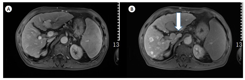
图 1 65岁男性患者的慢性乙型肝炎和Child-Pugh A肝硬化行钆塞酸增强肝脏MRI检查
在肝胆相图像上, A: 门静脉相对于肝实质呈低信号(评分2); B: 胆汁造影剂排泄至总胆管(白色箭头,评分2), 肝实质相对于肾脏呈高信号(评分2). 本例中3个FLIS参数的和是6.
Figure 1. Gadolinium seric acid enhanced liver MRI in a 65-year-old male patient with chronic hepatitis B and Child-Pugh A cirrhosis.

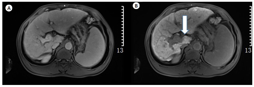
图 2 55岁的男性患者的慢性乙型肝炎和Child-Pugh B肝硬化行钆塞酸增强肝MRI检查
该患者经历了肝代偿。在肝胆期图像上, A: 门静脉相对于肝实质呈高信号(评分0); B: 胆汁对比物排泄至胆总管(白色箭头,评分2), 肝实质相对于肾脏呈等信号(评分1). 本例中3个FLIS参数的和为3.
Figure 2. A 55-year-old male patient with chronic hepatitis B and Child-Pugh B cirrhosis underwent gadolinium seric acid-enhanced liver MRI.

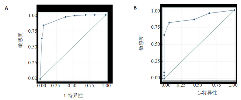
图 3 预测CLD或CP A肝硬化的特异性和敏感度
Figure 3. Specificity and sensitivity for predicting CLD or CP A cirrhosis.
表 1 FLIS参数的相关性以及FLIS与CP评分系统的相关性
Table 1. Correlation of three FLIS parameters and the correlation be-tween FLIS and CP scoring system
参数 r P Child-Pugh评分 - - 肝实质增强质量评分 -0.68 < 0.001 胆汁造影剂排泄质量评分 -0.60 < 0.001 门静脉标志质量评分 -0.82 < 0.001 FLIS,以上3个分数之和 -0.80 < 0.001 FLIS: 功能性肝脏成像评分.  下载: 导出CSV
表 2 与肝功能失代偿发生相关的单因素和多因素Cox比例风险分析
Table 2. Univariate and multivariate Cox proportional risk analyses associated with hepatic decompensation
临床和MR特征 单变量 多变量 风险比 P 校正风险比 P FLIS评分 ≥5 1 1 < 5 50.0(6.2~400.4) < 0.001 50.0(6.2~400.4) < 0.001 总胆红素 < 34 1 ≥34 5.1(0.6~43.7) 0.14 白蛋白 ≥35 1 < 35 23.7(5.2~107.7) < 0.001 凝血酶原时间/国际标准化比值 < 1.7 1 ≥1.7 7.1(0.9~59.1) 0.07
下载: 导出CSV
-
[1] 尤云峰, 王明亮, 曾蒙苏. 肝内门静脉-体静脉分流的MRI表现[J]. 中国临床解剖学杂志, 2020, 38(4): 396-400. https://www.cnki.com.cn/Article/CJFDTOTAL-ZLJZ202004007.htm [2] Poetter-Lang S, Bastati N, Messner A, et al. Quantification of liver function using gadoxetic acid- enhanced MRI[J]. Abdom Radiol (NY), 2020, 45(11): 3532-44. doi: 10.1007/s00261-020-02779-x [3] Huang MQ, Shen SL, Cai HS, et al. Regional liver function analysis with gadoxetic acid- enhanced MRI and virtual hepatectomy: prediction of postoperative short-term outcomes for HCC[J]. Eur Radiol, 2021, 31(7): 4720-30. doi: 10.1007/s00330-020-07606-x [4] 陈川梅, 魏忠荣, 杨柠娜, 等. MRI及CT诊断肝脏局灶性结节状脂肪浸润的价值及误诊分析[J]. 影像研究与医学应用, 2019, 3(21): 191-3. https://www.cnki.com.cn/Article/CJFDTOTAL-YXYY201921124.htm [5] 李秀梅, 陈群林, 陈晓丹, 等. Gd-BOPTA增强MRI肝胆期肝实质强化程度与肝纤维化分期的相关性[J]. 中国介入影像与治疗学, 2016, 13 (12): 728-32. https://www.cnki.com.cn/Article/CJFDTOTAL-JRYX201612006.htm [6] Lee S, Kim KW, Jeong WK, et al. Gadoxetic acid-enhanced MRI as a predictor of recurrence of HCC after liver transplantation[J]. Eur Radiol, 2020, 30(2): 987-95. doi: 10.1007/s00330-019-06424-0 [7] Park HJ, Lee SS, Park B, et al. Radiomics analysis of gadoxetic acid-enhanced MRI for staging liver fibrosis[J]. Radiology, 2019, 292(1): 269. [8] T. Denecke, I.G. Steffen, S. Agarwal, 等. 肝细胞腺瘤在钆塞酸增强MR成像上的表现[J]. 国际医学放射学杂志, 2012, 35(5): 507-8. https://www.cnki.com.cn/Article/CJFDTOTAL-GWLC201205119.htm [9] 宣浩波, 祝跃明, 沈健, 等. 肝细胞特异性对比剂Gd-EOB-DTPA在肝转移瘤MRI诊断中的应用[J]. 中国现代医生, 2012, 50(16): 94-5, 97, 161. https://www.cnki.com.cn/Article/CJFDTOTAL-ZDYS201216045.htm [10] Elkilany A, Geisel D, Müller T, et al. Gadoxetic acid-enhanced MRI in primary sclerosing cholangitis: added value in assessing liver function and monitoring disease progression[J]. Abdom Radiol (NY), 2021, 46(3): 979-91. [11] Grieser C. Editorial comment: advances in the evaluation of liver imaging and function with gadoxetic acid-enhanced MRI[J]. Eur Radiol, 2021, 31(11): 8374-5. [12] Shimada K, Isoda K, Hirokawa Y, 等. 比较Gd-EOB-DTPA增强MRI与扩散加权成像对肝内小转移灶的检出[J]. 国际医学放射学杂志, 2011, 34(1): 93. https://www.cnki.com.cn/Article/CJFDTOTAL-GWLC201101086.htm [13] Soussan M, Aubé C, Bahrami S, 等. 偶发性局灶性实性肝脏病变: 对比增强超声及MRI诊断作用[J]. 国际医学放射学杂志, 2010, 33(5): 499. https://www.cnki.com.cn/Article/CJFDTOTAL-GWLC201005100.htm [14] Hectors SJ, Kennedy P, Huang KH, et al. Fully automated prediction of liver fibrosis using deep learning analysis of gadoxetic acid-enhanced MRI[J]. Eur Radiol, 2021, 31(6): 3805-14. [15] Shinmura R, Matsui O, Kadoya M, 等. 肝细胞癌交界性病变的恶性富血供病灶的检测: 动态MDCT、动态MRI和SPIO增强MRI的对照研究[J]. 国际医学放射学杂志, 2008, 31(6): 514. https://www.cnki.com.cn/Article/CJFDTOTAL-GWLC200806111.htm [16] 罗维华, 赵新湘, 王燕, 等. 无周围胆管扩张的肝内周围型胆管细胞癌MRI表现及病理分析[J]. 临床放射学杂志, 2013, 32(4): 519-21. https://www.cnki.com.cn/Article/CJFDTOTAL-LCFS201304020.htm [17] Rhee H, Cho ES, Nahm JH, et al. Gadoxetic acid-enhanced MRI of macrotrabecular-massive hepatocellular carcinoma and its prognostic implications[J]. J Hepatol, 2021, 74(1): 109-21. [18] Jhaveri KS, Babaei Jandaghi A, Thipphavong S, et al. Can preoperative liver MRI with gadoxetic acid help reduce open-close laparotomies for curative intent pancreatic cancer surgery?[J]. Cancer Imaging, 2021, 21(1): 45. [19] Elkilany A, Fehrenbach U, Auer TA, et al. A radiomics- based model to classify the etiology of liver cirrhosis using gadoxetic acid-enhanced MRI[J]. Sci Rep, 2021, 11(1): 10778. -

 点击查看大图
计量
- 文章访问数: 170
- HTML全文浏览量: 141
- PDF下载量: 7
- 被引次数: 0