


Value of serum creatine kinase, soluble vascular endothelial growth factor receptor-1 combined with MRI in evaluating placenta accrete
-
摘要:
目的 分析血清肌酸激酶(CK)、可溶性血管内皮生长因子受体-1(sFlt-1)联合MRI征象检查评估胎盘植入的价值。 方法 选择本院2015年4月~2021年4月收治的108例可疑胎盘植入患者作为研究对象,依据产后病理诊断结果将患者分为胎盘植入组(n=30)及非胎盘植入组(n=78)。检测所有孕妇产前血清CK和sFlt-1水平,并进行MRI检查。比较两组血清CK、sFlt-1水平,MRI检查异常征象(胎盘内暗带、胎盘内血管、子宫膨大、子宫边界中断)发生率。采用Pearson相关性分析血清CK、sFlt-1水平与胎盘植入的相关性;采用Spearman相关性分析胎盘内暗带、胎盘内血管、子宫膨大、子宫边界中断与胎盘植入的相关性;采用ROC曲线分析血清CK、sFlt-1水平联合MRI检查评估胎盘植入的临床价值。 结果 胎盘植入组的血清CK水平高于非胎盘植入组,sFlt-1水平低于非胎盘植入组(P < 0.05);胎盘植入组MRI检查出现胎盘内暗带、胎盘内血管、子宫膨大、子宫边界中断的发生率高于非胎盘植入组(P < 0.05)。相关性分析显示,血清CK水平、胎盘内暗带、胎盘内血管、子宫膨大、子宫边界中断与胎盘植入与胎盘植入呈正相关关系(r=0.503、0.511、0.416、0.422、0.436,P < 0.05),血清sFlt-1水平与胎盘植入呈负相关关系(r=-0.486,P < 0.05)。ROC曲线分析结果显示,血清CK、sFlt-1水平联合MRI检查评估胎盘植入的曲线下面积为0.945,敏感度为90.0%,特异性为93.6%,明显优于各指标单独评估。 结论 血清CK、sFlt-1水平及胎盘内暗带、胎盘内血管、子宫膨大、子宫边界中断等MRI征象均与胎盘植入的发生密切相关,血清CK、sFlt-1水平联合MRI检查对胎盘植入具有显著的临床价值。 Abstract:Objective To analyze the value of serum creatine kinase (CK), soluble vascular endothelial growth factor receptor-1 (sFlt-1) combined with MRI examination in evaluating placenta accreta. Methods A total of 108 patients with suspected placenta accreta who were admitted to our hospital from April 2015 to April 2021 were selected. According to the results of postpartum pathological diagnosis, the patients were divided into placenta accreta group (n=30) and non-placenta accreta group (n=78). Prenatal serum CK and sFlt-1 levels of all pregnant women were detected, and MRI was performed. The serum CK and sFlt-1, and the incidence of abnormal signs on MRI (intraplacental dark zone, intraplacental blood vessels, uterine enlargement, and uterine border interruption) were compared between the two groups. Pearson correlation was used to analyze the correlation between serum CK and sFlt-1 levels and placenta accreta. Spearman correlation was used to analyze the correlation between placental dark zone, intraplacental blood vessels, uterine enlargement, uterine border interruption and placenta accreta. The clinical value of placenta accreta was evaluated by ROC curve of serum CK and sFlt-1 levels combined with MRI examination. Results The serum CK in the placenta accreta group was higher than that in the non-placenta accreta group, and the sFlt-1 was lower than that in the non-placenta accreta group (P < 0.05). The uterine enlargement and the incidence of uterine border interruption were higher than those in the non-placenta accreta group (P < 0.05). Correlation showed that serum CK, intraplacental dark zone, intraplacental blood vessels, uterine enlargement, and uterine border disruption were positively correlated with placenta accreta (r=0.503, 0.511, 0.416, 0.422, 0.436, P < 0.05). Serum sFlt-1 level was negatively correlated with placenta accreta (r=-0.486, P < 0.05). The results of ROC curve analysis showed that the AUC of serum CK and sFlt-1 levels combined with MRI in evaluating placenta accreta was 0.945, the sensitivity was 90.0%, and the specificity was 93.6%, which was significantly better than the evaluation of each index alone. Conclusion Serum CK and sFlt-1 levels and MRI signs such as dark zone in placenta, intraplacental blood vessels, uterine enlargement and uterine border interruption are closely related to the occurrence of placenta accreta. Serum CK and sFlt-1 levels combined with MRI have significant clinical value in placenta accreta. -

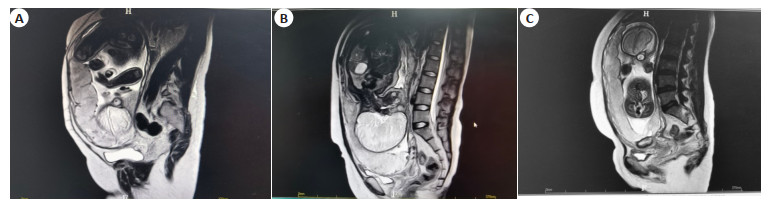
图 1 胎盘植入患者的影像学表现
A:患者1; B:患者2; C:患者3.
Figure 1. Imaging manifestations of patients with placenta implantation.

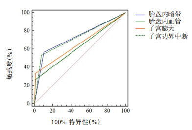
图 2 MRI异常征象评估胎盘植入的ROC曲线
Figure 2. ROC curve of abnormal MRI signs in evaluating placenta implantation.

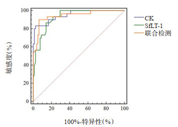
图 3 血清CK、sFlt-1水平联合MRI异常征象评估胎盘植入的ROC曲线
Figure 3. ROC curve of serum CK and sFlt-1 levels combined with abnormal MRI signs in evaluating placenta implantation.
表 1 两组产前血清CK、sFlt-1水平比较
Table 1. Comparison of prenatal serum CK and sFlt-1 levels between the two groups (Mean±SD)
组别 CK(U/L) sFlt-1(ng/mL) 胎盘植入组(n=30) 137.66±27.25 2.32±0.34 非胎盘植入组(n=78) 76.84±22.72 2.97±0.32 t 11.774 9.292 P < 0.001 < 0.001 CK: 肌酸激酶; sFlt-1: 可溶性血管内皮生长因子受体-1  下载: 导出CSV
表 2 两组术前MRI检查异常征象发生情况比较
Table 2. Comparison of the occurrence of abnormal signs of preoperative MRI examination in the two groups [n(%)]
MRI检查异常征象 胎盘植入组(n=30) 非胎盘植入组(n=78) χ2 P 胎盘内暗带 17(56.67) 8(10.26) 26.232 < 0.001 胎盘内血管 8(26.67) 1(1.28) 18.277 < 0.001 子宫膨大 10(33.33) 1(1.28) 24.331 < 0.001 子宫边界中断 16(53.33) 6(7.69) 27.825 < 0.001
下载: 导出CSV
表 3 血清CK、sFlt-1水平及MRI检查异常征象与胎盘植入的相关性分析
Table 3. Correlation analysis of serum CK and sFlt-1 levels and MRI examination for abnormal findings and placental implantation
项目 CK sFlt-1 胎盘内暗带 胎盘内血管 子宫膨大 子宫边界中断 胎盘植入 r 0.503 -0.486 0.511 0.416 0.422 0.436 P < 0.05 < 0.05 < 0.05 < 0.05 < 0.05 < 0.05
下载: 导出CSV
表 4 血清CK、sFlt-1水平联合MRI检查对胎盘植入的评估效能
Table 4. Serum levels of CK and sFlt-1 combined with MRI examination assessed the efficacy of placental implantation
检验结果变量 曲线下面积 标准误 P 95% CI 下限 上限 血清CK水平 0.952 0.022 < 0.001 0.893 0.984 血清sFlt-1水平 0.929 0.023 < 0.001 0.863 0.970 胎盘内暗带 0.732 0.049 < 0.001 0.638 0.813 胎盘内血管 0.627 0.042 0.002 0.529 0.718 子宫膨大 0.660 0.044 < 0.001 0.563 0.749 子宫边界中断 0.728 0.049 < 0.001 0.634 0.809 联合检测 0.945 0.025 < 0.001 0.884 0.980
下载: 导出CSV
-
[1] 付晓敏, 漆洪波. 胎盘植入研究进展[J]. 中国实用妇科与产科杂志, 2014, 30(1): 27-30. https://www.cnki.com.cn/Article/CJFDTOTAL-ZGSF201401010.htm [2] Bartels HC, Horsch A, Cooney N, et al. Living beyond placenta accreta spectrum: parent's experience of the postnatal journey and recommendations for an integrated care pathway[J]. BMC Pregnancy Childbirth, 2022, 22(1): 397. doi: 10.1186/s12884-022-04726-8 [3] 郑蔚然, 杨馨蕊, 孙瑾, 等. 前置胎盘附着于剖宫产子宫瘢痕部位伴或不伴胎盘植入对妊娠结局的影响[J]. 中华妇产科杂志, 2021, 56(12): 861-7. [4] 杨燕, 杨燕, 徐学翠, 等. 超声在胎盘植入性疾病分级诊断中的价值[J]. 医学研究生学报, 2021, 34(5): 486-9. https://www.cnki.com.cn/Article/CJFDTOTAL-JLYB202105008.htm [5] Xia H, Ke SC, Qian RR, et al. Comparison between abdominal ultrasound and nuclear magnetic resonance imaging detection of placenta accreta in the second and third trimester of pregnancy[J]. Medicine, 2020, 99(2): e17908. doi: 10.1097/MD.0000000000017908 [6] 何峰艺, 廖锦元. MRI诊断胎盘植入研究进展[J]. 中国医学影像技术, 2020, 36(5): 784-7. https://www.cnki.com.cn/Article/CJFDTOTAL-ZYXX202005041.htm [7] 张茂春, 张红薇, 陈娇, 等. 产前超声征象评分联合肌酸激酶对前置胎盘合并胎盘植入的诊断价值分析[J]. 现代生物医学进展, 2018, 18 (18): 3563-7. https://www.cnki.com.cn/Article/CJFDTOTAL-SWCX201818037.htm [8] 高素娟, 刘彬, 曹艳敏, 等. 子宫动脉灌注栓塞治疗胎盘植入的效果及对患者血清VEGF和SFlt-1水平的影响[J]. 广西医科大学学报, 2017, 34(9): 1299-302. https://www.cnki.com.cn/Article/CJFDTOTAL-GXYD201709010.htm [9] 周冰峰, 阚长利, 侯敬, 等. 孕妇血清VEGF和sFlt-1联合多普勒超声诊断胎盘植入效果[J]. 中国计划生育学杂志, 2021, 29(1): 114-7. https://www.cnki.com.cn/Article/CJFDTOTAL-JHSY202101029.htm [10] 沈铿, 马丁. 妇产科学[M]. 3版. 北京: 人民卫生出版社, 2015. [11] 曹满瑞, 杜牧, 黄怡, 等. 胎盘植入的MRI征象分析[J]. 中华放射学杂志, 2012, 46(7): 629-32. https://www.cnki.com.cn/Article/CJFDTOTAL-XYXZ202208020.htm [12] Li N, Hou R, Liu CX, et al. Integration of transcriptome and proteome profiles in placenta accreta reveals trophoblast overmigration as the underlying pathogenesis[J]. Clin Proteomics, 2021, 18(1): 31. [13] Varlas VN, Bors RG, Birsanu S, et al. Maternal and fetal outcome in placenta accreta spectrum (PAS) associated with placenta previa: a retrospective analysis from a tertiary center[J]. J Med Life, 2021, 14(3): 367-75. [14] Ophir E, et al. Creatine kinase as a biochemical marker in diagnosis of placenta increta and percreta[J]. Am J Obstet Gynecol, 1999, 180(4): 1039-40. [15] 陈宏霞, 曹伍兰. 血清肌酸激酶及同工酶在胎盘植入中的诊断价值[J]. 中国实用妇科与产科杂志, 2008, 24(9): 686-7. https://www.cnki.com.cn/Article/CJFDTOTAL-ZGSF200809020.htm [16] Schwickert A, Chantraine F, Ehrlich L, et al. Maternal serum VEGF predicts abnormally invasive placenta better than NT-proBNP: a multicenter case-control study[J]. Reprod Sci, 2021, 28(2): 361-70. [17] Bus P, Scharpfenecker M, Van Der Wilk P, et al. The VEGF-A inhibitor sFLT-1 improves renal function by reducing endothelial activation and inflammation in a mouse model of type 1 diabetes [J]. Diabetologia, 2017, 60(9): 1813-21. http://www.nstl.gov.cn/paper_detail.html?id=1abb7a1e797b056e163cc1e015faeb1c [18] Wang N, Shi DD, Li N, et al. Clinical value of serum VEGF and sFlt-1 in pernicious placenta previa[J]. Ann Med, 2021, 53(1): 2041-9. [19] 柳月霞, 王东旭, 魏菊红, 等. 妊娠晚期母体血清VEGF、PlGF和sFlt-1对胎盘植入性疾病的预测价值[J]. 中国计划生育学杂志, 2019, 27 (11): 1548-51. https://www.cnki.com.cn/Article/CJFDTOTAL-JHSY201911035.htm [20] 吴华臣, 李丽华, 刘燕君, 等. 彩色多普勒超声检查联合AFP、CK监测对产前胎盘植入诊断的价值研究[J]. 临床和实验医学杂志, 2018, 17 (24): 2673-6. https://www.cnki.com.cn/Article/CJFDTOTAL-SYLC201824030.htm [21] 张艳, 袁玉红. 彩色多普勒超声检查联合血清VEGF、sFlt-1检测在胎盘植入产前诊断中的应用价值[J]. 中国妇幼保健, 2018, 33(7): 1622-4. https://www.cnki.com.cn/Article/CJFDTOTAL-ZFYB201807062.htm [22] 范田依, 庞静, 王艳蕾, 等. 超声结合血清肌酸激酶、同工酶检测对胎盘植入的诊断价值[J]. 中国计划生育学杂志, 2020, 28(12): 2059-62. https://www.cnki.com.cn/Article/CJFDTOTAL-JHSY202012033.htm [23] Morrow D, Hatch E, Hamm K, et al. Flk-1/KDR mediates ethanolstimulated endothelial cell Notch signaling and angiogenic activity [J]. J Vasc Res, 2014, 51(4): 315-24. [24] 孔德会, 刘翠芳, 刘云, 等. MRI多征象联合在胎盘植入中的诊断价值[J]. 放射学实践, 2017, 32(3): 271-4. https://www.cnki.com.cn/Article/CJFDTOTAL-FSXS201703018.htm -

 点击查看大图
计量
- 文章访问数: 162
- HTML全文浏览量: 115
- PDF下载量: 9
- 被引次数: 0