

 Find Duplicates
 Check Document
 Submission(new)
 Experts Office
 Editorial Office
2023 Vol. 46, No. 2
Display Method:
2023, 46(2): 187-194.
doi: 10.12122/j.issn.1674-4500.2023.02.01
Abstract:
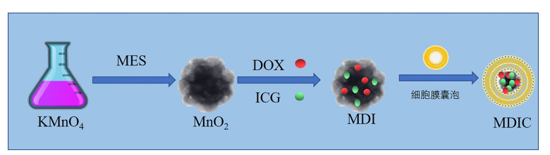
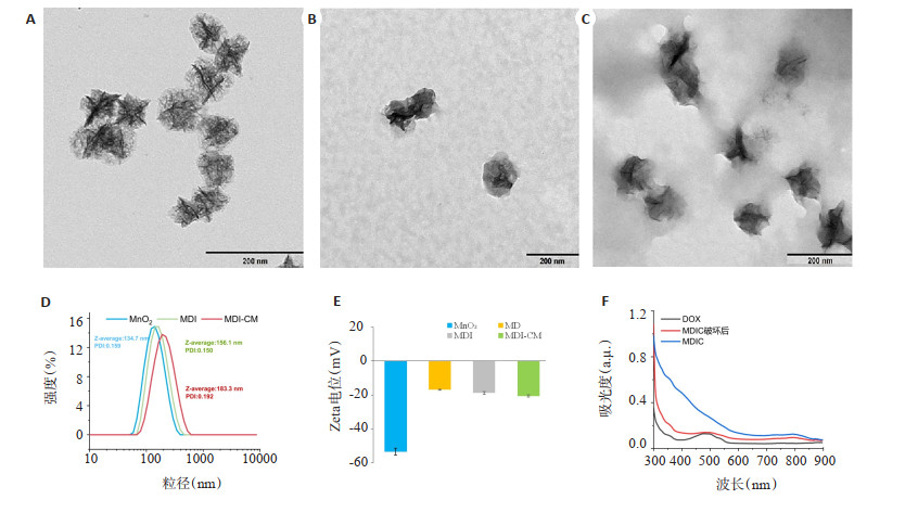
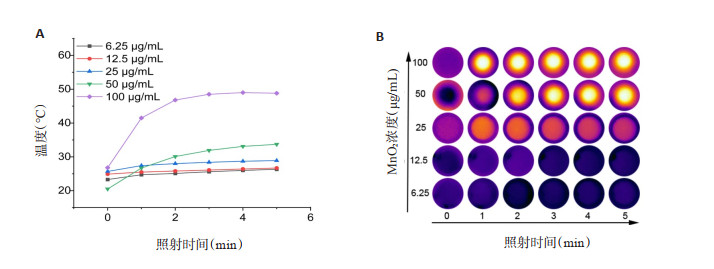
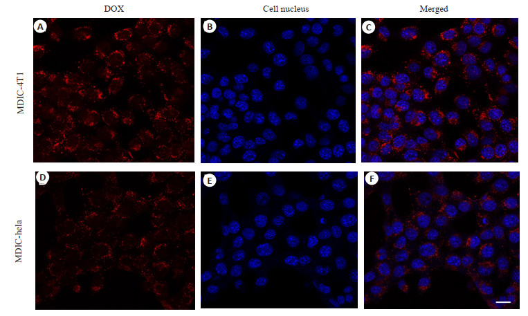
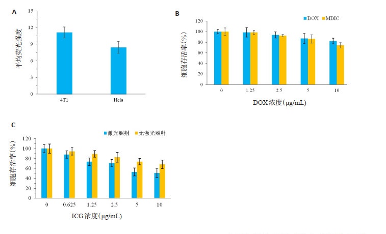
Objective To prepare homologous targeting therapeutic integrated nanoprobe MnO2@DOX@ICG@CM(MDIC), observe the homologous targeting ability of MDIC nanoprobe, and explore its multimodal imaging value and the synergistic effect of photothermal combined chemotherapy. Methods The cell membrane of breast cancer 4T1 was obtained by chemical lysis and ultrasonic homogenization. The MnO2 nanoprobe coated with doxorubicin DOX and photosensitizer indocyanine green was prepared by liposome extruding method. The appearance morphology, particle size and surface potential of MDIC were characterized, and its photoacoustic imaging and magnetic resonance imaging capabilities were studied. The photothermal heating properties of MDIC nanoprobes with different concentrations were observed. MTT assay was used to evaluate the antitumor effect of photothermal-chemotherapy synergistic therapy on 4T1 cells. Results MDIC nanoprobes were successfully prepared with flower-like structure, average particle size of 183.3 nm, and surface potential of -20.6±0.77 mV. The membrane was successfully coated under transmission electron microscope. After laser irradiation of MDIC, the solution temperature increases with the increase of nanoprobe concentration, which has a good photothermal heating effect. The results of photoacoustic and magnetic resonance imaging showed that MDIC had good imaging contrast ability. Laser confocal microscopy showed that MDIC had good homology targeting. The results of cytotoxicity test showed that photothermalchemotherapy combined therapy had better cancer cell killing effect. Conclusion A homologous targeting photoacoustic/ magnetic resonance multimodal imaging contrast probe MDIC was successfully prepared. The probe has good photothermal heating efficiency and anti-tumor effect in vitro. It can significantly enhance the photoacoustic imaging effect and magnetic resonance T1 enhancement effect.
2023, 46(2): 195-201.
doi: 10.12122/j.issn.1674-4500.2023.02.02
Abstract:
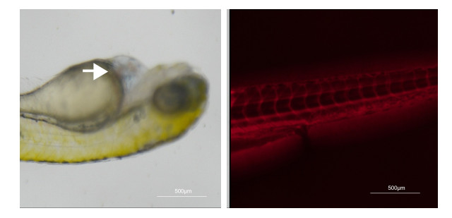
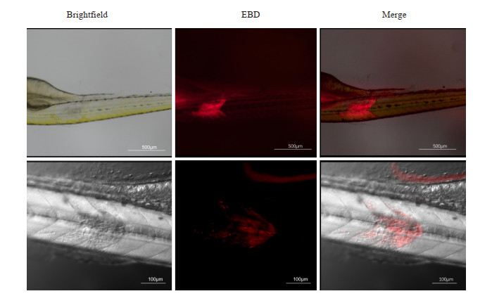
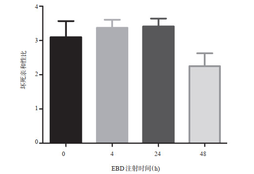
Objective To explore the feasibility of making a muscle necrosis model in zebrafish, and to apply this model to investigate the necrosis affinity of Evans Blue dye (EBD) by in vivo fluorescence imaging. Methods In order to make a zebrafish muscle necrosis model, 10 nL of absolute alcohol was injected into the dorsal muscle of zebrafish larvae 3-7 d post fertilization by using the microinjection pump instrument. The distribution of EBD in the muscle necrosis model group and the control group were dynamically observed by fluorescence and laser confocal microscopy at 0, 4, 24 and 48 h after injection of 5 nL 0.1% EBD at the common cardinal vein. The fluorescence intensity, range and ratio of EBD in both the necrotic and normal muscle area were quantitative analyzed. Results Zebrafish model of muscle necrosis was successfully established with a high rate. Fluorescence and laser confocal microscopy showed that there was a large amount of EBD red fluorescence accumulation in the necrotic muscle area, which was consistent with the necrotic muscle in bright field. Dynamic observation showed that the red fluorescence intensity gradually increased, and gradually decreased after reaching the peak at 24 h. The difference in fluorescence intensity between necrotic and normal muscle was statistically significant (P < 0.05). At 4 h and 24 h, the difference was the most significant, which were 58.30±5.14, 17.36±1.16 and 54.20±4.25, 15.96±0.79, respectively. However, no significant differences were found in the necrosis affinity ratio among different time points (P>0.05). Conclusion The proposed model of muscle necrosis in zebrafish is simple and convenient, which can be used as a new experimental platform for studying necrosis avid agents. This study preliminary showed that EBD may selectively accumulate in the necrotic muscle of zebrafish, and it could be a potential necrosis avid contrast agent.

2023, 46(2): 202-209.
doi: 10.12122/j.issn.1674-4500.2023.02.03
Abstract:
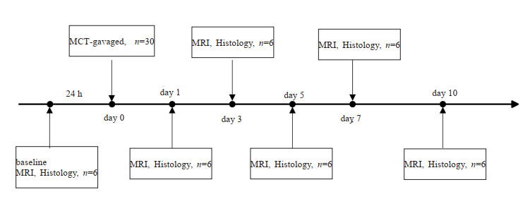
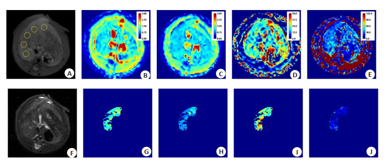
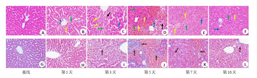
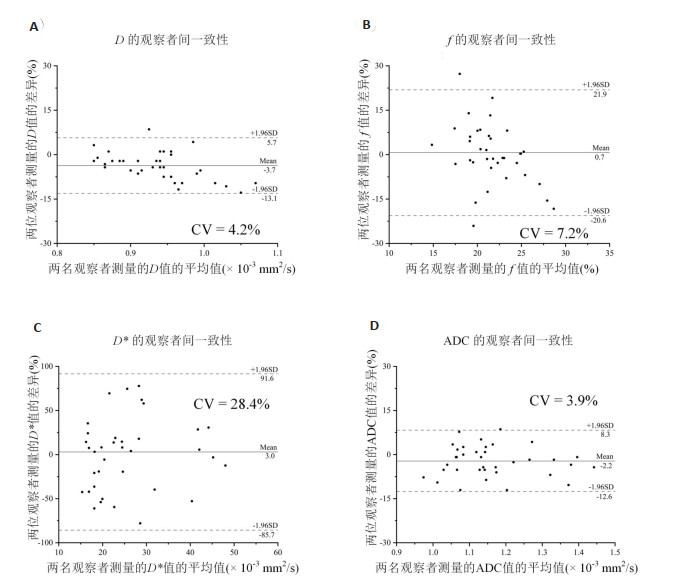
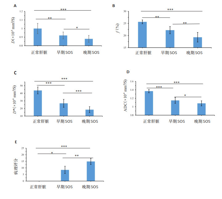
Objective To investigate the feasibility of introvoxel incoherent motion (IVIM) MRI parameters in evaluating the progression of microcirculatory disturbances in a monocrotaline-induced sinusoidal obstruction syndrome (SOS) rat model. Methods Thirty-six Sprague-Dawley rats were randomly divided into SOS group (n=30) and baseline group (n=6). Rats in SOS group were gavaged with monocrotaline at a dose of 160 mg/kg. Six rats were chosen to perform IVIM MRI scan at 1st, 3rd, 5th, 7th and 10th days after monocrotaline administration, respectively. Pure molecular diffusion (D), perfusion fraction (f), pseudodiffusion (D*), apparent diffusion coefficient (ADC) were measured. After MRI scan, the rats in SOS group were sacrificed for histological evaluation. Rats in baseline group without any intervention, and IVIM MRI was performed one day before MCT administration in SOS group. After the IVIM MRI scan, the rats in baseline group were sacrificed for histological evaluation. The liver samples were categorized into normal, "early", and"late"SOS according to the fibrotic degree. The correlation between MRI parameters and pathological scores as well as receiver operating characteristic curves were analyzed. Results D, f, D* and ADC values decreased on days 1, 3 and 5, increased on days 7 and 10 during SOS progression, and were negatively correlated with pathological scores. Compared with those of normal livers, the D (P < 0.01), f (P < 0.01), D* (P < 0.001) and ADC (P < 0.001) of"early"SOS significantly decreased, and those of"late"SOS significantly decreased (P < 0.001). The correlation coefficient between f (r=-0.732) and pathological score was better than that of D (r=-0.539), D* (r=-0.550), and ADC (r=-0.554). The AUC of D and f (AUC=0.8 and 0.85) were much higher than those of D* and ADC (AUC=0.74 and 0.73) in detecting"late"SOS. Conclusion IVIM MRI analysis can quantitatively characterize the different stages of hepatic SOS. In the progression of SOS injury, IVIM MRI quantitative parameters can provide important information for clinical monitoring of SOS.
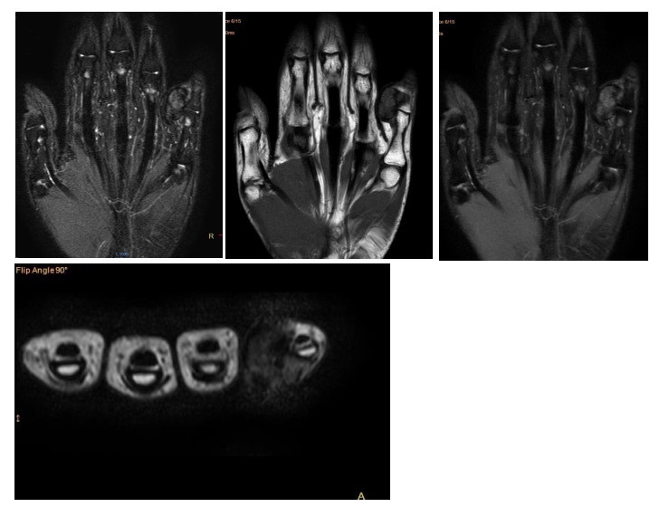
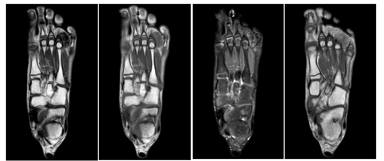
2023, 46(2): 210-213.
doi: 10.12122/j.issn.1674-4500.2023.02.04
Abstract:
Objective To investigate the MR features and pathological basis of localized tenosynovial giant cell tumor in hands and feet. Methods Ten patients with pathologically confirmed localized tenosynovial giant cell tumor in hands and feet in the Third Affiliated Hospital of Southern Medical University from April 2018 to July 2022 were retrospectively analyzed. The patients included 5 males and 5 females with a median age of 26.5 years. All patients underwent preoperative MR scanning and postoperative pathological examinations. MR findings of the lesions were analyzed by two experienced doctors, including the location, shape, size, boundary, signal intensity patterns, involvement range, enhancement patterns and bone abnormalities. The pathological basis of MR features was explored too. Results Six cases occurred in the hand and 4 cases in the feet of the 10 patients. The maximum axial diameter was about 24 mm, and the maximum extension diameter was about 26 mm. The lesions were hypo-or isointense at T1WI, mainly hypo-or isointense at T2WI, and mainly hyperintense at PDWI. Most of the lesions (n=8) showed strip or patchy hypointensity on all sequences, with mild to moderate inhomogeneous enhancement. Adjacent bone compressed was observed in 3 cases and bone eroded in 1 case. Pathological examinations showed that the masses were hard, mainly composed of mononuclear cells, and hemosiderin was seen between the cells. Conclusion Localized tenosynovial giant cell tumor in hands and feet shows relatively specific MR findings, which reflects its pathological features.
2023, 46(2): 214-219.
doi: 10.12122/j.issn.1674-4500.2023.02.05
Abstract:
Objective To investigate the clinical manifestations, laboratory and imaging features of patients with anti-GABA-B receptor encephalitis. Methods Three patients with anti-GABA-B receptor encephalitis were selected from our hospital from May 2021 to November 2022 with complete clinical data and in accordance with the diagnostic criteria of autoimmune peripheral encephalitis in the Consensus of Chinese Experts on the Diagnosis and Treatment of Autoimmune Encephalitis (2022 edition). Clinical manifestations, cerebrospinal fluid (CSF), immunological, electroencephalogram and imaging examinations of 3 patients of anti-GABA-B receptor encephalitis were analyzed retrospectively. Results All the 3 patients suffered from acute onset with epilepsy as first symptom or main clinical manifestation, accompanied by cognitive dysfunction and mental behavior abnormality of different degrees. All the 3 patients were complicated with tumors, including multiple myeloma, lung cancer and nasopharyngeal carcinoma. Immunological examinations of CSF and serum indicated that CSF and/or serum anti-GABA-B receptor antibody were positive. Electroencephalogram were abnormal, showing epileptic discharge, scattered distribution or diffuse slow wave. Imaging examinations showed that the lesion involved unilateral hippocampus, brain CT showed low density, MRI showed long T1 signal, long T2 signal, high T2 FLAIR signal, high DWI signal, slight high or equal ADC signal. Conclusion Patients with anti-GABA-B receptor encephalitis suffered from epilepsy as main manifestation and are prone to tumor. We reported that anti-GABA-B receptor encephalitis combined with nasopharyngeal carcinoma for the first time, which expands the disease spectrum.
2023, 46(2): 220-226.
doi: 10.12122/j.issn.1674-4500.2023.02.06
Abstract:
Objective To investigate the clinical application of endoscopic ultrasonography guided fine needle aspiration (EUS-FNA) biopsy in pancreatic space occupying lesions. Methods Fifty-five patients who underwent EUS-FNA examination for suspected pancreatic space-occupying lesions in the First Affiliated Hospital of Bengbu Medical College from January 2018 to January 2022 were screened, with an average age of 62.91±11.21 years, including 29 males and 26 females. The general clinical data, lesion characteristics, pancreatic duct conditions, puncture times and postoperative complications of patients undergoing EUS-FNA were analyzed, with the histopathological results of puncture samples as the gold standard. We evaluated the sensitivity, specificity, and accuracy of EUS-FNA in diagnosing pancreatic space-occupying lesions, as well as the clinical factors affecting the accuracy. Results EUS-FNA was successfully performed in all these 55 patients, the puncture success rate was 100%, and puncture pathology was obtained in all patients. Postoperative adverse reactions occurred in 2 cases, including abdominal pain in 1 case and hyperamylasemia in 1 case. All patients recovered well after routine clinical treatment. Forty-eight cases (87.27%) were definitely diagnosed by EUS-FNA, and 7 cases (12.73%) were not definitely diagnosed. The diagnostic sensitivity was 92.31% (48/52), specificity was 100% (3/3), accuracy was 92.73% (51/55), positive predictive value was 100% (48/48), negative predictive value was 42.86% (3/7). The accuracy of EUS-FNA was related to the patient's age and pancreatic duct dilatation (P < 0.05), but not gender, blood glucose, tumor markers, lesion characteristics, lesion size, location, times of puncture needles, cytological examination methods (P > 0.05). Conclusion EUS-FNA has high sensitivity, specificity and accuracy in the diagnosis of pancreatic space occupying lesions. The complications are rare and mild, which provides an effective diagnostic method for clinical practice.
2023, 46(2): 227-231.
doi: 10.12122/j.issn.1674-4500.2023.02.07
Abstract:
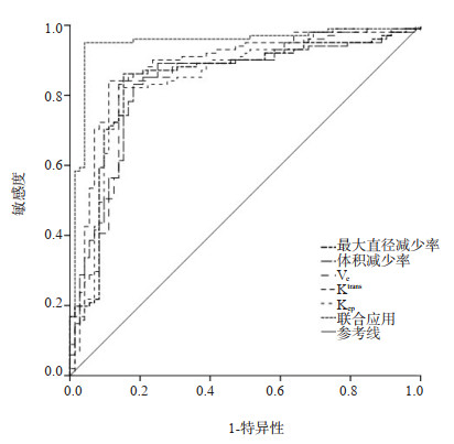
Objective To analyze the construction and evaluation of radiotherapy effect prediction model for lung cancer patients based on tumor volume. Methods A total of 173 patients with lung cancer treated in our hospital from January 2019 to December 2021 were selected as the research objects. The subjects received radiotherapy regimen for intervention treatment. Imaging examination was performed before treatment and 1 month after radiotherapy to read the lesion volume, calculate the maximum diameter reduction rate and volume reduction rate before and after treatment. We scaned and analyzed the lesion areas Ve, Ktrans and Kep. After treatment, the clinical efficacy of the patients was evaluated. Pearson correlation test was used to analyze the correlation between maximum diameter reduction rate and volume reduction rate. Logistic regression model was used to analyze the efficacy evaluation model of Ve, Ktrans and Kep combined application to predict patients' tumor volume. ROC curve was drawn to analyze the value of each index alone and combined application to predict patients' efficacy. Results The maximum diameter reduction rate and volume reduction rate in CR+PR group were significantly lower than those in SD+PD group (P < 0.05). There was a significant positive correlation between the maximum diameter reduction rate and volume reduction rate and the clinical efficacy of lung cancer patients (P < 0.05). Ve, Ktrans and Kep in CR+PR group were significantly higher than those in SD+PD group (P < 0.05). The multiple regression prediction model of MRI combined with tumor volume and maximum diameter was log (P)=0.685× maximum diameter reduction rate+0.651×physical examination reduction rate+0.604×Ve+ 0.612×Ktrans+0.644×Kep+0.849; The value of using MRI index combined with tumor volume and maximum diameter to predict the clinical efficacy of patients was significantly higher than that of using each index alone (P < 0.05). Conclusion The evaluation of tumor volume and MRI scan in patients with lung cancer can effectively analyze the prognosis quality of patients with lung cancer.
2023, 46(2): 232-237.
doi: 10.12122/j.issn.1674-4500.2023.02.08
Abstract:
Objective To investigate the diagnostic and differential diagnostic value of CT enterography for colon-type Crohn's disease and ulcerative colitis. Methods Thirty-six patients with colonic Crohn's disease and 36 patients with ulcerative colitis admitted to the First Affiliated Hospital of Bengbu Medical College and Jiangsu Provincial Hospital of Nanjing University of Traditional Chinese Medicine from January 2019 to January 2022 were selected for CT enterography examination. The imaging features of both were observed. The patients with Crohn's disease and ulcerative colitis were retrospectively analyzed by combining laboratory indices and clinical manifestations. Results Among the 36 patients with Crohn's disease, CT enterography images showed 21 cases of uneven thickening of the intestinal wall, 22 cases of intestinal stricture, 7 cases of fistula formation, and 21 cases of involvement of the ascending colon were higher than those of patients with ulcerative colitis (P < 0.05). Among the 36 patients with ulcerative colitis, 18 cases of mucus stools and 22 cases of bloody stools were higher than those of patients with Crohn's disease (P < 0.05). The mean age of onset was older in patients with ulcerative colitis than in patients with colonic Crohn's disease(P < 0.05). The mean values of C-reactive protein and erythrocyte sedimentation rate in patients with Crohn's disease were 28.17 mg/L and 32.25 mm/h, respectively. The mean values of C-reactive protein and erythrocyte sedimentation rate in patients with ulcerative colitis were 17.67 mg/L and 22.95 mm/h, respectively. The mean values of C-reactive protein and erythrocyte sedimentation rate were not statistically significant for the differentiation of Crohn's disease from ulcerative colitis (P > 0.05). Conclusion The CT enterography imaging features can be used to differentiate colon-type Crohn's disease from ulcerative colitis more visually. It provides a more reliable basis for treatment plan design by combining clinical, endoscopic manifestations and pathological diagnosis.
2023, 46(2): 238-242.
doi: 10.12122/j.issn.1674-4500.2023.02.09
Abstract:
Objective To explore the diagnostic value of ultrasound-guided fine needle aspiration (US-FNA) combined with contrast-enhanced ultrasonography (CEUS) and TI-RADS classification criteria for thyroid benign and malignant nodules. Methods From January 2020 to June 2021, 110 patients with thyroid nodules (totally 134 nodules) in Hainan Western Central Hospital were selected as the research objects.All patients were underwent US-FNA examination and CEUS examination.The results of individual examination and combined examination were compared, and the sensitivity, specificity, accuracy, positive predictive value and negative predictive value were compared. Results The sensitivity, specificity, accuracy, positive predictive value and negative predictive value of US-FNA examination were 96.23%, 85.71%, 94.03%, 96.23% and 85.71%, CEUS examination were 92.45%, 42.86%, 82.09%.85.96% and 60.00%, the combined inspection were 100.00%, 28.57%, 85.07%, 77.94% and 100.00%.The comparison of the sensitivity, accuracy, positive predictive value, and negative predictive value of the three methods were not statistically significant (P>0.05), but the specificity of US-FNA examination were significantly higher than that of CEUS examination and combined examination (P<0.05). Conclusion The US-FNA examination is highly specific in the diagnosis of benign and malignant thyroid nodules.The combined examination of US-FNA and CEUS has not effectively improved the accuracy and sensitivity of the detection.
2023, 46(2): 243-250.
doi: 10.12122/j.issn.1674-4500.2023.02.10
Abstract:
Objective To compare the pathological features and CT features of pulmonary space occupying in AIDS complicated with lung cancer, tuberculosis complicated with lung cancer and simple lung cancer. Methods Thirty-six patients with AIDS combined with lung cancer, 36 patients with tuberculosis combined with lung cancer and 36 patients with simple lung cancer admitted to our hospital from November 2021 to June 2022 were selected as the research objects.General data, clinical symptoms, pathological characteristics, and CT features of lung space occupying lesions were collected, and the above data of the three groups of patients were compared. Results The proportions of fever and poor appetite in the AIDS combined lung cancer group and the tuberculosis combined lung cancer group were higher than those in the simple lung cancer group (P < 0.05).The proportion of anemia and fungal infection in the group of AIDS with lung cancer was higher than that in the group of tuberculosis with lung cancer and the group of simple lung cancer (P < 0.05).There was no significant difference in other clinical symptoms among the three groups (P>0.05).The proportion of adenocarcinoma in the AIDS combined lung cancer group was higher than that in the tuberculosis combined lung cancer group and the simple lung cancer group (P < 0.05).The proportion of squamous cell carcinoma was lower than that in the tuberculosis combined lung cancer group and the simple lung cancer group (P < 0.05).There were no significant difference in pulmonary pathological classification between the tuberculosis combined lung cancer group and the simple lung cancer group (P>0.05).There were no significant difference in TNM staging among the three groups (P>0.05).The proportion of fibrous shadow in CT manifestations of HIV/AIDS patients with lung cancer was higher than that in pulmonary tuberculosis patients with lung cancer and lung cancer alone (P < 0.05).The CT findings of pulmonary space occupying in patients with pulmonary tuberculosis combined with lung cancer were higher than those in patients with simple lung cancer (P < 0.05).The percentages of miliary shadow and satellite focus were higher than those in patients with AIDS combined with lung cancer and patients with simple lung cancer (P < 0.05). Conclusion There are differences in pathological features and CT features of pulmonary space occupying between AIDS combined with lung cancer, tuberculosis combined with lung cancer and simple lung cancer.Clinical evaluation of patients with different types of lung cancer can be made according to the pathological features and CT features of pulmonary space occupying.

2023, 46(2): 251-256.
doi: 10.12122/j.issn.1674-4500.2023.02.11
Abstract:
Objective To explore the application value of acoustic palpation tissue imaging (VTI) and acoustic palpation tissue quantitative imaging (VTIQ) in C-TIRADS classification combined with acoustic radiation force pulse imaging technology in differential diagnosis of thyroid papillary carcinoma in C-TIRADS 3-5 thyroid nodules. Methods A total of 194 patients with thyroid nodules classified as C-TIRADS 3-5 in our hospital with 201 nodules were selected for VTIQ and VTI examination to obtain the average value of shear wave velocity and VTI image of the lesions.The ROC curve of the subjects was constructed based on the gold standard of pathology, and AUC was calculated to obtain the best diagnostic threshold of each diagnostic method.The diagnostic efficacy of three groups of independent diagnosis and C-TIRADS+VTI and C-TIRADS+VTIQ combined diagnosis were analyzed.The binary Logistic regression prediction model of C-TIRADS combined with VTIQ and VTI was constructed.The diagnostic efficacy of the three combined diagnostic groups was calculated and analyzed.The AUCs under the curve of each group were compared by Z-test. Results Eighty-one benign nodules and 120 malignant nodules were confirmed by clear cytopathology and histopathology.C-TIRADS grade was positively correlated with the malignant rate of thyroid nodules (r=0.624, P < 0.01).Compared with the application of C-TIRADS alone (0.806), the AUC of C-TIRADS VTI group, C-TIRADS VTIQ group and C-TIRADS VTI VTIQ group increased to a certain extent, and the combination of the three groups increased the most significantly (0.908), the difference was statistically significant (P < 0.05).The kappa value of the consistency between the three combined diagnosis and pathology was higher than that of C-TIRADS alone.The sensitivity, specificity and accuracy of C-TIRADS alone in the diagnosis of benign and malignant thyroid nodules were 90.0%, 70.4% and 82.0%, respectively.The sensitivity, specificity and accuracy of C-TIRADS VTIQ VTI combined prediction model group in diagnosis of benign and malignant thyroid nodules were 91.7%, 80.2% and 86.1%, respectively, and the accuracy of diagnosis of malignant thyroid nodules was 89.2%. Conclusion The VTI and VTIQ in acoustic radiation force pulse technology enhance the diagnostic performance of C-TIRADS for benign and malignant thyroid nodules.Acoustic radiation force pulse technology can be used as a supplement to conventional ultrasound to improve the ability to differentiate thyroid papillary carcinoma in nodules classified as 3-5 in a non-invasive way.

2023, 46(2): 257-261.
doi: 10.12122/j.issn.1674-4500.2023.02.12
Abstract:
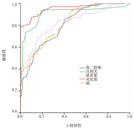
Objective To explore preliminary evaluation of pericoronal adipose tissue changes in patients with coronary atherosclerosis by texture analysis. Methods We selected 220 patients with coronary artery CT angiography from Hainan Hospital of PLA General Hospital, including 110 in the normal group and 110 in the coronary atherosclerosis group (including calcified, non-calcified and mixed plaques).The texture features of pericoronal adipose tissue around the target plaque were analyzed.We analyzed MPR images of the proximal three main coronary arteries (including right coronary artery, left anterior descending branch and circumflex branch), selected five texture feature parameters: angular second moment, contrast, correlation, inverse difference moment, and entropy.Mann-Whitney U was used to test differences in texture characteristics between groups. Results All the texture feature parameters showed significant differences between the two groups (PASM < 0.001, Pcontrast < 0.001, Pcorelation < 0.001, PIDM < 0.001, Pentropy < 0.001).There were differences in the texture features of pericoronary adipose tissue between the normal group and the coronary atherosclerosis group, and each texture parameter has good diagnostic value.ROC analysis confirmed that the area under the curve of the five texture features was greater than 0.8, which had good diagnostic value. Conclusion The texture features of pericoronary adipose tissue differ between the normal group and the coronary atherosclerosis group.Therefore, the texture feature analysis can be based on coronary CT angiography examination, preliminary evaluation of pericoronal adipose tissue changes in patients with coronary atherosclerosis.
2023, 46(2): 262-266.
doi: 10.12122/j.issn.1674-4500.2023.02.13
Abstract:
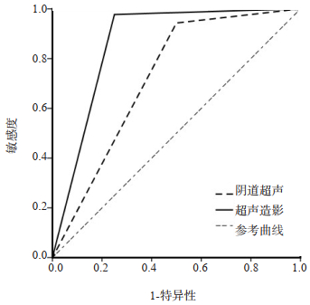
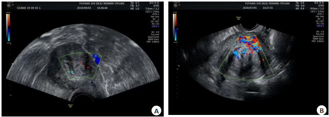
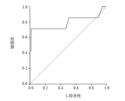
Objective To analyze the diagnostic efficacy of transvaginal ultrasound combined with contrast enhanced ultrasound for first-trimester caesarean scar ectopic pregnancy. Methods Ninety-two women with suspected scar pregnancy in our hospital from December 2019 to December 2021 were enrolled. The lower uterine segment thickness was measured using transvaginal ultrasound combined with contrast enhanced ultrasound. The imaging findings and the pathological results were compared between two examinations were compared. ROC curve was used to evaluate the diagnostic efficacy of lower uterine segment thickness on scar pregnancy. Results The mean lower uterine segment thickness was 7.03 ± 0.17 mm measured by transvaginal ultrasound, 7.25±0.14 mm by contrast enhanced ultrasound and 7.42±0.15 mm by pathological examination. The scar thickness in contrast enhanced ultrasound was significantly thicker than that in transvaginal ultrasound (P < 0.05). Comparison of the two types of images showed that 83 of the 92 patients diagnosed by transvaginal ultrasound had clearly observed the implantation of embryos at the scar and were diagnosed as scar pregnancy, while the implantation position of the other 9 patients could not be determined and scar pregnancy could not be ruled out. Contrast enhanced ultrasound showed that 86 patients were diagnosed with scar pregnancy, including 57 cases of cyst type, 29 cases of mass type, and 6 cases of myometrial trophoblastic tissue implantation. Pathological examination results showed that 88 cases of scar pregnancy, 2 cases of isthmic pregnancy, 2 cases of low embryo implantation site. Transvaginal ultrasound had a diagnostic accuracy of 92.39%, sensitivity of 94.31% and specificity of 50.00%, while contrast-enhanced ultrasound had a diagnostic accuracy of 96.74%, sensitivity of 97.73% as well as specificity of 75.00%, respectively. The AUCs of transvaginal ultrasound and contrast-enhanced ultrasound for the diagnosis of scar pregnancy were 0.722 and 0.864. The overall diagnostic efficacy of contrast enhanced ultrasound was significantly better than that of transvaginal ultrasound. Conclusion Taking pathological findings as golden standard, contrast- enhanced ultrasound has a better uterine scar thickness measurement compared with transvaginal ultrasound, which can clearly observe embryo implantation site and the relationship with the myometrium, achieves better diagnostic accuracy, sensitivity and specificity for scar pregnancy.
2023, 46(2): 267-270.
doi: 10.12122/j.issn.1674-4500.2023.02.14
Abstract:
Objective To explore the diagnostic significance of echocardiographic of coronary artery related blood flow velocity combined with segmental wall motion in patients with atherosclerotic heart disease with no significant change in ST-T segment detected by electrocardiogram. Methods A retrospective study was conducted on patients admitted to the Department of Cardiology in our hospital from January 2020 to June 2021 who were confirmed by coronary angiogrophy as coronary heart disease with vascular stenosis greater than 50% but not completel occlusion. The patients were performed with electrocardiogram examination. Forty patients without significant ST-T segment changes were selected as coronaty artery disease group. Meanwhile, 30 patients with vascular stenosis less than 50% confirmed by coronary angiography were selected as control group. All patients underwent echocardiography during admission to observe coronary blood flow and ventricular wall motion. Results The diastolic blood flow velocity in the coronaty artery disease group was significantly higher than that in the control group (P < 0.05). Echocardiography showed that the accuracy of diastolic segmental peak flow velocity in the diagnosis of coronaty artery disease was 92.3%, the sensitivity was 82.4%, the specificity was 90%, the critical value was 94.5 cm/s, the sensitivity of segmental wall motion in the diagnosis of coronary heart disease was 55.9%, the specificity was 86.7%, and the sensitivity and specificpatity of combined diagnosis of coronary heart disease were 84.2% and 92.5%. Conclusion Echocardiographic coronary flow velocity combined with segmental ventricular wall motion can improve the diagnostic rate of patients with coronary heart disease without obvious ST-T change. It is beneficial to reduce the misdiagnosis rate of clinical misdiagnusis of coronary heart disease patients.
2023, 46(2): 271-275.
doi: 10.12122/j.issn.1674-4500.2023.02.15
Abstract:
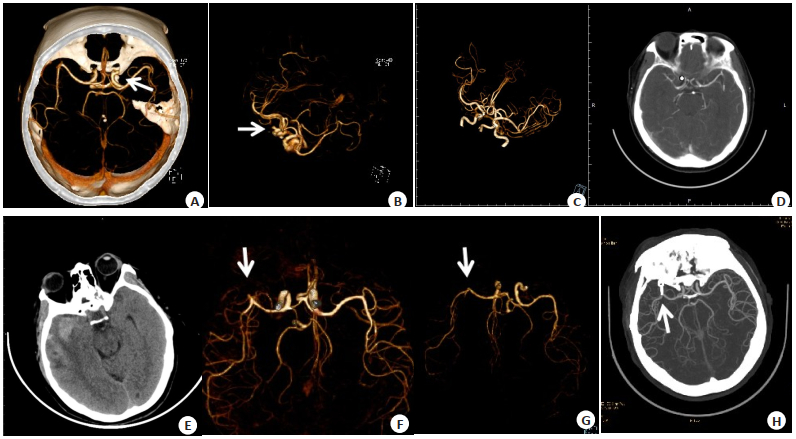
Objective To explore the diagnostic efficiency of head and neck computerized tomography angiography (CTA) for intracranial aneurysms and its application value in surgical guidance. Methods A total of 129 patients with suspected intracranial aneurysms admitted to the hospital were enrolled as the research objects from June 2018 to September 2021. All patients underwent head and neck CTA and magnetic resonance angiography (MRA). Taking digital subtraction angiography (DSA) as the golden standard, application value of head and neck CTA and MRA in the diagnosis and surgical guidance of intracranial aneurysms was compared. Results Taking DSA diagnosis as the golden standard, there were 80 cases confirmed with intracranial aneurysms in the 129 patients. The diagnostic accuracy, sensitivity, specificity, positive predictive value and negative predictive value of head and neck CTA and MRA were 92.24%, 95.06%, 87.50%, 92.77%, 91.30% and 88.37%, 91.46%, 82.97%, 90.36%, 84.78%, respectively. There was no significant difference in detection sites of lesions between head and neck CTA and MRA (P > 0.05). The number of tumors with diameter < 3 mm by head and neck CTA was more than that by MRA (P < 0.05). The Kappa values of head and neck CTA and MRA were 0.850 and 0.747, respectively. The diagnosis consistency between head and neck CTA and DSA was higher. Conclusion The diagnostic value of head and neck CTA is higher for intracranial aneurysms, which is of better guidance roles in surgical treatment.
2023, 46(2): 276-280.
doi: 10.12122/j.issn.1674-4500.2023.02.16
Abstract:
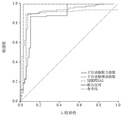
Objective To analyze the application value of ultrasonic measurement of uterine artery blood flow parameters combined with the detection of serum Serpin family member 1(SERPINA1) as a potential marker in the diagnosis of uterine fibroids. Methods A total of 67 patients with uterine fibroids who were treated in our hospital from February 2018 to May 2022 were selected as the observation group, and 103 patients with adenomyosis were selected as the control group. The ultrasound examination was performed to record the uterine artery resistance index (RI), uterine artery pulsatility index (PI), uterine artery systolic blood flow velocity, and uterine artery diastolic blood flow velocity. We detected the serum SERPINA1 protein level of the subjects. We used the logistic regression model to analyze significant differences in the combined application of each index for diagnosis. The model was evaluated and ROC curve was drawn to analyze the value of each index alone and in combination in predicting uterine fibroids. Results The RI of the observation group was significantly lower than that of the control group (0.53±0.13 vs 0.69±0.08), and the PI was significantly higher than that of the control group (1.56±0.21 vs 1.25±0.19) (P < 0.05). The level of SERPINA1 in the blood of the observation group was significantly higher than that of the control group (138.59±20.28 vs 46.71±4.39), and the difference was statistically significant (P < 0.05). The uterine artery blood flow parameters combined with SERPINA1 were used to diagnose uterine fibroids. The prediction model is Log(P)=0.658×uterine artery resistance index+0.617×uterine artery pulsatility index+0.642×SERPINA1+0.809; the AUCs of RI, PI, and SERPINA1 used alone to predict uterine fibroids were all greater than 0.85. The combination of three indicators was used to predict uterine fibroids. The AUC of the application to predict uterine fibroids was significantly higher than that of each index alone(P < 0.05). Conclusion Ultrasound uterine artery blood flow parameters combined with SERPINA1 detection can effectively diagnose and evaluate uterine fibroids, and have high clinical application value.
2023, 46(2): 281-285.
doi: 10.12122/j.issn.1674-4500.2023.02.17
Abstract:
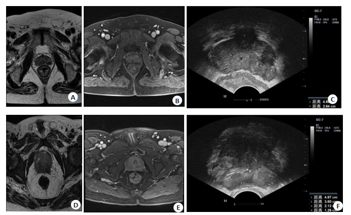
Objective To observe the application value of transrectal ultrasound (TRUS) and dynamic contrast-enhanced magnetic resonance imaging (DCE-MRI) combined with serum prostate specific antigen (PSA) detection in the differential diagnosis of benign and malignant prostate tumors. Methods A total of 102 patients with prostate tumors admitted to the hospital were enrolled from January 2019 to October 2022. All the patients underwent TRUS and DCE- MRI examination. Taking the diagnostic results of needle biopsy as the golden standard, level of serum PSA in patients with benign and malignant prostate tumors was observed. The value of TRUS, DCE- MRI, PSA and combined detection in the differential diagnosis of benign and malignant prostate tumors was compared. Results Among the 102 patients with prostate tumors, needle biopsy showed that there were 38 cases with malignant tumors and 64 cases with benign tumors. The sensitivity, specificity and accuracy of TRUS, DCE-MRI and PSA in the differential diagnosis of benign and malignant prostate tumors were 63.16%, 76.56%, 71.57%; 73.68%, 78.13%, 76.47% and 89.47%, 70.31%, 76.47%, respectively. Needle biopsy showed that PSA level and DCE- MRI parameters in patients with malignant tumors were significantly higher than those with benign tumors (P < 0.05). The sensitivity and accuracy of combined detection were 97.37% and 88.24%, significantly higher than those of single single (P < 0.05). Conclusion Compared with patients with benign prostate tumors, level of serum PSA is higher in patients with malignant tumors. The combined detection of TRUS, DCE-MRI and PSA can significantly improve differential diagnosis efficiency of benign and malignant prostate tumors.
2023, 46(2): 286-292.
doi: 10.12122/j.issn.1674-4500.2023.02.18
Abstract:
Objective To compare MRI and CT in the diagnosis of hepatocellular carcinoma tumors and to conduct high-precision radiotherapy planning. Methods Twenty-six patients with unresectable liver metastases (n=8), hepatocellular carcinoma (n=10) and cholangiocarcinoma (n=8) from July 2020 to July 2022 were selected. All patients underwent MRI and three-phase CT scans of diagnostic quality at the time of radiotherapy planning, and intrahepatic anatomical reference points were identified. In the CT and MRI series that best visualized the tumor, liver and tumor volume were delineated and intrahepatic anatomical reference points were identified. Deformable registration was used to register CT and MRI livers. Results The number of CT lesions was different from that of MRI in 5 cases of liver cancer. MRI lesions were more in 3 cases and CT lesions were more in 2 cases. After liver deformation registration, the average distance between CT tumor surface and MRI tumor surface in the population was 3.7(2.2-21.3) mm. The median percentage of tumor surface area difference of 5 mm was 26%(38%-86%). The median coincidence rate was 81%(77%-86%)for metastases, 78%(44%-86%) for hepatocellularcarcinoma, and 69%(25%-85%) for cholangiocarcinoma. Conclusion The tumor volume of hepatocellular carcinoma diagnosed by MRI is significantly different from that diagnosed by CT, and it is more common in primary hepatocellular carcinoma.
2023, 46(2): 293-296.
doi: 10.12122/j.issn.1674-4500.2023.02.19
Abstract:
Objective To investigate the differences of humoral immune function and chest CT findings between Mycoplasma pneumoniae (MP) complicated with Streptococcus pneumoniae (SP) pneumonia and MP alone in children. Methods Seventy children with pneumonia admitted to our hospital from March 2020 to March 2022 were selected, including 46 cases of MP infection and 24 cases of MP combined with SP infection. The humoral immunity indexes and CT manifestations of the children were detected, and the differences between humoral immunity and CT manifestations of the two different infection types were analyzed. Results There was no significant difference in lesion distribution between the two groups (P > 0.05). Compared with the SP group, the incidence of bronchial vascular bundle thickening, bronchial wall thickening, reticular shadow, ground glass shadow and fan-shaped slice shadow in MP combined with SP group was significantly lower, and the incidence of consolidation shadow was significantly higher(P < 0.05). In MP combined with SP group, the thickness of pleural effusion was significantly thicker and the maximum transverse diameter of lymph nodes was significantly larger (P < 0.05). There were no significant differences in IgA, IgG and IgM levels between the two groups (P > 0.05). Conclusion Compared with SP infection alone, the humoral immune dysfunction caused by MP combined with SP infection in children with pneumonia is not obvious. but there is a big difference in CT image signs, which is helpful for differential diagnosis.
2023, 46(2): 297-301.
doi: 10.12122/j.issn.1674-4500.2023.02.20
Abstract:
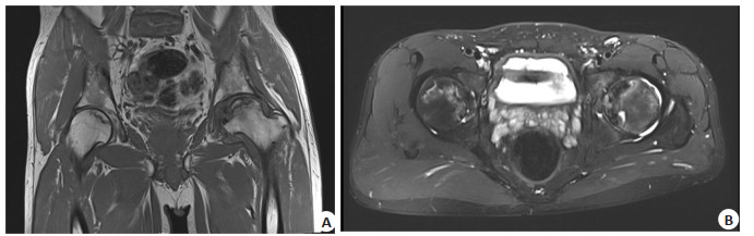
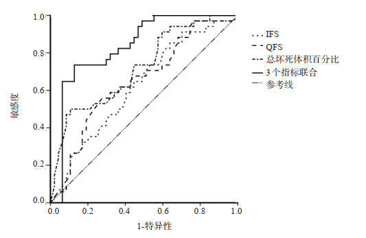
Objective To explore the clinical value of MRI parameters related to ischial femur impingment [ischial femur space (IFS), quadrate femoral space (QFS)] combined with the percentage of necrosis volume in predicting the collapse and osteonecrosis of the femoral head. Methods Sixty-seven patients (81 hips) with osteonecrosis of the femoral head admitted to our hospital from March 2013 to June 2022 were selected. According to the collapse of the femoral head (X-ray film), the patients were divided into 38 cases (47 hips) in the non-collapse group and 29 cases (34 hips) in the collapse group. General data, IFS, QFS, quadrants and total necrotic volume were compared between the two groups. Logistic regression model was used to study the influencing factors of ONFH and collapse. Spearman test was used for correlation analysis. The combined prediction value of IFS, QFS and percentage of total necrotic volume in the ONFH and collapse was evaluated by ROC curve. Results There were no significant differences in age, gender and etiology between the two groups (P > 0.05), but there were significant differences in bone marrow edema, ARCO stage and necrotic morphology between the two groups (P < 0.05). Compared with the non-collapse group, IFS, QFS, quadrants and total necrotic volume in the collapse group were significantly increased (P < 0.05), but there was no statistical significance in necrotic volume in the PIM quadrant (P > 0.05). Logistic regression model showed that ARCO stage Ⅱ, peripheral necrosis type, bone marrow edema, IFS, QFS and percentage of total necrosis volume were risk factors for femoral head necrosis collapse (P < 0.05). Spearman test indicated that IFS, QFS and percentage of total necrotic volume were positively correlated with necrotic collapse of femoral head (P < 0.05). The ROC curve showed that the combined prediction of IFS, QFS and percentage of total necrosis volume (AUC=0.832) was higher than that of the single application of each index. The prediction sensitivity and specificity were 73.52% and 87.22%, and the critical point were 16.98 mm, 14.65 mm and 11.12%. Conclusion IFS and QFS combined with total necrotic volume percentage of MRI related parameters of ischifemoral impinging have high efficacy in predicting femoral head necrotic collapse.
2023, 46(2): 302-306.
doi: 10.12122/j.issn.1674-4500.2023.02.21
Abstract:
Objective To investigate the diagnostic value of diffusion weighted imaging (DWI) in active ulcerative colitis (UC). Methods The clinical and imaging data of 80 patients with UC who were treated in the hospital from 2018 to the present were retrospectively analyzed. With the diagnostic results of intestinal endoscopy as the golden standard, the patients were divided into active group (UC in active phase, n=73) and inactive group (UC in inactive phase, n=7). All patients underwent CT and MRI examinations. The CT and MRI image features, apparent diffusion coefficient (ADC) values of the two groups were analyzed. ROC curve was used to evaluate the diagnostic value of ADC value in active UC. Results MRI findings mainly included colorectal mucosa and submucosa with high signal or slightly higher signal on T1WI, high signal or isointensity on T2WI. All diseased intestinal segments showed different degrees of increased signals on DWI. The ADC value of active group was significantly lower than that of inactive group (P < 0.05). The ROC curve analysis results showed that the AUC value of ADC value for diagnosing active UC was 0.795 (95% CI: 0.691-0.878, P < 0.05), indicating high diagnostic performance. With the diagnostic results of intestinal endoscopy as the golden standard, the sensitivity, accuracy and negative predictive value of DWI for diagnosing active UC were higher than those of CT (P < 0.05). Conclusion CT and MRI findings of UC are characteristic. DWI is helpful for differential diagnosis of active UC, and it can be used as an effective method for diagnosing the stage of UC.
2023, 46(2): 307-310.
doi: 10.12122/j.issn.1674-4500.2023.02.22
Abstract:
Objective To evaluate the efficacy of rectal cancer after chemotherapy by texture analysis of magnetic resonance T2-weighted images. Methods A total of 117 patients with rectal cancer who were clinically and pathologically diagnosed in our hospital from January 2019 to April 2021. The patients were divided into complete remission group (n=38) and incomplete remission group (n=79), they were scanned with 3.0T MRI before and after chemotherapy. Omni Kineitics software was used to extract texture features of subjects, and the kurtosis, variance, entropy and energy parameters of texture features were calculated. ROC curves were drawn to analyze the texture of magnetic resonance T2-weighted images, and the rectal value of pathological complete response after cancer chemotherapy. Results After treatment, the variance and entropy of the subjects in the complete remission group were significantly lower than those in the incomplete remission group (1534±312 vs 2312±586, 5.43±0.41 vs 6.42±0.29), the kurtosis and energy were significantly higher than those in the incomplete remission group (4.85±0.66 vs 3.72±0.49, 0.016±0.002 vs 0.013±0.02), and the difference was statistically significant (P < 0.05). Conclusion Texture analysis using magnetic resonance T2-weighted images can evaluate the efficacy of chemotherapy and predict the pathological complete response of rectal cancer after chemotherapy.
2023, 46(2): 311-315.
doi: 10.12122/j.issn.1674-4500.2023.02.23
Abstract:
Objective To assess the diagnostic value of musculoskeletal ultrasound and MRI in the diagnosis degenerative osteoarthritis of the knee (KOA). Methods A total of 126 patients with unilateral KOA who were treated in our hospital from January 2017 to December 2019 were selected and all underwent arthroscopy. Musculoskeletal ultrasound, MRI and arthroscope imaging were performed on all patients included in the study. Using arthroscopic findings as the gold standard, the diagnostic value of ultrasound and MRI examinations was assessed for joint effusion, synovial hyperplasia, suprapatellar bursa effusion, articular cartilage damage, osteophytes, meniscal damage and ligament damage, respectively. Results A total of 126 patients underwent musculoskeletal ultrasound, MRI and arthroscopy successfully. With arthroscopic results as the gold standard, ultrasonography and MRI were in good agreement with arthroscopic results in the detection of synovial hyperplasia, suprapatellar sac effusion, meniscus injury, ligament injury, tendon rupture and popliteal cyst in patients with KOA (Kappa values of ultrasound: 0.81, 0.85, 0.79, 0.79, 1.00, 1.00; Kappa values of MRI: 0.76, 0.93, 0.86, 0.89, 1.00, 1.00, Kappa values > 0.75). In the diagnosis of synovial hyperplasia and hyperosteogeny in KOA, the diagnostic efficacyof ultrasound was better than that of MRI (AUC=0.978, 0.972, P < 0.05). In the diagnosis of joint effusion, suprapatellar sac effusion, articular cartilage damage and ligament injury, MRI had better diagnostic performance than ultrasound (AUC=0.991, 0.995, 0.969, 0.961, P < 0.05). In the diagnosis of ligament injury, there was no statistical difference between ultrasound and MRI (P > 0.05). Among 126 patients, 57 patients were found to have joint space stenosis by ultrasound examination, and the average joint space was about 3.54±0.53 mm, while 52 patients were found to have joint space stenosis by MRI examination, and the average joint space was about 3.62±0.61 mm. There was no significant difference between the two measurements (P > 0.05). Conclusion Musculoskeletal ultrasound and MRI examination can better identify the characteristics of KOA and have high clinical value.
2023, 46(2): 316-320.
doi: 10.12122/j.issn.1674-4500.2023.02.24
Abstract:
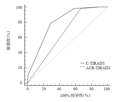
Objective To explore the value of Chinese version of thyroid imaging reporting and data system (C-TIRADS) by comparing with the American College of Radiology (ACR) TIRADS in the differential diagnosis between benign and malignant thyroid nodules. Methods A total of 166 patients with thyroid nodules (195 nodules) who underwent thyroid ultrasonography and confirmed by pathology from August 2021 to July 2022 were retrospectively collected. The pathological results were used as the gold standard to evaluate the malignant proportion of different classifications of C‑TIRADS and TIRADS respectively. The ROC curves of the two systems for diagnosing benign and malignant thyroid nodules were drawn, and the diagnostic efficacy of the two systems were compared. Results The actual malignant proportion of the two TIRADS risk stratification levels were consistent with the malignant rate recommended by the guidelines. The AUC of C-TIRADS in the diagnosis between benign and malignant thyroid nodules was 0.796 (95% CI: 0.741-0.852), which was significantly higher than 0.658 (95% CI: 0.587-0.724) of ACR-TIRADS (P < 0.05). The optimal critical points of C‑TIRADS and ACR‑TIRADS were 4C and 5, respectively. There was no significant difference in sensitivity between the two classification systems (94.12% vs 95.10 %, P > 0.05), but the specificity of C‑TIRADS was significantly higher than that of ACR‑TIRADS (56.99% vs 35.48%, P < 0.05). Conclusion For the identification of thyroid nodules, C-TIRADS has higher diagnostic efficiency and higher specificity than ACR-TIRADS.
2023, 46(2): 321-325.
doi: 10.12122/j.issn.1674-4500.2023.02.25
Abstract:
Objective To investigate the clinical value and significance of transesophageal echocardiography (TEE) in evaluating the volume and function of left atrial appendage in patients with non- valvular atrial fibrillation. Methods A total of 138 patients with non-valvular atrial fibrillation who underwent TEE in Xindu District People's Hospital, Chengdu from August 2016 to August 2020 were defined as the atrial fibrillation group. Forty-seven individuals who underwent TEE during the same period because of other cardiovascular diseases were defined as the control group. Multiplane TEE was used to observe left atrial appendage. The volume parameters of left atrial appendage were obtained by Simpson's biplane method. Three-dimensional left atrial volume images of all subjects were collected by transthoracic echocardiography, and imported into Qlab9.0 software to obtain left atrial volume and change parameters. The volume parameters and hemodynamic parameters of left atrium and left atrial appendage in the two groups were comparatively analyzed. The thrombosis and spontaneous contrast in patients with different left atrial appendage function were analyzed. Results Left atrial ejection fraction, left atrial appendage ejection fraction, volume change rate of left atrial appendage, emptying maximum speed of left atrial appendage and left atrial appendage emptying index standardized by body surface area in the atrial fibrillation group were lower than those in the control group. The maximum left atrial volume, minimum left atrial volume, maximum volume of left atrial appendage and minimum volume of left atrial appendage were greater than those in the control group (P < 0.05). In the atrial fibrillation group, there were 89 cases (64.49%) with normal left atrial appendage function, 31 cases (22.46%) with slight decline in left atrial appendage function, and 18 cases (13.04%) with moderate to severe decline in left atrial appendage function. The status of thrombosis and spontaneous contrast showed statistically significant differences in patients with different status of left atrial appendage function (P < 0.05). The thrombosis rate in patients with moderate to severe decline was higher than that in patients with mild decline or patients with normal function (P < 0.05). The severity of spontaneous contrast in patients with moderate to severe decline was higher than that in patients with normal function (P < 0.05). Conclusion TEE is helpful for evaluating the volume and function of left atrial appendage in patients with non-valvular atrial fibrillation. The greater the left atrial appendage function decline, the higher the risk of thrombosis and spontaneous contrast.
2023, 46(2): 326-330.
doi: 10.12122/j.issn.1674-4500.2023.02.26
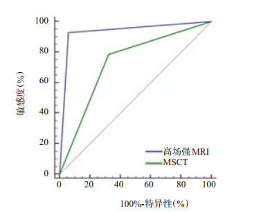
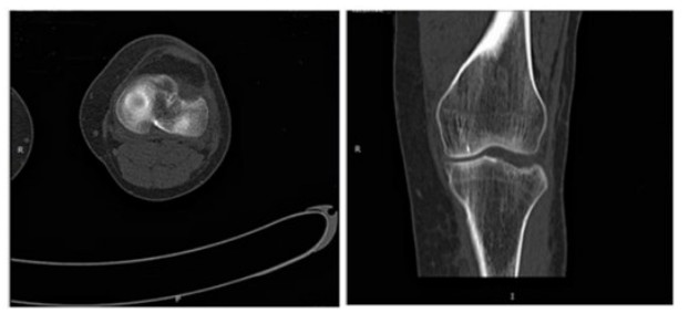
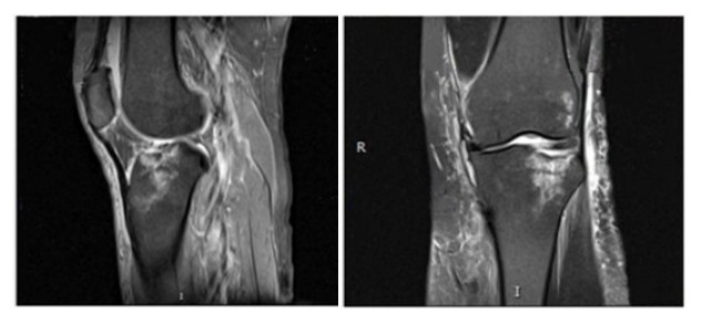
Abstract:
Objective To investigate the diagnostic value of high field intensity MRI and multi-slice CT (MSCT) for occult fracture of knee joint. Methods A total of 90 patients with suspected occulted fracture of knee joint admitted to our hospital from January 2019 to January 2022 were studied. All patients underwent arthroscopy, high field intensity MRI and MSCT examination. With arthroscopy as the gold standard, compare the examination results of high field intensity MRI and MSCT on occult fracture of knee joint with the arthroscopic detection, diagnostic value, fracture collapse degree and horizontal dislocation degree. Results Among 90 suspected occult fracture of knee joint, 56 cases were diagnosed by arthroscopy. The detection rate of high field intensity MRI was higher than that of MSCT (92.86% vs 78.57%, P < 0.05). The sensitivity, specificity, positive predictive value, negative predictive value, accuracy and area under curve (92.86%, 94.12%, 96.30%, 88.89%, 93.33%, 0.935) of high field intensity MRI in the diagnosis of latent fracture of knee joint were higher than those of MSCT (78.57%, 67.65%, 80.00%, 65.71%, 74.44%, 0.731, P < 0.05). High field intensity MRI showed higher rates of fracture collapse degree and horizontal dislocation degree (64.44%, 73.33%) than MSCT (43.33%, 47.78%, P < 0.05). Conclusion Compared with MSCT, high-field MRI had a higher detection rate and diagnostic value for occult fracture of knee joint, and had a better indication of the degree of collapse and horizontal dislocation, which could provide reliable information for the clinical diagnosis of occult fracture of knee joint.
2023, 46(2): 331-336.
doi: 10.12122/j.issn.1674-4500.2023.02.27
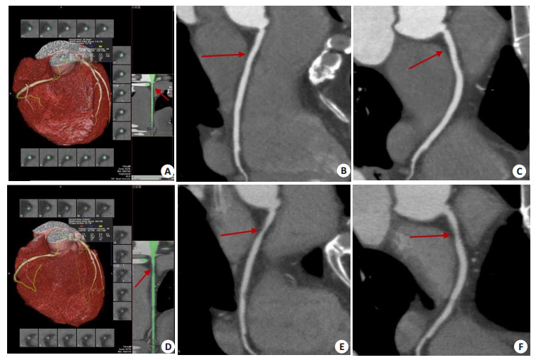
Abstract:
Objective To explore the application value of 320-slice CT coronary angiography in evaluating the changes of unstable plaque load of coronary artery during the follow-up period. Methods Seventy-eight inpatients with coronary heart disease in Huaibei Miners General Hospital from August 2013 to December 2020 were selected as the research objects. During the follow-up period, 320-slice CT was used to examine their coronary angiography. At the same time, age, BMI, smoking history, hypertension and diabetes were recorded in detail. The blood lipid index and high-sensitivity C-reactive protein were measured. According to the results of two CT coronary angiography examinations during the review period, 18 patients with negative CT coronary angiography results were taken as the normal control group, and the rest patients found at least one coronary artery segment with plaque at the end of the follow-up period, which was regarded as the plaque progression group (n=60). The χ2 test and Logistic regression analysis were used to compare the risk factors of plaque progression. At the same time, the coronary artery stenosis, plaque properties and load changes of 78 patients during the follow- up period were compared. Result There was no significant difference in the average follow- up interval between patients with plaque and normal control group (P > 0.05). Compared with the normal group, the proportion of smokers in the plaque group was significantly different (P < 0.05). Among other risk factors, there were no significant differences between the two groups (P > 0.05). The increase of gender, age, smoking, hypertension, diabetes, obesity, the abnormal indexes of some blood lipids [total cholesterol, triglyceride, apolipoprotein A1, apolipoprotein B, lipoprotein (a)] and high-sensitivity C-reactive protein were increased the risk of increased coronary artery plaque burden, and caused stenosis of the lumen. In 78 patients, the stenosis degree of each vascular branch after follow-up were significantly worse than those before follow-up, and the differences were statistically significant (P < 0.05). After follow-up, the proportion of calcified plaque (26.80%) was significantly lower than that before follow-up (56.52%), while the proportions of non-calcified plaque and mixed plaque (42.27% and 30.93%, respectively) were significantly higher than those before follow-up (23.19% and 11.59%) (P < 0.05). After follow-up, the right coronary artery, left anterior descending artery, circumflex artery and total plaque load of 78 patients were significantly higher than those before follow-up, and the differences were statistically significant (P < 0.05). Conclusion For patients with coronary heart disease risk factors, CT coronary angiography can better evaluate the coronary artery stenosis and the changes of plaque properties and loads during follow- up, which is of great significance for predicting the development and changes of their condition.
2023, 46(2): 337-341.
doi: 10.12122/j.issn.1674-4500.2023.02.28
Abstract:
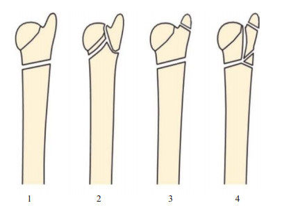
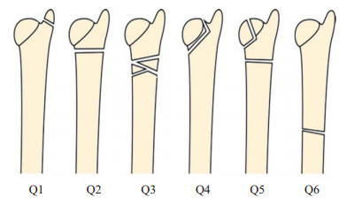
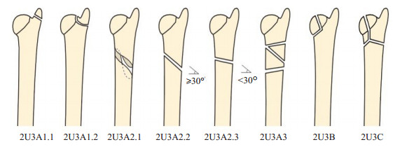
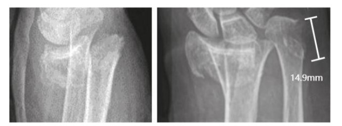
Objective To compare the reliability and reproducibility of three different imaging classification systems for clinical guidance of distal ulnar fractures. Methods Sixty-one patients who visited our hospital for distal ulnar fractures from 2015 to 2020 were included with the age of 63.3 ± 17.1 years old. X-ray images of each patient were independently classified by 3 physicians using the Biyani, International Society for the Study of Internal Fixation/Orthopaedic Trauma Association (AO/OTA) 2007 and AO/OTA 2018 classification systems, and repeated assessment after 2 weeks. Interand intra observer agreement was assessed using consistency evaluation (Kappa value) analysis. Results Inter-observer agreement was 0.44 and intra-observer agreement was 0.58 for the Biyani classification. Inter-observer agreement was 0.40 and intra-observer agreement was 0.52 for the AO/OTA 2007 classification. Inter-observer agreement was 0.43 and intra-observer agreement was 0.53 for the AO/OTA 2018 classification and 0.53 for intra-observer agreement (P < 0.05). The intra- and inter-observer agreement grades were moderate, except for the inter-observer agreement grade of the AO/OTA 2007 classification, which was fair. Except for inter-observer agreement rating with an average using the AO/OTA 2007 classification, the intra-and inter-observer agreement ratings were moderate. Conclusion The consistency difference between the 3 classifications is not significant, but the clinical guidance value is poor. The development of specialized classification method dedicate to distal ulnar fractures based on Biyani classification will improve the accuracy, reliability and reproducibility of the classification and help to better guide clinical treatment.
2023, 46(2): 342-346.
doi: 10.12122/j.issn.1674-4500.2023.02.29
Abstract:
Objective To investigate the diagnostic value of semi-quantitative parameters of 99mTc-3PRGD2 SPECT/CT imaging in suspicious breast lesions, and to analyze its correlation with clinicopathology according to the molecular classification of breast cancer. Methods The case data of 70 patients with suspected breast lesions who were examined in our hospital from December 2020 to August 2022 were collected. According to the pathological results, the correlation between Luminal positive, Her-2 positive and triple negative molecular subtypes of breast cancer and T/N value was analyzed.The relationship between the expression of ER, PR, Her-2 and Ki67 and the imaging indexes were analyzed. Results The pathological examination results of 70 patients confirmed 51 cases (72.86%) of breast cancer. The pathological classification was 22 cases of Luminal positive type, 20 cases of Her-2 positive type and 9 cases of triple negative type.There were 24 cases (27.14%) of benign breast lesions.The T/N value of breast cancer patients was higher than that of benign lesions (3.96±0.82 vs 1.16±0.32), and the difference was statistically significant (t=14.426, P < 0.05). The diagnostic specificity was 84.21% (16/19), the sensitivity was 82.35% (42/51), the accuracy was 82.86 % (58/70), and the area under the ROC curve was 0.834 (0.755-0.913). The diagnostic threshold was 1.56, that is, when the T/N value was≥1.56, it was judged as malignant lesions. The T/N value of Her-2 positive type was higher than that of triple negative type (P < 0.05). The T/N value of Her-2 positive patients was higher than that of Her-2 negative patients (P < 0.05). There was no significant difference in T/N value between patients with positive and negative expression of ER, PR and Ki67 (P>0.05). Conclusion The semi-quantitative index of 99mTc-3PRGD2 SPECT/CT imaging has higher sensitivity in the diagnosis of benign and malignant breast lesions, and the T/N value of Her-2 positive patients is significantly increased. It is helpful for early detection and diagnosis of breast lesions, and can guid in predicting the molecular classification and treatment of breast cancer.
2023, 46(2): 347-351.
doi: 10.12122/j.issn.1674-4500.2023.02.30
Abstract:
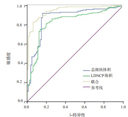
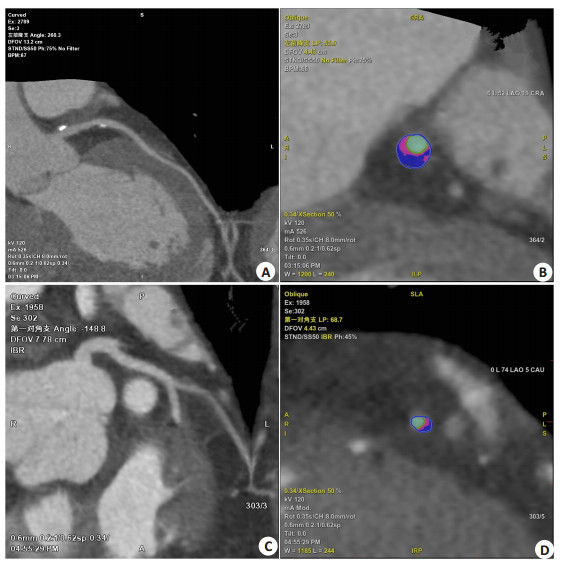
Objective To analyze the clinical value of coronary CT angiography (CTA) plaque quantitative parameters on predicting myocardial ischemia events in patients with coronary heart disease. Methods A total of 256 patients with newly diagnosed coronary heart disease during the period from January 2020 and June 2022 were included as the research subjects. All patients underwent coronary CTA examination. The plaque quantitative parameters were detected. According to fractional flow reverse, the patients were divided into myocardial ischemia group and non-myocardial ischemia group. The coronary CTA plaque quantitative parameters were compared between the two groups. Multivariate linear regression analysis was used to analyze the relationship between plaque quantitative parameters and myocardial ischemic injury. ROC curve was drawn to evaluate the predictive value of plaque quantitative parameters on myocardial ischemic injury. Results The total plaque volume, non-calcified plaque volume, volume of low-density non-calcified plaque (LDNCP), plaque length and diameter stenosis in myocardial ischemia group were larger or higher than those in non-myocardial ischemia group (P < 0.05) while the calcified plaque volume and fractional flow reverse were smaller than that in non-myocardial ischemia group (P < 0.05). Multivariate linear regression analysis showed that total plaque volume and LDNCP volume were the independent influencing factors of myocardial ischemia in patients with coronary heart disease (P < 0.05). ROC curve showed that the sensitivity, specificity and area under curve of total plaque volume and LDNCP volume in combination on predicting myocardial ischemic injury were 94.30%, 77.80% and 0.948, respectively. Conclusion The changes in coronary CTA plaque quantitative parameters are related to myocardial ischemic injury in patients with coronary heart disease. Total plaque volume and LDNCP volume can be used as predictors of myocardial ischemic events.
2023, 46(2): 352-356.
doi: 10.12122/j.issn.1674-4500.2023.02.31
Abstract:
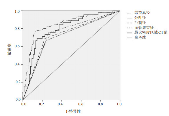
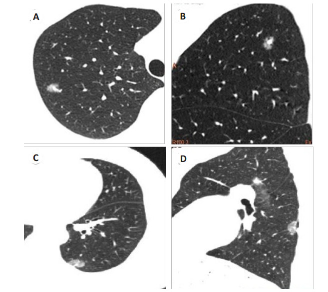
Objective To explore the value of CT in the differential diagnosis of lung invasive adenocarcinoma (IAC) and minimally invasive adenocarcinoma (MIA). Methods A total of 120 patients with lung ground glass nodules who underwent surgical treatment in our hospital from January 2020 to December 2021 were selected. Among the patients, 75 cases with IAC diagnosed by biopsy or postoperative pathology were enrolled as IAC group, and 45 cases with MIA were included in MIA group. The CT imaging data of the two groups were compared, and ROC curve was used to evaluate the value of CT in differentiating IAC from MIA. Results There were no statistical differences in nodule location, clear/fuzzy edge, pleural indentation sign and lesion CT density between IAC group and MIA group (P>0.05). However, the maximum nodule diameter and CT value at the maximum density area in IAC group were higher than those in MIA group (P < 0.05), and the proportions of irregular nodule morphology, lobulation sign, spiculation sign and vascular convergence sign were also higher in IAC group than those in MIA group (P < 0.05). The maximum nodule diameter, lobulation sign, spiculation sign, vascular convergence sign and CT value at the maximum density area were the related risk factors leading to the differences in pathological results (P < 0.05). ROC results showed that when the Youden index of the maximum nodule diameter was the maximum (0.636), the corresponding cut-off value, AUC, sensitivity and specificity in differentiating IAC from MIA were 1.31 cm, 0.846, 75.56% and 88.00%, respectively. The AUC, sensitivity and specificity in differentiating IAC from MIA were 0.713, 66.67% and 76.00% of lobulation sign, were 0.731, 68.89% and 77.33% of spiculation sign, and were 0.744, 75.56% and 73.33% of vascular convergence sign respectively. When the Youden index of CT value at the maximum density area was the maximum at 0.542, the corresponding cut-off value was -531.75 Hu, and the AUC, sensitivity and specificity in the differential diagnosis of IAC and MIA were 0.801, 68.89% and 85.33%, respectively. Conclusion CT technology has a high guiding role in the differential diagnosis of IAC and MIA. Nodule diameter, CT value at the maximum density area, lobulation sign, spiculation sign and vascular convergence sign are of great value for the clinical differentiation of IAC and MIA.
2023, 46(2): 357-361.
doi: 10.12122/j.issn.1674-4500.2023.02.32
Abstract:
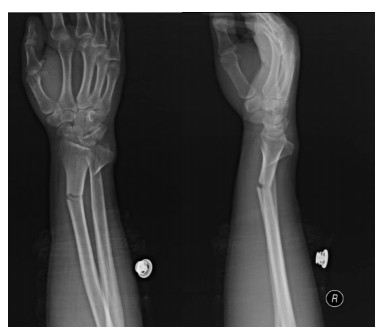
Objective To explore the effect of different surgical methods on traumatic distal radius fracture based on wrist function, elbow function and imaging parameters. Methods We selected 80 patients with traumatic distal radius fractures who were treated in our department from January 2015 to January 2022. All patients were injured by indirect violence and were unilateral closed fractures. According to the type and degree of fractures, the patients were divided into two groups according to different surgical methods. Among them, 38 patients in group A were treated by open reduction and internal fixation with steel plates through the dorsal approach; 42 patients in group B were treated with open reduction and internal fixation with steel plate through the volar approach. The wrist joint function scores and elbow joint function scores of the two groups were compared. The imaging parameters (distal radius height, palmar inclination angle, ulnar deflection angle) related to the reduction of the fracture end in the two groups were measured and recorded with the medical image information processing system of our hospital. Logistic regression model was used to analyze the influencing factors of poor joint function recovery after traumatic distal radius fracture fixation. Results The scores of pain, function and activity in both groups were higher than those before operation (P < 0.05), and the scores of wrist function in group B were higher than those in group A (P < 0.05). The scores of range of motion, pain, stability and function of joints in both groups were higher than those before operation (P < 0.05), and those of elbow joints in group B were higher than those in the control group (P < 0.05). The height of distal radius, palmar obliquity and ulnar declination angle in both groups were higher than those before operation (P < 0.05), and the palmar obliquity in the study group was higher than that in the control group (P < 0.05). According to the wrist joint function score and elbow joint function score combined with imaging review, 58 patients recovered well and 22 patients did not. The univariate analysis showed that age, the last review palm angle and the mode of operation were related to the poor recovery of joint function after the fixation of traumatic distal radius fracture (P < 0.05), while the gender and fracture type were not related to the poor recovery of joint function after the fixation of traumatic distal radius fracture (P>0.05). The Logistic regression analysis showed that age, the last review palm angle, and the mode of operation were all independent influencing factors for poor recovery of joint function after fixation of traumatic distal radius fracture (P < 0.05). Conclusion The treatment of traumatic distal radius fracture by volar approach open reduction and internal fixation with steel plate is more effective. It can promote the recovery of wrist joint function and elbow joint function, improve the imaging parameters related to the reduction of the fracture end after surgery, and effectively improve the prognosis of patients.
2023, 46(2): 362-365.
doi: 10.12122/j.issn.1674-4500.2023.02.33
Abstract:
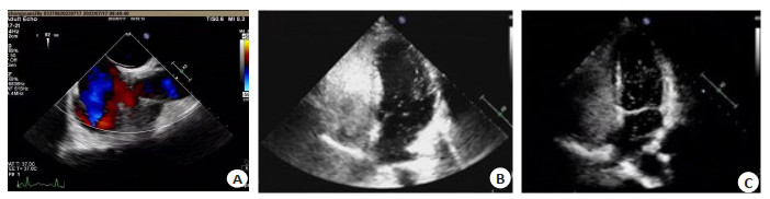
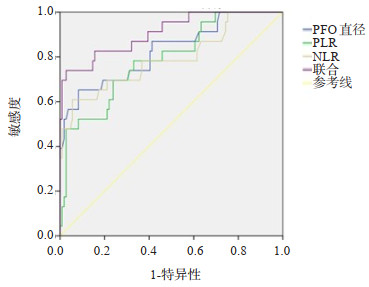
Objective To investigate the the application of transesophageal echocardiography (TEE) combined with platelet/lymphocyte ratio (PLR) and neutrophil/lymphocyte ratio (NLR) in the evaluation of cryptogenic stroke in patients with patent foramen ovale (PFO). Methods Thirty-two patients with patent foramen ovale admitted in our hospital from August 2019 to August 2021 were selected, including 23 patients with cryptogenic stroke. All patients received TEE detection, PFO diameter was measured, and blood routine tests were performed. PLR and NLR ratios were calculated according to the counts of platelets, lymphocytes, and neutrophils. The differences of PLR and NLR ratios between PFO with cryptogenic stroke and non cryptogenic stroke were compared. Spearman was used to analyze the correlation between PFO diameter, PLR, NLR and PFO with cryptogenic stroke. The evaluation value of PFO diameter, PLR and NLR detection in patent foramen ovale combined with cryptogenic stroke was analyzed by receiver operating characteristic curve. Results TEE test showed that the diameter of PFO in patients with PFO without cryptogenic stroke was significantly lower than that with cryptogenic stroke (P < 0.05), and angiography showed different levels of right-to-left shunts at the atrial level. PFO patients with cryptogenic stroke PLR, NLR ratio was significantly higher than that in patients without cryptogenic stroke (P < 0.05). Spearman analysis showed that PFO diameter, PLR, NLR ratio were positively correlated with the risk of PFO combined with cryptogenic stroke (r=0.385, 0.429, 0.378, P < 0.05). PFO The area under the curve of diameter combined with PLR and NLR to assess the risk of PFO combined with cryptogenic stroke was significantly higher than the area under the curve of PLR and NLR single index (P < 0.05). Conclusion TEE can better distinguish the diameter of PFO, and the combination of PLR and NLR can help to evaluate the risk of PFO complicated with cryptogenic stroke.
2023, 46(2): 366-370.
doi: 10.12122/j.issn.1674-4500.2023.02.34
Abstract:
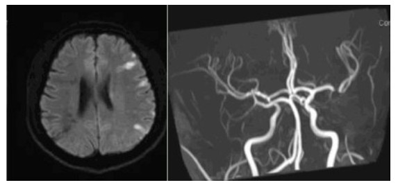
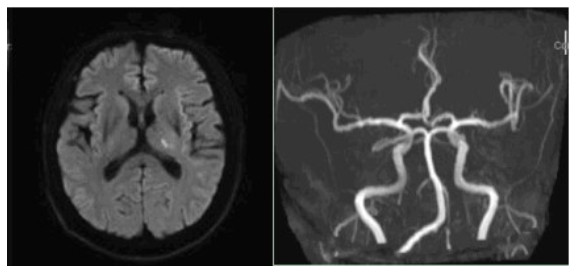
Objective To investigate the correlation between plasma homocysteine(Hcy) level and TOAST classification in patients with cerebral infarction by imaging examination. Methods A total of 736 cerebral infarction patients were subtypes according to the TOAST classification standard based on imaging, including 333 cases of large atherosclerosis, 374 cases of small artery occlusion, 9 cases of cardiogenic embolism, 11 cases of other etiological type and 9 cases of unknown cause, which were grouped according to gender. The correlation between Hcy level and TOAST subtypes were determined separately. Results The samples of the three subgroups of cardiogenic embolism type, other etiology type and unknown cause type were too small to have statistical significance. The level of Hcy in male large atherosclerosis group was significantly higher than that in small artery occlusion group (13.65±9.65 μmol/L vs 11.27±5.52 μmol/L, P < 0.05). Conclusion There is no significant correlation between Hcy level and TOAST score in the overall patients. According to gender analysis, Hcy level is significantly correlated with large atherosclerosis group in male patients with cerebral infarction, suggesting that higher plasma Hcy level in males is more likely to cause cerebral infarction by promoting atherosclerosis of the great arteries.
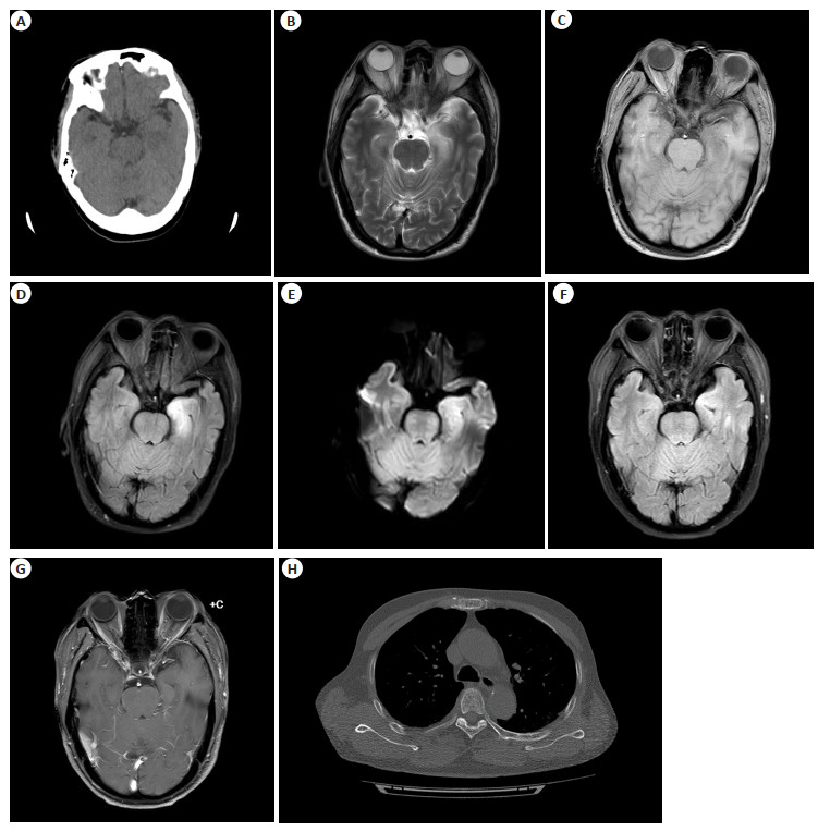
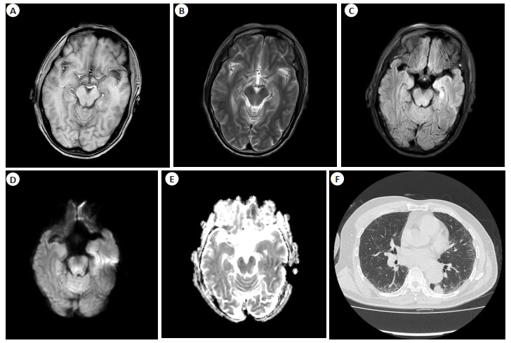
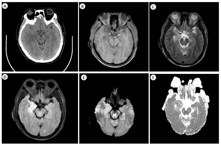
2023, 46(2): 371-374.
doi: 10.12122/j.issn.1674-4500.2023.02.35
Abstract:
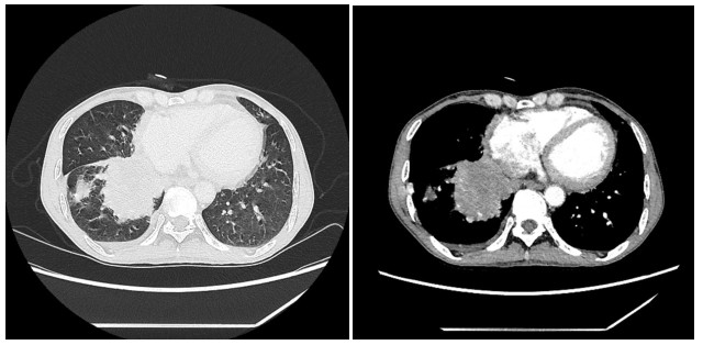
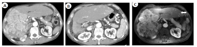
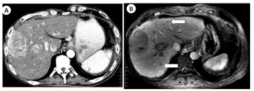
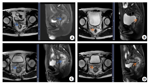
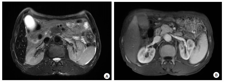
Pancreatic iron overload is a rare disease, presented with diabetes as the first symptom and with altered pancreatic morphology. Clinically, the lack of awareness and diagnostic criteria for pancreatic iron overload often leads to misdiagnosis of pancreatic disease, as well as unnecessary economic, physical, and mental burden. Here, we report a case of myelodysplastic syndromes complicated by pancreatic iron overload that was clinically misdiagnosed as pancreatic cancer, aiming at raising awareness and reducing the rate of misdiagnosis.
Pancreatic iron overload is a rare disease, presented with diabetes as the first symptom and with altered pancreatic morphology. Clinically, the lack of awareness and diagnostic criteria for pancreatic iron overload often leads to misdiagnosis of pancreatic disease, as well as unnecessary economic, physical, and mental burden. Here, we report a case of myelodysplastic syndromes complicated by pancreatic iron overload that was clinically misdiagnosed as pancreatic cancer, aiming at raising awareness and reducing the rate of misdiagnosis.
2023, 46(2): 375-380.
doi: 10.12122/j.issn.1674-4500.2023.02.36
Abstract:
Uterine fibroids are the most common benign tumors of female genitalia, most of which occurred in of 30 to 50 years old childbearing-aged women. The traditional treatment of uterine fibroids is surgical resection, including hysterectomy and myomectomy. However, the surgical trauma is severe and costs a long term of recovery. With the development of minimally invasive medical technology and the improvement of women's cognition, more and more patients diagnosed with uterine fibroids choose to receive minimally invasive treatment with the hope of better curative effect and the preservation of fertility. Radiofrequency ablation is one of the most commonly used minimally invasive methods for the treatments of uterine fibroids which safety and effectiveness has been clinically proven. After operation, the symptoms of patients are relieved, the life quality is improved, and the tumor recurrence rate is low. In this paper, radiofrequency ablation methods of Uterine fibroids are classified into four methods by both puncture paths and ultrasound-guided methods: transabdominal ultrasound-guided, laparoscopic ultrasound-guided, transvaginal ultrasound-guided and transcervical ultrasound-guided. In addition, these four methods are briefly reviewed from principle, method, ablation path, effectiveness, safety and influence on fertility ability.
Uterine fibroids are the most common benign tumors of female genitalia, most of which occurred in of 30 to 50 years old childbearing-aged women. The traditional treatment of uterine fibroids is surgical resection, including hysterectomy and myomectomy. However, the surgical trauma is severe and costs a long term of recovery. With the development of minimally invasive medical technology and the improvement of women's cognition, more and more patients diagnosed with uterine fibroids choose to receive minimally invasive treatment with the hope of better curative effect and the preservation of fertility. Radiofrequency ablation is one of the most commonly used minimally invasive methods for the treatments of uterine fibroids which safety and effectiveness has been clinically proven. After operation, the symptoms of patients are relieved, the life quality is improved, and the tumor recurrence rate is low. In this paper, radiofrequency ablation methods of Uterine fibroids are classified into four methods by both puncture paths and ultrasound-guided methods: transabdominal ultrasound-guided, laparoscopic ultrasound-guided, transvaginal ultrasound-guided and transcervical ultrasound-guided. In addition, these four methods are briefly reviewed from principle, method, ablation path, effectiveness, safety and influence on fertility ability.