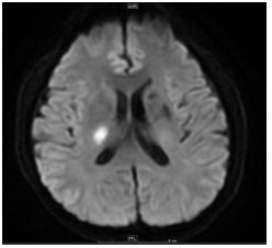
Clinical value of MRI radiomics model in prognosis of patients with acute cerebral infarction
-
摘要:
目的 研究讨论扩散加权成像(DWI)序列联合T2 Flair影像组学模型对急性基底节区脑梗死患者非溶栓治疗预后的临床价值。 方法 纳入2021年滨州医学院附属医院神经内科经过头颅MRI扫描的患者78例作为研究对象,患者均经临床症状及辅助检查证实为急性基底节区脑梗死。基于研究对象入院时和非溶栓治疗1周后出院时的改良Rankin量表评分变化,分成预后好组、预后差组。采用3D Slicer软件手动勾画DWI图像及T2 Flair图像高信号的梗死部位区域,采用Radiomics插件对所勾取的影像资料进行组学特征提取,结合患者出入院时的改良Rankin量表评分分别行单因素分析;再通过Logistic回归曲线算法进行多因素分析,利用ROC曲线分析各种因素的预测效能,评估研究对象的预后价值。 结果 DWI、T2 Flair序列双重组合影像组学模型,训练组的曲线下面积(AUC)为0.9189,测试组为0.7846。吸烟和组学积分是急性基底节区脑梗死非溶栓治疗后预后不良试组AUC为的独立危险因素,训练组ROC临床模型的AUC为0.6497,测试组为0.5468。训练组Nomogram模型的AUC为0.9297,测试组AU为0.8154。Nomogram模型与临床模型的差异、Nomogram模型与影像组学模型的差异均有统计学意义(P<0.05)。 结论 联合DWI、T2 Flair序列的影像组学模型可以评估急性脑梗死患者非溶栓治疗的预后,Nomogram模型比临床预测模型以及组学模型的预测效能更好。 -
关键词:
- 改良Rankin量表评分 /
- 弥散加权成像 /
- 影像组学 /
- 急性基底节区脑梗死 /
- 预后
Abstract:Objective To explore the clinical value of the diffusion weighted imaging (DWI) and T2 Flair radiomics model in prognosis of patients with acute cerebral infarction in the basal ganglia region after non-thrombolytic therapy. Methods Seventy-eight patients with acute cerebral infarction in the basal ganglia region which were given the brain MRI scan from the Department of Neurology of Binzhou Medical University Hospital in 2021 were included. They were diagnosed as acute cerebral infarction by the clinical symptoms and auxiliary examinations. According to the changes of modified Rankin scale scores at admission and discharge, the patients who did not accept thrombolytic therapy for one week were divided into good prognosis group and poor prognosis group. Firstly, 3D Slicer was used to delineate the infarct region of high signal intensity in DWI images and T2 Flair images. The Radiomics module was used to collect the radiomics features from the delineated images. Combined with modified Rankin scale scores of patients on admission and discharge, the data were analyzed by univariate analysis and then carried on multivariate analysis respectively by multivariate logistic regression algorithm. The predictive efficacy of various factors was analyzed by ROC curve to assess the prognostic value of the study subjects. Results By establishing the combined radiomics model of 3.0T MRI-DWI sequence and T2 Flair sequence, the area under the ROC curve (AUC) of the prediction model in the training group was 0.9189 respectively, and the prediction model in the validation group was 0.7846. Smoking and rad-scores were found to be independent risk factors for poor prognosis after nonthrombolytic therapy in patients with acute cerebral infarction. In clinical model, the AUC of the training group and the test group were 0.6497 and 0.5468, respectively. The nomogram model was constructed based on these models, and the AUC of the training group and the test group were 0.9297 and 0.8154, respectively. The differences between nomogram model and clinical model, nomogram model and radiography model were statistically significant (P<0.05). Conclusion The radiomics model combined with DWI and T2 Flair sequence can evaluate the prognosis of non- thrombolytic therapy in patients with acute cerebral infarction in the basal ganglia region. Nomogram model has better prediction efficiency than clinical prediction model and omics model. -
表 1 临床模型与Nomogram预测效能对比(De-long检验)
Table 1. Comparison of predictive efficacy of clinical models and nomograms (De-long test)
组别 AUC临床模型 AUCNomogram Z P 训练组 0.597 0.93 5.928 < 0.001 测试组 0.559 0.815 2.406 0.016 -
[1] 中华医学会神经外科学分会, 国家卫健委脑卒中筛查与防治工程委员会, 海峡两岸医药卫生交流协会神经外科分会缺血性脑血管病学组. 大面积脑梗死外科治疗指南[J]. 中华医学杂志, 2021, 101(45): 3700-11. [2] 肖文, 张建军, 潘宁, 等. 不同阶段大面积脑梗死头颅CT、MRI检查影像学征象及其预后评估价值[J]. 中国CT和MRI杂志, 2022, 20(8): 22-3, 34. https://www.cnki.com.cn/Article/CJFDTOTAL-CTMR202208006.htm [3] 吴铁成, 冯中全, 钱伟军, 等. 磁共振血管成像联合增强磁共振成像在大面积脑梗死慢性期预后评估中的价值[J]. 实用医技杂志, 2022, 29(7): 686-9. https://www.cnki.com.cn/Article/CJFDTOTAL-SYYJ202207003.htm [4] 陈星隆. 大面积脑梗死慢性期应用MRA联合PWI检查在患者预后评估中的价值[J]. 现代医用影像学, 2022, 31(7): 1243-6. https://www.cnki.com.cn/Article/CJFDTOTAL-XDYY202207014.htm [5] 潘小玲, 张美霞, 胡传琛, 等. 急性脑梗死静脉溶栓后远隔部位脑出血的临床特征及预后[J]. 浙江医学, 2022, 44(9): 965-9. https://www.cnki.com.cn/Article/CJFDTOTAL-ZJYE202209013.htm [6] Huang DX, Li SK, Dai ZZ, et al. Novel gradient echo sequencebased amide proton transfer magnetic resonance imaging in hyperacute cerebral infarction[J]. Mol Med Rep, 2015, 11(5): 3279-84. [7] Yin J, Chang H, Wang DM, et al. Fuzzy C-means clustering algorithm-based magnetic resonance imaging image segmentation for analyzing the effect of edaravone on the vascular endothelial function in patients with acute cerebral infarction[J]. Contrast Media Mol Imaging, 2021, 2021: 4080305. [8] Kanou S, Nakahara S, Asaki M, et al. Initial medical protocol efforts using both CT and MRI/MRA for acute cerebral infarction[J]. Am J Emerg Med, 2022, 61: 199-204. [9] Shang W, Zhang Y, Xue L, et al. Evaluation of collateral circulation and short-term prognosis of patients with acute cerebral infarction by perfusion-weighted MRI[J]. Ann Palliat Med, 2022, 11(4): 1351-9. [10] 包婉秋, 彭霞, 向橙, 等. 影像组学在神经系统非肿瘤性疾病中的应用及研究进展[J]. 中国中西医结合影像学杂志, 2021, 19(4): 395-7. https://www.cnki.com.cn/Article/CJFDTOTAL-JHYX202104023.htm [11] 王亚楠, 吴思缈, 刘鸣. 中国脑卒中15年变化趋势和特点[J]. 华西医学, 2021, 36(6): 803-7. https://www.cnki.com.cn/Article/CJFDTOTAL-HXYX202106017.htm [12] 张广波, 殷小芳, 徐丽华, 等. 高危非致残性脑梗死患者出院6个月内卒中复发的危险因素[J]. 中国神经免疫学和神经病学杂志, 2022, 29(3): 230-3. https://www.cnki.com.cn/Article/CJFDTOTAL-ZSMB202203011.htm [13] 姚利和, 陈军, 谷有全, 等. 脑梗死的危险因素分析[J]. 甘肃医药, 2022, 41(1): 22-5. https://www.cnki.com.cn/Article/CJFDTOTAL-GSYY202201008.htm [14] 梁菊萍, 杨旸, 董继存. 急性脑梗死患者流行病学调查及危险因素[J]. 中国老年学杂志, 2021, 41(12): 2484-7. https://www.cnki.com.cn/Article/CJFDTOTAL-ZLXZ202112007.htm [15] 王强, 余丹, 梁霁, 等. 急性脑梗死患者血浆中AIM2、IL-1β和IL-18的表达及意义[J]. 中南大学学报: 医学版, 2021, 46(2): 149-55. https://www.cnki.com.cn/Article/CJFDTOTAL-HNYD202102008.htm [16] Zheng XL, Zhang Y, Man Y, et al. Clinical features, risk factors, and early prognosis for wallerian degeneration in the descending pyramidal tract after acute cerebral infarction[J]. J Stroke Cerebrovasc Dis, 2021, 30(2): 105480. [17] 刘德全, 韩海荣, 伊鹏飞. 急性脑梗死患者出院后复发情况及危险因素调查分析[J]. 临床医学工程, 2021, 28(2): 255-6. https://www.cnki.com.cn/Article/CJFDTOTAL-YBQJ202102064.htm [18] 蒋孝宗, 马兰, 张守成, 等. 弥散加权成像联合ABCD2评分对短暂性脑缺血发作患者90 d内卒中的预测价值研究[J]. 实用心脑肺血管病杂志, 2020, 28(2): 48-52. https://www.cnki.com.cn/Article/CJFDTOTAL-SYXL202002011.htm [19] 党小珂, 张慧, 马明. DWI联合PWI在急性脑梗死患者预后评估中的应用[J]. 医药论坛杂志, 2022, 43(10): 98-101. https://www.cnki.com.cn/Article/CJFDTOTAL-HYYX202210027.htm [20] 季荣文, 李彬, 魏巍, 等. DWI联合SWI在脑梗死治疗后转归中的影像价值分析[J]. 中国CT和MRI杂志, 2022, 20(5): 51-3. https://www.cnki.com.cn/Article/CJFDTOTAL-CTMR202205019.htm [21] 申小亮, 赵本好, 陈三丽. 急性脑梗死临床诊断中MRI、多层螺旋CT与数字减影血管造影的比较[J]. 分子影像学杂志, 2021, 44(4): 691-4. doi: 10.12122/j.issn.1674-4500.2021.04.23 [22] Jiang L, Peng M, Chen H, et al. Diffusion-weighted imaging (DWI) ischemic volume is related to FLAIR hyperintensity-DWI mismatch and functional outcome after endovascular therapy[J]. Quant Imaging Med Surg, 2020, 10(2): 356-67. [23] van Timmeren JE, Cester D, Tanadini-Lang S, et al. Radiomics in medical imaging "how-to" Guide and critical reflection[J]. Insights Imaging, 2020, 11(1): 1-16. -


 下载:
下载:


