

 Find Duplicates
 Check Document
 Submission(new)
 Experts Office
 Editorial Office
2022 Vol. 45, No. 6
column
Display Method:
2022, 45(6): 799-803.
doi: 10.12122/j.issn.1674-4500.2022.06.01
Abstract:
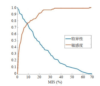
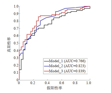
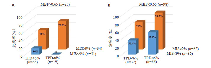
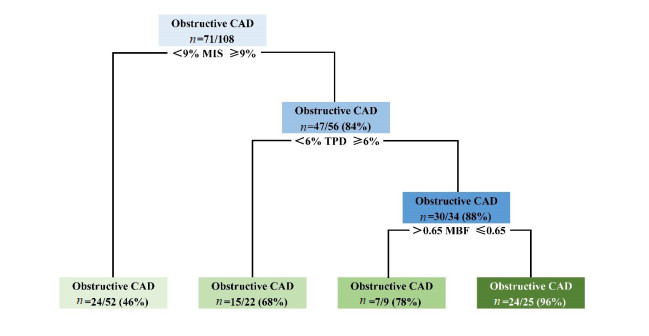
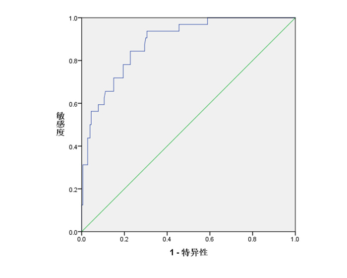
Objective To investigate whether quantitative PET myocardial perfusion-metabolism mismatch can further improve the diagnosis accuracy in predicting obstructive coronary artery disease (CAD). Methods A total of 97 patients with suspected obstructive CAD were retrospectively collected, and randomly divided into training set (61 patients) and validation set (36 patients) according to 5∶3. Obstructive CAD was defined as coronary artery ≥75% stenosis. Univariate and multivariate logistic regression analysis were used for model training and performance evaluation. The ROC curve analysis was used to determine the best trade-off for diagnosing obstructive CAD and compare the differences between models. Results The areas under the ROC curve (AUC) of three univariate logistic regression models were 0.735, 0.758 and 0.823, respectively, and the best trade-off of MIS was 9%. The AUC values of three multivariate logistic regression models were 0.766, 0.823 and 0.839, respectively. The model including TPD/MBF/MIS achieved the highest AUC and it was significantly higher than the control model including TPD/MBF (P=0.0034). The decision tree based on this model had an accuracy of up to 96% in the validation set. Conclusion PET myocardial perfusion- metabolism mismatch is an effective predictor of obstructive CAD, which can effectively improve the diagnostic accuracy and has important clinical value for the rapid and accurate differential diagnosis of suspected cases.
2022, 45(6): 804-809.
doi: 10.12122/j.issn.1674-4500.2022.06.02
Abstract:
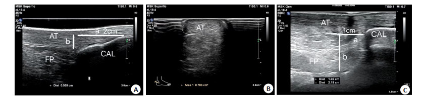
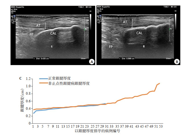
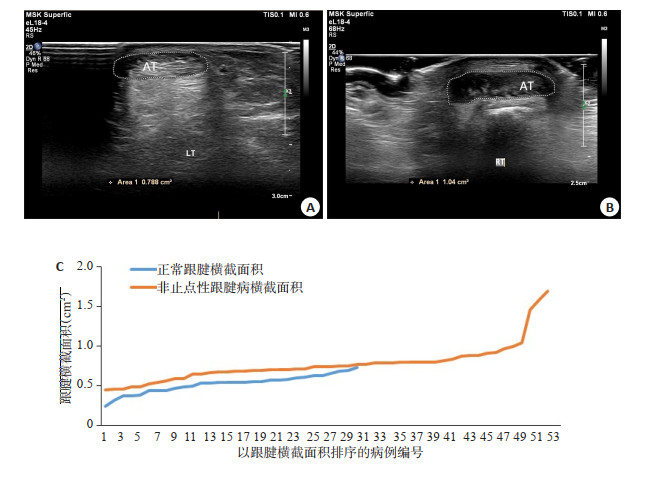
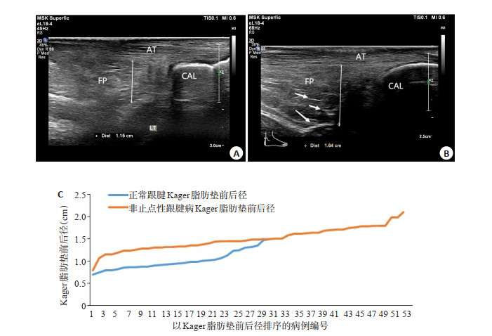
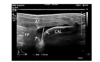
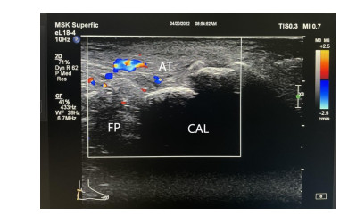
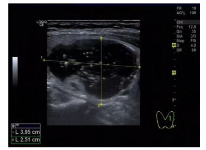
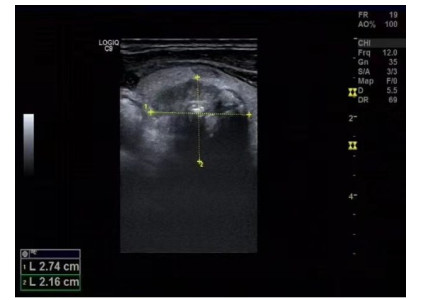
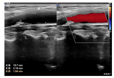
Objective To summarize the musculoskeletal ultrasonographic features of noninsertional Achilles tendinopathy. Methods Ultrasound results of 82 Achilles tendons in 60 cases were collected from November 2020 to April 2022, and 15 cases including 30 feet were set as healthy control group (group A), 45 cases including 52 feet as noninsertional Achilles tendon group (group B). Musculoskeletal ultrasound and color Doppler flow imaging were used to observe and compare the features of the noninsertional Achilles tendon: (1) The thickness of Achilles tendon at 2 cm above the posterior calcaneal tubercle; (2) The cross-sectional area of Achilles tendon at 2cm above the Achilles tendon insertion point; (3)The anterior-posterior diameter of Kager fat pad at 1cm above the upper edge of calcaneus; (4)The detection rate of retrocalcaneal bursal effusion; (5)The detection rate of blood flow signal in Achilles tendon. The features of noninsertional Achilles tendinopathy were summarized. Results (1) The thickness of Achilles tendon at 2 cm above the posterior calcaneal tubercle: group A was less than group B (0.43± 0.06 cm vs 0.55±0.17 cm, P < 0.05). (2)The cross-sectional area of Achilles tendon at 2 cm above the Achilles tendon insertion point: group A was less than group B (0.52±0.11 cm2 vs 0.74±0.23 cm2, P < 0.05). (3)The anterior-posterior diameter of Kager fat pad at 1 cm above the upper edge of calcaneus: group A was smaller than group B (1.01±0.21 cm vs 1.49±0.26 cm, P < 0.05). (4) The detection rate of retrocalcaneal bursal effusion: negative in group A and 38.46% in group B, the difference was statistically significant (P < 0.05). (5)The detection rate of blood flow signal in Achilles tendon: group A was not detected and 51.92% in group B, the difference was statistically significant (P < 0.05). Conclusion Noninsertional Achilles tendinopathy was characterized by thickening of Achilles tendon thickness, increasing of cross- sectional area of Achilles tendon, widening of anterior-posterior diameter of Kager fat pad, increasing of retrocalcaneal bursal effusion and the blood flow signal in Achilles tendon.

2022, 45(6): 810-819.
doi: 10.12122/j.issn.1674-4500.2022.06.03
Abstract:
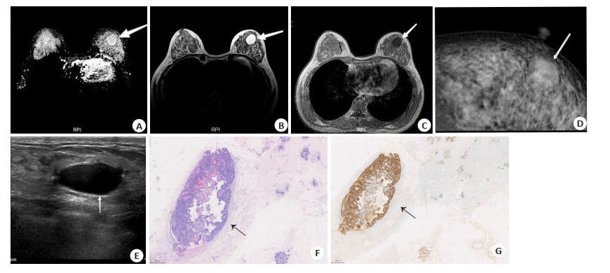
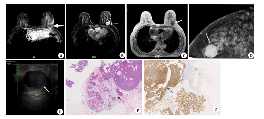
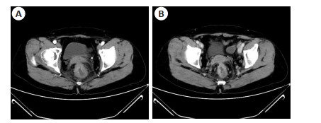
Objective To investigate the ultrasound (US), mammography (MG) and MRI profiles of the pathological subtypes of papillary carcinoma of the breast, to compare and analyze the differences of clinical and imaging features and clinical significance of the pathological subtypes of papillary carcinoma. Methods The clinical and imaging data of 47 patients with pathologically confirmed breast papillary carcinoma from February 2012 to December 2021 were collected. According to the latest tumor histological classification of World Health Organization, the patients were divided into four groups: papillary ductal carcinoma in situ (pDCIS), encapsulated papillary carcinoma (EPC), solid papillary carcinoma (SPC) and invasive papillary carcinoma (IPC) groups. The clinical features (n=47), ultrasound (n=47), MG (n=45) and MRI (n=14) of different pathological groups were compared. According to the BI-RADS classification criteria, BI-RADS classification ≥4A was considered as a positive diagnosis, and the misdiagnosis rate of the three imaging methods was analyzed. Results Clinical features: Palpable mass was more common than nipple discharge in all groups. pDCIS (9/16, 56.3%) had the highest proportion of nipple discharge, and IPC (9/11, 81.8%) had the highest proportion of palpation of mass. US imaging features: The proportion of irregular mass shape in IPC subgroup was also significantly higher than that in EPC subgroup (P=0.023). The proportion of unclear mass boundary in IPC subgroup was much higher than that in SPC group (P=0.025). Compared with pDCIS, EPC subgroup had a higher proportion of cystic solid mass (P=0.048). MG imaging features: The SPC subgroup had the highest proportion of irregular mass shape (4/6, 66.7%), and the EPC subgroup had the highest proportion of regular mass shape (5/6, 83.3%). The proportion of the SPC subgroup was higher than that of the pDCIS subgroup (P=0.015). MRI imaging features: The ADC of SPC group was lower than that of other subgroups (ADC=0.37×10⁻³ mm2/s, P=0.005), and EPC group was lower than pDCIS subgroup (P=0.017). The misdiagnosis rates of US, MG and MRI were 46.8%, 37.8% and 14.3%, respectively, and the misdiagnosis rate of US was significantly higher than that of MRI (P=0.029). Conclusion The imaging features of papillary breast carcinoma subgroups overlap. Although both US and MRI can show the morphological characteristics of tumors, the misdiagnosis rate of US is significantly higher than that of MRI. MG is more sensitive to the type and distribution of tumor calcification, and MRI is more effective in detecting tumor spread and concurrent tumors. The blood flow signal in the mass shown by US, the margin and morphology of the mass shown by US and MG, and the apparent diffusion coefficient of MRI are different in the histological subtypes of breast papillary carcinoma.
2022, 45(6): 820-824.
doi: 10.12122/j.issn.1674-4500.2022.06.04
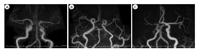
Abstract:
Objective To study the correlation between stenosis degree and prognosis in patients with middle cerebral artery stenosis by PETRA-MRA imaging. Methods Seventy-five patients with middle cerebral artery atherosclerotic stenosis admitted to our hospital from May 2021 to June 2022 were selected as the subjects. According to the Rankin Scale (mRS score > 2 indicates poor prognosis), the patients were divided into poor prognosis group (n=15) and good prognosis group (n=57). The differences of MCA blood flow signal score and magnetic sensitivity pseudo- film score in patients with different stenosis degree and prognosis were compared. Results For the diagnosis of middle cerebral artery sclerosis patients, the degree of stenosis and lesion in the lesion site of patients diagnosed by TOF-MRA was significantly higher than that by PETRA-MRA (P < 0.05). The scores of magnetic sensitivity pseudo-film and blood flow signal of the lesions of patients diagnosed by TOF-MRA were higher than those of PETRA-MRA (P < 0.05). There were statistically significant differences in the length of lesion site and the degree of stenosis among patients with different degrees of stenosis (P < 0.05). There was no statistical significance in the magnetic sensitivity pseudo-film scores and blood flow signal scores of patients with different stenosis degrees (P > 0.05). The length of lesion site and stenosis degree in patients with good prognosis group were significantly lower than those in control group (P < 0.05). There was no statistical significance in the magnetic sensitivity pseudo-film scores and blood flow signal scores of patients with different prognosis (P > 0.05). Conclusion The image quality of PETRA-MRA is good to evaluate the stenosis degree and prognosis of patients with middle cerebral artery atherosclerotic stenosis.
2022, 45(6): 825-827.
doi: 10.12122/j.issn.1674-4500.2022.06.05
Abstract:
Objective To investigate the clinical value of ultrasound "firefly" technique and X-ray mammography for ductal carcinoma in situ (DCIS) of the breast. Methods A total of 98 patients with suspected breast intraductal carcinoma in situ admitted from December 2019 to December 2021 were selected as the research objects. All 98 patients underwent surgical treatment in our hospital. Before operation, ultrasound "firefly" technology and X-ray molybdenum target examination were performed. The detection of intraductal carcinoma in situ of breast was statistically analyzed. Results All 98 cases of breast intraductal carcinoma in situ were single lesions, 59 cases were benign lesions, 39 cases were malignant lesions. The sensitivity of ultrasound "firefly" in the diagnosis of breast ductal carcinoma in situ was higher than that of X-ray mammography (71.8% vs 61.5%), and the specificity was lower than that of X-ray mammography (66.1% vs 76.3%). There was no significant difference in the diagnostic value of ultrasound "firefly" in breast DCIS (P > 0.05). The sensitivity, specificity and accuracy of ultrasound "firefly" technique combined with X-ray mammography in the diagnosis of breast DCIS were 92.3%, 91.5% and 91, 8%, respectively, which were higher than those of ultrasound "firefly" technique alone (71.8%, 66.1% and 68.4%) and X-ray mammography alone (61.5%, 76.3% and 70.4%), the difference was statistically significant (P < 0.05). Conclusion Ultrasound "firefly" technology combined with X-ray mammography has high sensitivity, specificity and accuracy in the diagnosis of breast DCIS, which is helpful to improve the diagnostic accuracy of breast DCIS and provide more value for clinical diagnosis.
2022, 45(6): 828-832.
doi: 10.12122/j.issn.1674-4500.2022.06.06
Abstract:
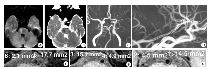
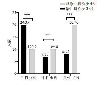
Objective To investigate basilar artery tortuosis and plaque characteristics in patients with acute pontine infarction by multimodal magnetic resonance technology. Methods Clinical, laboratory and imaging data of 35 patients with acute pontine infarction and 40 patients with non-acute pontine infarction were collected. The multimodal magnetic resonance scanning was performed to measure the angle of basilar artery tortuation and plaque characteristics (peritubular peritubular area at stenosis, lumen and wall area, reference peritubular and luminal area, plaque load, remodeling mode and remodeling rate). The univariate analysis and multivariate binary logistic regression were used to analyze the risk factors of acute pontine infarction. Results Univariate analysis showed that there were statistically significant differences in plaque load, remodeling mode and remodeling rate between groups (P < 0.05), but no significant differences in basilar artery detour, peritubular periduct at stenosis, lumen, wall area, reference peritubular and luminal area (P > 0.05). Multivariate binary logistic regression analysis showed that plaque load was an independent protective factor for acute pontine infarction (OR=0.001, 95%CI=0.000-0.306) and the remodeling mode was an independent risk factor for acute pontine infarction (OR=2.514, 95% CI=1.380-4.580). Conclusion Plaque load and remodeling patterns can affect the occurrence of acute pontine infarction. With a decrease in basilar plaque load, the incidence of acute pontine infarction decreases. Positive remodeling of the basilar artery can easily lead to acute pontine infarction.
2022, 45(6): 833-837.
doi: 10.12122/j.issn.1674-4500.2022.06.07
Abstract:
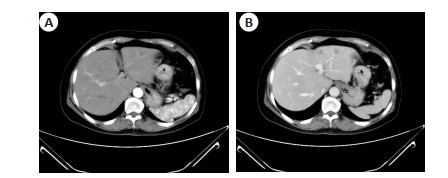
Objective To investigate the value of localization and qualitative diagnosis of malignant biliary obstruction by magnetic resonance cholangiopancreatography (MRCP) and diffusion-weighted imaging (DWI) combined with enhanced CT. Methods Patients with suspected malignant biliary obstruction admitted to our hospital from June 2015 to December 2020 were selected. All patients received MRCP, DWI, CT enhanced scanning examination and pathological diagnosis. We took pathological diagnosis as the "gold standard", and the morphology of MRCP in all patients was compared. The value of MRCP+ DWI alone and enhanced CT scan in the localization and qualitative diagnosis of biliary obstruction were compared. Results Histopathological results showed that among the 80 patients, 33 (41.25%) had benign extrahepatic biliary obstruction, including 27 bile duct stones, 6 cholangitis strictures. There had 47 malignant extrahepatic biliary obstruction, accounting for 58.75%, including 34 cases of bile total duct cancer, 4 cases of ampullary carcinoma, and 9 cases of pancreatic head carcinoma. MRCP imaging features showed that patients with benign obstruction were mainly "withered branches shape", and patients with malignant obstruction were mainly "soft vine shape". Among the two detection methods, MRCP + DWI combined with enhanced CT scanning had higher accuracy in diagnosing biliary obstruction than MRCP + DWI (P < 0.05). The diagnostic coincidence rates of MRCP + DWI for biliary obstruction in bile duct stones, cholangitic stenosis, cholangiocarcinoma, and pancreatic head cancer were lower than those for MRCP + DWI combined with enhanced CT scanning. The diagnostic coincidence rate of the two methods for ampullary carcinoma was 100%. The overall coincidence rate of qualitative diagnosis of MRCP + DWI combined with enhanced CT scan was higher than that of MRCP + DWI (P < 0.05). Conclusion MRCP + DWI combined with enhanced CT scanning can accurately locate malignant biliary obstruction and improve the accuracy of qualitative diagnosis of malignant biliary obstruction.
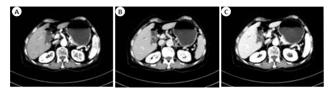
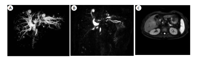
2022, 45(6): 838-841.
doi: 10.12122/j.issn.1674-4500.2022.06.08
Abstract:
Objective To investigate the imaging features of magnetic resonance enterography in the diagnosis of radiation enteritis (RE). Methods The imaging data of magnetic resonance enterography in 108 patients with suspected RE after gynecological pelvic tumor radiotherapy in our hospital from January 2016 to June 2019 were analyzed. We put the pathological examination results of patients' enteroscopy and follow- up data as the diagnosis reference of RE. The imaging characteristics of RE in magnetic resonance enterography was summarized. We analyzed the value of magnetic resonance enterography in diagnosing RE. Results All patients underwent colonoscopy in the later stage and were followed up. According to the examination results and follow-up, a total of 81 of 108 patients were diagnosed as RE, and 27 of them were excluded from RE. Taking the final diagnosis as the "gold standard", statistics showed that the sensitivity and specificity of magnetic resonance enterography in diagnosing RE were 87.65% and 74.07%. RE had "concentric circle" layered high signal performance in enhanced MR enterography and DWI scanning images. The intestinal wall showed enhanced signal, and there were changes around the intestinal tube and thickening of the intestinal wall. Conclusion RE has special imaging features in MR enterography, which can be used for clinical diagnosis of RE.
2022, 45(6): 842-846.
doi: 10.12122/j.issn.1674-4500.2022.06.09
Abstract:
Objective To explore the diagnostic value of multi-slice spiral CT (MSCT) combined with serum CA199 and CEA for colorectal cancer (CRC) and liver metastases. Methods A total of 110 patients with CRC and 110 patients with benign intestinal lesions who were pathologically confirmed in the hospital from January 2019 to December 2021 were retrospectively selected as CRC group and benign lesion group, respectively. MSCT data and levels of serum CA199 and CEA in the two groups were compared and analyzed. Based on the results of pathological diagnosis, the diagnostic value of MSCT combined with serum CA199 and CEA in the diagnosis of CRC. The diagnostic value of for liver metastasis were analyzed. Results There were significant differences in serum CA199 and CEA levels in patients with colorectal cancer, and higher index levels in CRC patients (P < 0.05). The consistency test showed that kappa values, sensitivity, specificity and accuracy rates of serum CA199, CEA, MSCT and their combination in the diagnosis of CRC were 0.491, 0.355, 0.667, 0.682; 84.54%, 72.73%, 91.82%, 72.73%; 64.55%, 62.73%, 74.55%, 95.45% and 74.55%, 67.73%, 83.18%, 84.09%, respectively. Serum CA199 and CEA levels of CRC patients with or without liver metastasis were significantly different, and the levels of serum CA199 and CEA were higher in CRC patients with liver metastasis (P < 0.05). The results of kappa consistency test showed that kappa values, sensitivity, specificity and accuracy rates of serum CA199, CEA, MSCT and their combination in the diagnosis of liver metastases were 0.408, 0.353, 0.445, 0.726; 84.00%, 72.00%, 92.00%, 72.00%; 69.41%, 71.76%, 68.24%, 96.47% and 72.73%, 71.82%, 73.64%, 90.91%, respectively. Conclusion MSCT, serum CA199 and CEA have their own advantages and disadvantages in the diagnosis of CRC. The combined detection is conducive to the diagnosis of liver metastases in CRC.
2022, 45(6): 847-852.
doi: 10.12122/j.issn.1674-4500.2022.06.10
Abstract:
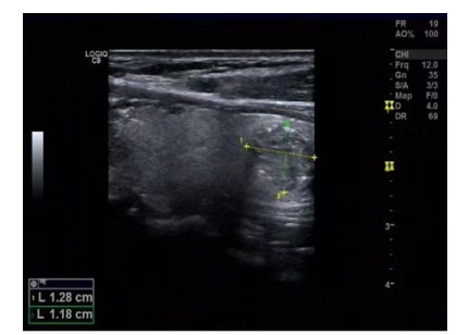
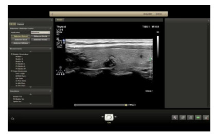
Objective To analyze the value of elastic contrast index (ECI) and shear wave elastography (SWE) quantitative parameters in predicting cervical lymph node metastasis of thyroid papillary carcinoma. Methods Sixty patients with thyroid papillary carcinoma admitted to our hospital from January 2018 to December 2021 were selected, including 21 patients with lymph node metastasis and 39 patients without lymph node metastasis. The differences of ultrasound images, ECI, SWEmax, SWEmin and SWEmean between the two groups were compared. We analyzed the predictive efficacy of ECI, SWEmax, SWEmin and SWEmean combined detection for lymphatic metastasis in patients with thyroid papilloma. Results There were statistically significant differences in aspect ratio, number of nodules, margin, calcification, relationship with capsule and blood flow signals between the two groups (P < 0.05). ECI in lymphatic metastasis group was significantly higher than that in non-lymphatic metastasis group (P < 0.05). SWEmax, SWEmin and SWEmean in lymphatic metastasis group were significantly lower than those in control group (P < 0.05). The specificity of ECI, SWEmax, SWEmin and SWEmean combined detection for lymphatic metastasis in patients with thyroid papilloma was significantly higher than that of single detection. According to ROC curve analysis, the area under curve of lymphatic metastasis in patients with thyroid papilloma by ECI, SWEmax, SWEmin and SWEmean combined detection were significantly higher than that by single detection. The truncation values of ECI, SWEmax, SWEmin and SWEmean were 4.61, 30.11 kPa, 21.98 kPa and 25.55 kPa, respectively. Conclusion ECI and SWE quantitative parameters have significant predictive value for cervical lymph node metastasis of thyroid papillary carcinoma, suggesting clinical promotion.
2022, 45(6): 853-858.
doi: 10.12122/j.issn.1674-4500.2022.06.11
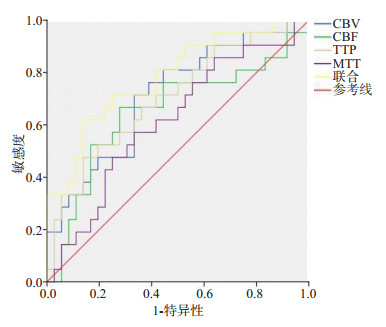
Abstract:
Objective To explore the effect of scalp acupuncture combined with butylphthalide on cerebral perfusion and cognitive function in severe middle cerebral artery stenosis hypoperfusion area. Methods A total of 96 patients with unilateral middle cerebral artery stenosis who were admitted to the department of Neurology outpatient department of our hospital from March 2019 to December 2020 were selected as the subjects. They were randomly divided into study group and control group according to random number table method, with 48 patients in each group. Both groups were given basic drug therapy: atorvastatin + clopidogrel, 1 tablet per day orally for 4 weeks. In the study group, the combination of head point penetration and butylphthalide soft capsule was added. CT perfusion imaging (CTP) was performed before and after treatment in 2 groups. Cerebral blood volume (CBV), cerebral blood flow (CBF), mean time to pass (MTT) and peak time (TTP) in affected area and symmetric non- affected area were observed. The ratio of affected area perfusion parameters to symmetric nonaffected area perfusion parameters, namely rCBV, rCBF, rMTT and rTTP were calculated. Serum nitric oxide, endothelin-1, thromboxane B2 and AngII were detected in fasting venous blood of two groups before and after treatment, vascular endothelial growth factor, brain-derived neurotrophic factor, neuron specific enolase, S100 β protein and the Simple Mental State Assessment Scale were used to evaluate the cognitive ability of the patients. Results After treatment, CBV and CBF in the study group were significantly higher than those before and after treatment (P<0.05). MTT, TTP, rCBV, rCBF, rMTT and rTTP were significantly lower than those in the control group (P<0.05). After treatment, the levels of nitric oxide, endothelin-1, vascular endothelial growth factor, brain- derived neurotrophic factor, neuron specific enolase and s100β in the two groups were significantly higher than before, and the study group was significantly higher than the control group. After treatment, thromboxane B2 and AngII in both groups were significantly lower than before, and the study group was significantly lower than the control group (P<0.05). After treatment, the indexes of cognitive function in the two groups were significantly higher than before, and the study group was higher than the control group (P<0.05). Conclusion Head point penetrating acupuncture combined with butylphthalide can effectively improve distal collateral circulation opening, increase cerebral blood flow, upregulate neurotrophic factors and promote cognitive function recovery in patients with severe middle cerebral artery stenosis.
2022, 45(6): 859-862.
doi: 10.12122/j.issn.1674-4500.2022.06.12
Abstract:
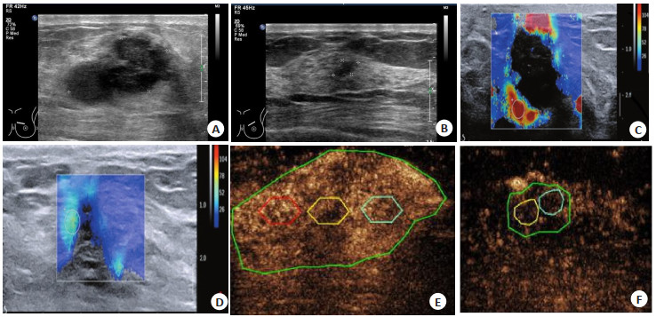
Objective To investigate the clinical significance of multimodal contrast ultrasonography for evaluating the efficacy of radiotherapy and chemotherapy in patients with radical mastectomy. Methods The clinical data of 140 patients with breast cancer undergoing radiotherapy and chemotherapy admitted to our hospital from July 2015 to January 2019 was retrospectively analyzed. The patients underwent contrast ultrasonography and ultrasonic elasticity-before and after chemotherapy. The postoperative pathology was used as the gold standard. The diagnostic value of the ultrasound contrast in radiotherapy and chemotherapy was analyzed. Results The curative effect of radiotherapy and chemotherapy evaluated by conventional ultrasound was 52.63%, the specificity was 87.50%, and the accuracy was 68.57%. The consistency between conventional radiography and pathology was poor by Kappa test (Kappa=0.388). The sensitivity, specificity and accuracy of contrast-enhanced ultrasound in evaluating chemotherapy efficacy were 94.7%, 98.4% and 96.4% respectively; The consistency analysis of contrast- enhanced ultrasound and pathology showed that Kappa=0.928, with good consistency. The ultrasound images of all patients showed high enhancement before radiotherapy and chemotherapy, while the effective patients after radiotherapy and chemotherapy showed low enhancement, the ineffective patients showed moderate and high enhancement (P < 0.05). After chemoradiotherapy, the regional blood flow, area under curve and strain ratio in effective patientswere were lower than those in ineffective patients, whereas the time to peak and elasticity scorewerehigher than the ineffective patients (P < 0.05). Conclusion Partial qualitative and quantitative indicators of multimodal contrast ultrasonography have some certain evaluation value for the efficacy of radiotherapy and chemotherapy in patients undergoing radical mastectomy.
2022, 45(6): 863-866.
doi: 10.12122/j.issn.1674-4500.2022.06.13
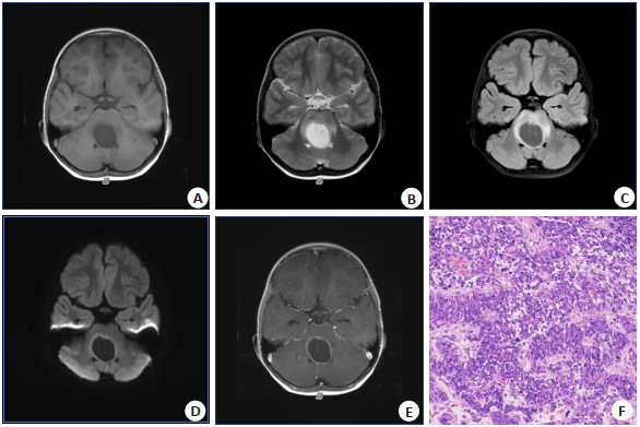
Abstract:
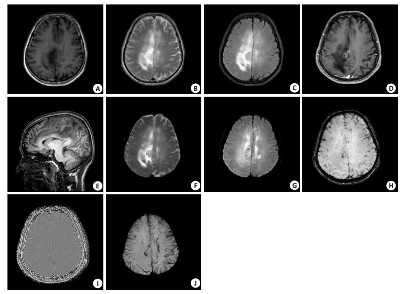
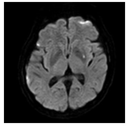
Objective To investigate the MRI features of pediatric diffuse midline gliomas, H3K27-altered. Methods A retrospective study was carried out to analyze 7 pediatric patients of diffuse midline gliomas, H3K27-altered confirmed by surgery and pathology in Shanghai Children's Hospital from July 2017 to November 2021. The clinical and MRI data were abstracted from their electronic medical records. All the 7 patients underwent plain and enhanced MRI scan before operation, and the MRI features of the tumors were observed and analyzed. Results All the patients showed a unilateral mass. 5 cases were located in brainstem, 1 case was located in thalamus, and 1 case was located in thoracic spinal cord. 4 cases were irregular and 2 cases were round-like with clear boundaries, but the case in thoracic spinal cord was strip with unclear boundary. 2 cases had cystic or necrosis, and all cases had no hemorrhage. In 6 cases, the solid components of tumor showed low signal on T1WI, slightly-high signal on T2WI and high signal on T2- FLAIR; The solid components of thoracic spinal cord tumor showed slightly higher signal on T1WI, high signal on T2WI and high signal on T2-FLAIR. 6 cases of DWI had diffusion restriction and 1 case had no diffusion restriction.MRI enhanced scans showed 6 cases had obvious uneven enhancement and 1 case had no obvious enhancement, of which 3 cases showed circular enhancement, 2 cases showed nodular enhancement and 1 case showed patchy enhancement. There was no obvious edema around the tumors. Distant metastasis and cerebrospinal fluid dissemination were no found in all cases. Conclusion There are certain MRI features of pediatric diffuse midline gliomas, H3K27-altered. The location of the tumor, diffusion restriction, and the mode and scope of enhancement will help to improve the level of imaging diagnosis and differential diagnosis of the disease.
2022, 45(6): 867-870.
doi: 10.12122/j.issn.1674-4500.2022.06.14
Abstract:
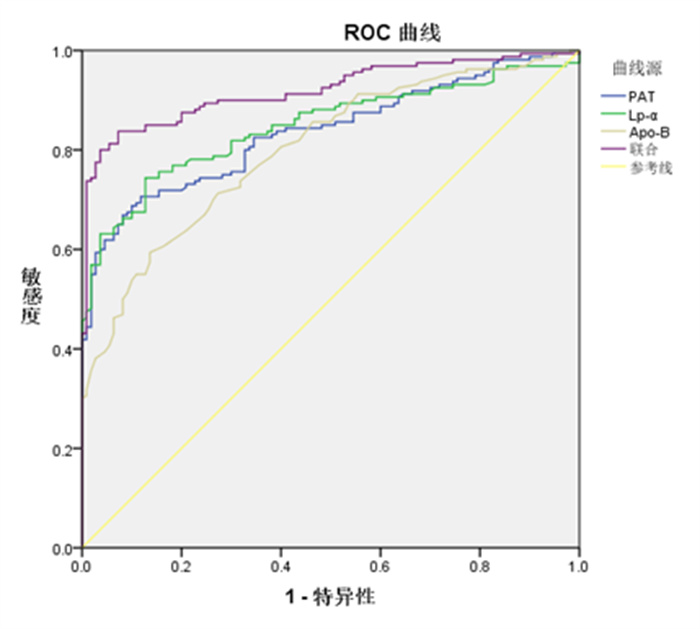
Objective To explore the diagnostic value of multi-slice spiral CT (MSCT) measurement of pericardial adipose tissue (PAT) combined with lipoprotein α (Lp-α) and apolipoprotein B (Apo-B) detection in patients with early coronary heart disease. Methods A total of 160 patients with early coronary heart disease who were treated in our hospital from March 2020 to April 2022 were selected as the observation group. The 110 healthy subjects were selected during the same period. We detected the levels of Lp-α and Apo-B, and used MSCT to measure PAT to analyze the value of the combined detection of these three indicators in the diagnosis and evaluation of coronary heart disease. Results The levels of PAT, Lp-α in the observation group were significantly higher than those in the control group (P < 0.05). Spearman correlation showed that the levels of PAT, Lp-α and Apo-B were positively correlated with the occurrence of coronary heart disease (P < 0.05). PAT, Lp-α, Apo-B levels were positively correlated with coronary SYNTAX score (P < 0.05). ROC curve showed that PAT, Lp-α, Apo-B levels combined diagnostic the area under the curve was 0.920, which was significantly higher than the area under the curve of each individual index (P < 0.05). Conclusion The PAT, Lp-α and Apo-B are elevated in patients with early coronary heart disease, which are closely related to the occurrence and severity of coronary heart disease. It has a high diagnostic value for coronary heart disease.
2022, 45(6): 871-875.
doi: 10.12122/j.issn.1674-4500.2022.06.15
Abstract:
Objective To investigate the influence of calcified plaque burden on the accuracy of coronary CT angiography (CCTA) in the diagnosis of lumen stenosis. Methods A total of 100 patients who underwent coronary angiography (CAG) and CCTA in the Department of Radiology at Shanghai Jiading District Central Hospital from February 2019 to January 2022 were selected as the research subjects. The calcification volume and calcification score were recorded in vascular segments. The patients were divided into four groups according to the calcification volume and calcification score, and the degree of vascular stenosis in each group was analyzed. The accuracy of CCTA in diagnosing lumen stenoses of different plaque calcification volume and scores was discussed. Results A total of 396 calcified coronary artery segments were selected from the 100 patients. Based on 15 segments, lumen stenosis ≥50% indicated significant stenosis. The sensitivity, specificity, accuracy, positive predictive value and negative predictive value were 99.03% (204/206), 83.16% (158/190), 91.41% (362/396), 86.44% (204/ 236) and 98.75% (158/160), respectively. Under the condition of calcification volume ≤25 mm2 and calcification score≤80 points, the sensitivity, specificity, accuracy, positive predictive value and negative predictive value all were 100%. Under the condition of 196 mm2<calcification volume≤1375 mm2, the sensitivity, specificity, accuracy, positive predictive value and negative predictive value were 85.71%, 36.36%, 75.47%, 83.72% and 40.00%, respectively. When the calcification score was higher than 200, the sensitivity, specificity, accuracy, positive predictive value and negative predictive value were 92.86%, 75.00%, 87.50%, 89.66% and 91.82%, respectively. The sensitivity, specificity, accuracy, positive predictive values and negative predictive values of CCTA to diagnose lumen stenosis of different calcification volumes and different calcification scores were significantly different (P<0.05). The sensitivity, specificity, accuracy, positive predictive value and negative predictive value were the highest to diagnose lumen stenosis of 0-25 mm2 calcification volume and 0-80 calcification score. Conclusion Different calcified plaque loads have different influence on the accuracy of CCTA in the diagnosis of lumen stenosis, which deserves attention in clinical practice.
2022, 45(6): 876-879.
doi: 10.12122/j.issn.1674-4500.2022.06.16
Abstract:
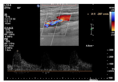
Objective To investigate the application value of shear wave elastography in the staging and thrombolytic efficacy of deep venous thrombosis (DVT) of lower extremities. Methods A total of 212 patients with DVT were selected as the research objects.The patients were divided into acute phase group (n=97), subacute phase group (n=68), chronic phase group (n=47) according to the time of onset and the results of color Doppler ultrasonography. According to treatment outcome, all patients received thrombolytic therapy and were divided into cure group (n=83), effective group (n=97), ineffective group (n=32). The shear wave elastography technique was used to measure the Young's modulus of patients with different stages and different therapeutic effects. The ROC curve was drawn to evaluate the diagnostic efficacy of Young's modulus of thrombolysis in patients with DVT after treatment. Results Before treatment, there were statistically significant differences in young's modulus of thrombus in patients with different stages of DVT (P<0.05), including acute stage group<subacute stage group<chronic stage group (P<0.001). After treatment, the young's modulus of thrombus in all groups was lower than before (P<0.05), and the difference was statistically significant (P<0.05), among which the acute stage<subacute stage<chronic stage (P<0.001). After treatment, young's modulus of thrombus in DVT patients with different prognosis had statistical significance (P<0.05), among which cured group<effective group<invalid group (P<0.001). ROC analysis results showed that when Youden index was 0.632, the cut-off value of Young's modulus of thrombolysis was 6.825 kPa, and the sensitivity, specificity and AUC for diagnosing ineffective thrombolytic therapy in DVT patients were 93.8%, 69.4% and 0.885 respectively. Conclusion The measurement of Young's modulus by shear wave elastography technique can be used as an auxiliary method to evaluate stage of DVT and the efficacy of thrombolysis.
2022, 45(6): 880-884.
doi: 10.12122/j.issn.1674-4500.2022.06.17
Abstract:
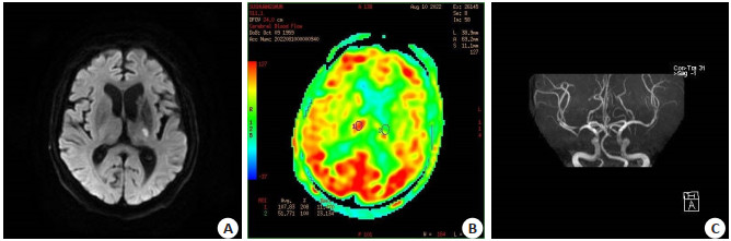
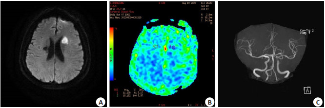
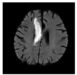
Objective To investigate the value of three-dimensional arterial spin labeling (3D-ASL) combined with diffusionweighted imaging (DWI) and magnetic resonance angiography (MRA) in assessment of ischemic penumbra (IP) in acute ischemic cerebral infarction. Methods A total of 62 patients with acute ischemic cerebral infarction were treated in the hospital from January 2021 to June 2022. All patients underwent 3D- ASL, DWI and MRA. The presence or absence of IP was determined according to the area of DWI hyperintensity at the maximum slice of the lesion and the abnormal perfusion area of ASL. According to the time from onset to first MRI, the patients were divided into the hyperacute phase (T<6 h) group and the acute phase (6 h≤T≤24 h) group. The apparent diffusion coefficient (ADC) values and cerebral blood flow (CBF) of central infarct area, IP area and contralateral area of IP were recorded. The degree of vascular stenosis was graded according to MRA images. Results The lesions of patients with acute ischemic cerebral infarction showed spotted or small pieces of high signals on DWI. 48 patients had significantly larger abnormal perfusion area on 3D-ASL than abnormal high signal area on DWI, which indicated the existence of IP area. MRA showed segmental stenosis of a intracranial main branch, significantly thinned or even disappeared distal blood vessels, and some blood vessels without obvious abnormalities. The CBF and ADC values of central infarct area, IP area, and contralateral area of IP increased in order (P<0.05). The rCBF and rADC values in hyperacute phase were higher than those in acute phase (P<0.05). MRA showed that vascular stenosis mainly was grade 0 in patients with IP, and mainly was grade 3 in patients without IP (P<0.05). Conclusion 3D-ASL combined with DWI and MRA can help to accurately assess the presence or absence of IP area and the degree of vascular stenosis in patients with acute ischemic cerebral infarction, which is beneficial to the selection of clinical treatment plan.
2022, 45(6): 885-890.
doi: 10.12122/j.issn.1674-4500.2022.06.18
Abstract:
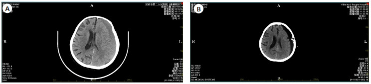
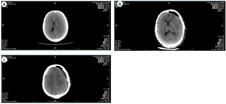
Objective To observe the curative effect and prognostic factors of patients with chronic subdural hematoma (CSDH) treated by intracranial minimally invasive evacuation under the guidance of CT density. Methods The clinical data of 134 CSDH patients treated in our hospital from December 2017 to December 2020 were analyzed retrospectively. According to the surgical methods, they were divided into observation group (n=74, minimally invasive evacuation of intracranial hematoma guided by CT density of lesions) and control group (n=60, conventional skull drilling and closed drainage). The perioperative indexes of the two groups were compared, and the neurological and motor functions [Chinese stroke scale (CSS), modified Barthel index (MBI)] and complications were recorded before and after operation. Markwalder's grade at 3 months after discharge was taken as the evaluation standard of prognosis, and the observation group was divided into poor prognosis group and good prognosis group. The risk factors affecting the prognosis of CSDH were analyzed by univariate and multivariate Logistic regression equation. Results There were no statistically significant differences in intraoperative bleeding, postoperative extubation time and hospital stay between the two groups (P>0.05). The operation time of the observation group was significantly longer (P<0.05), and the hematoma clearance rate was significantly higher 30 days after surgery (P<0.05). CT images show that before the treatment to the top of the left frontal temporal occipital low signal crescent images, local placeholder effect obvious, compression of brain tissue, midline structure shift, after treatment of chronic subdural hematoma on the first day after drilling drainage, drainage tube indwelling, subdural effusion pneumatosis performance, reduce local placeholder effect, midline structure shift better; There was no statistical significance in preoperative CSS and MBI scores between the two groups (P>0.05). The CSS score of the two groups at 30 days after surgery was significantly lower than that before surgery, while the MBI score was significantly higher than that before surgery(P<0.05). The CSS score of the observation group was lower and THE MBI score was higher after surgery(P<0.05). There was no death in the two groups within 3 months of follow-up, and there was no statistical significance in the incidence of complications (P>0.05). Age, brain atrophy and on admission GCS score in a set of good prognosis and bad prognosis way between the difference was statistically significant (P<0.05). Gender, symptoms to hospital time, hematoma volume, history of drinking, smoking history, hematoma, postoperative drainage, CT value of anticoagulant drugs in good prognosis group were not significantly different from the bad way prognosis (P>0.05). Age>65 years old and GCS score<13 at admission were independent risk factors of prognosis of CSDH after minimally invasive intracranial hematoma removal guided by CT density value (P<0.05). Conclusion Minimally invasive evacuation of intracranial hematoma guided by CT density of lesions is effective in the treatment of CSDH, and its prognosis is related to age and GCS score at admission.
2022, 45(6): 891-896.
doi: 10.12122/j.issn.1674-4500.2022.06.19
Abstract:
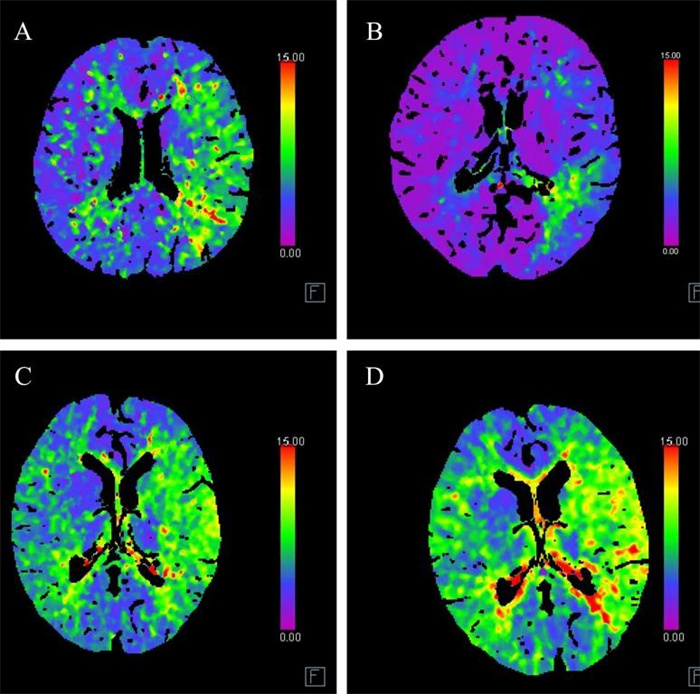
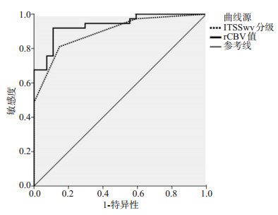
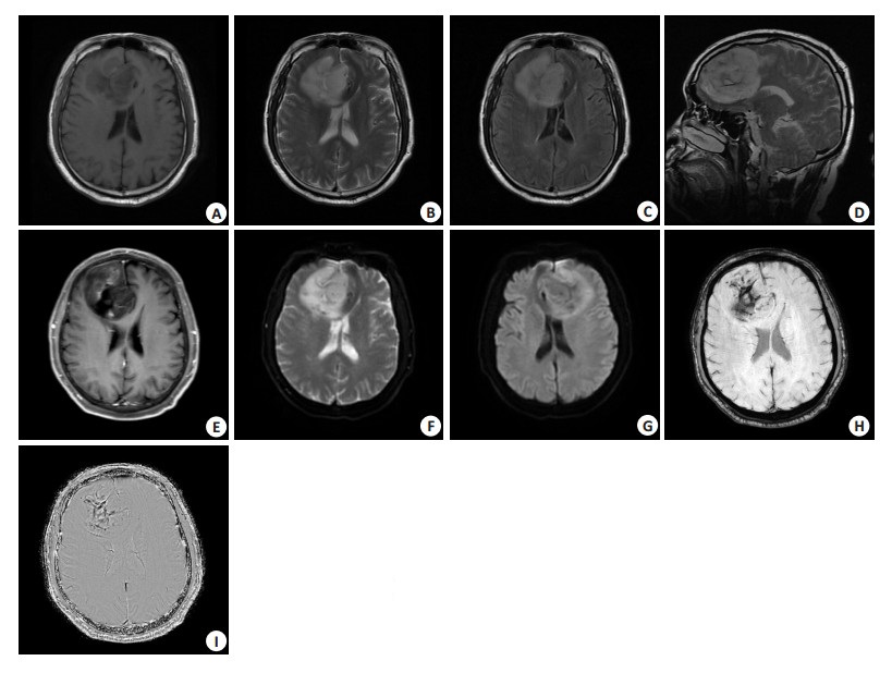
Objective To investigate the application of susceptibility weighted imaging (SWI) in the differential diagnosis, preoperative pathological grading and surgical guidance of glioma. Methods The clinical data of 64 patients with glioma, 15 patients with single brain metastases and 15 patients with intracranial lymphoma confirmed by surgery and histopathological examination in the People's Hospital of Xinjiang Uygur Autonomous Region from October 2018 to October 2021 were retrospectively analyzed. The pathological results were used as the gold standard. The patients with glioma were divided into low-grade group (n=27) and high-grade group (n=37) according to the WHO tumor classification criteria. All patients were detected by SWI, and the intratumoral susceptibility signal intensity (ITSS) grading evaluation and the relative cerebral blood volume (rCBV) value of the tumor parenchymal area and the peritumoral edema area were measured. The differences of different tumor types were compared, and the ROC curve was used to evaluate its differential value between high-grade and low-grade gliomas. Results Comparison of ITSS grade and rCBV value in tumor parenchyma between high-grade gliomas and solitary metastases (P > 0.05); the rCBV value in peritumoral edema area of high-grade glioma was higher than that of single brain metastasis (P < 0.05). The ITSS grade of high-grade glioma was lower than that of lymphoma, and the vascular score was higher than that of lymphoma (P < 0.05). ITSS grade of high grade glioma was higher than that of low grade glioma, rCBV value of tumor parenchyma was higher than that of low grade glioma (P < 0.05). Spearman correlation analysis showed that glioma grade was positively correlated with ITSS grade and rCBV value (r=0.728, 0.851, P < 0.005). The area under the high-level ROC curve of ITSS grading evaluation of glioma was 0.894, and the sensitivity, specificity and accuracy were 81.08%, 85.19% and 82.81%, respectively. The area under the ROC curve of rCBV was 0.937, and the sensitivity, specificity and accuracy were 91.89%, 88.89 % and 90.63%, respectively. Compared with conventional sequence, SWI sequence showed tumor boundary more clearly and showed tumor microangiopathy information in 64 patients with glioma. The rCBV values in glioma parenchyma were higher than those in peritumoral edema (P < 0.05). Conclusion SWI is helpful for the differential diagnosis of high-grade gliomas, and has high evaluation efficiency for low-grade and high-grade gliomas, which can guide intraoperative glioma resection to a certain extent.
2022, 45(6): 897-901.
doi: 10.12122/j.issn.1674-4500.2022.06.20
Abstract:
Objective To investigate the effect of local area Ta treatment applied with Qihua San on hematoma and pain degree after ultrasound-guided minimally invasive rotary mastectomy. Methods According to the random number table method, 118 cases of patients with ultrasound-guided minimally invasive mastectomy treated in our hospital from January 2019 to January 2022 were divided into observation group and control group, with 59 cases in each group. The control group was treated with conventional western medicine, and the observation group was local applied with Qihua San Ta treatment on the basis of conventional western medicine. We analyzed the effect of treatment on the degree of hematoma and pain. Results The time of hematoma and pain resolution in observation group was significantly shorter than that in control group (P < 0.05). The visual analog scale (VAS) score was different at different time after treatment (P < 0.05), the VAS score of the observation group was lower than that of the control group (P < 0.05), and the variation trend of VAS score was different between the observation group and the control group (P < 0.05). The levels of TNF-α, IL-6, PGE2 and substance P in observation group were significantly lower than those in control group (P < 0.05). The ADL score in observation group was significantly higher than that in control group (P < 0.05), and the PSQI score was significantly lower than that in control group (P < 0.05). Conclusion Traditional Chinese medicine Qihua San local Ta treatment for ultrasound guided minimally invasive rotary mastectomy patients can reduce the levels of inflammatory factors and pain factors, promote the rapid subside of hematoma and pain, and improve the postoperative sleep quality and the ability of daily living.
2022, 45(6): 902-907.
doi: 10.12122/j.issn.1674-4500.2022.06.21
Abstract:
Objective To explore the application value of one-stop CT angiography (CTA) + CT cerebral perfusion imaging (CTP) in the diagnosis of acute cerebral infarction with cerebral vascular stenosis or occlusion and in the evaluation of cerebral blood perfusion. Methods 86 patients with acute cerebral infarction from January 2021 to May 2022 were selected as the research subjects, and all of them were examined by CTA + CTP. According to the degree of cerebral vascular stenosis, the patients were divided into mild-to-moderate group (moderate stenosis, mild stenosis, n=60) and severe group (cerebral vascular occlusion, severe stenosis, n=26). The evaluated value of cerebral blood perfusion indicators [cerebral blood flow (CBF), cerebral blood volume (CBV), mean transit time (MTT), time to peak (TTP)] on the degree of cerebral vascular stenosis was analyzed. The patients were classified into good collateral circulation group (n=36) and poor collateral circulation group (n=21) according to the degree of collateral circulation, and the evaluated value of cerebral vascular perfusion indicators on opening degree of collateral circulation was analyzed. Results The incidence rates of cerebral vascular occlusion, severe stenosis, moderate stenosis and mild stenosis in 86 patients with cerebral infarction were 10.47% (9/86), 19.77% (17/86), 36.05% (31/86) and 33.72% (29/86) respectively. The CBV and CBF of the affected side of the mild-to-moderate group and severe group were smaller than those of the healthy side, and the TTP and MTT were longer than those of the healthy side (P < 0.05). The CBV and CBF of the healthy side in severe group were smaller than those in mild-to-moderate group (P < 0.05). The decreases of CBV and CBF and the increases of TTP and MTT of the affected side compared with the healthy side were greater in severe group than those in mild-to-moderate group (P < 0.05). The AUC of the combined detection of CTP indicators on evaluating the degree of cerebral vascular stenosis was greater than that of CBV, CBF and other indicators alone (P < 0.05). The CBF in poor collateral circulation group was higher than that in good collateral circulation group while the CBV was lower than that in good collateral circulation group, and MTT and TTP were shorter than those in good collateral circulation group (P < 0.05). The AUC of the combined detection of CTP indicators was greater than that of each indicator alone in evaluating the opening degree of collateral circulation (P < 0.05). Conclusion CTA+CTP can evaluate the cerebral blood perfusion in patients with acute cerebral infarction, and can be used to evaluate the degree of cerebral vascular stenosis and the degree of collateral circulation opening.
2022, 45(6): 908-911.
doi: 10.12122/j.issn.1674-4500.2022.06.22
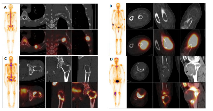
Abstract:
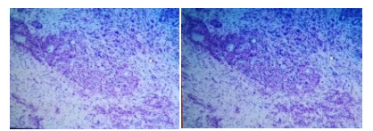
Objective To explore the imaging features of aneurysmal bone cyst in whole- body bone imaging and SPECT/CT tomosynthesis imaging, so as to improve the diagnostic accuracy of aneurysmal bone cyst. Methods A retrospective analysis was performed on patients who underwent whole-body bone imaging and SPECT/CT tomosynthesis imaging, pathologically diagnosed with aneurysmal bone cyst in our hospital from January 2016 to May 2022. We summarized its imaging features in whole-body bone imaging and SPECT/CT tomosynthesis imaging. Results Twenty-four patients underwent whole-body bone imaging and SPECT/CT tomosynthesis imaging in our hospital and were pathologically diagnosed as aneurysmal bone cysts, including 19 primary cases and 5 secondary cases, and mostly teenagers. It distributed in the long bones of the extremities, but also in the ribs and pelvic bones. The lesions were mainly expansile bone destruction with different degrees of increased bone metabolism in the corresponding parts. Conclusion Whole-body bone imaging combined with SPECT/CT tomosynthesis imaging can better display the imaging features and bone metabolism of aneurysmal bone cysts, which is helpful for the qualitative diagnosis of aneurysmal bone cysts.
2022, 45(6): 912-916.
doi: 10.12122/j.issn.1674-4500.2022.06.23
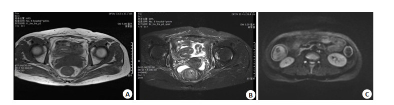
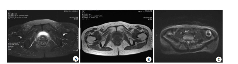
Abstract:
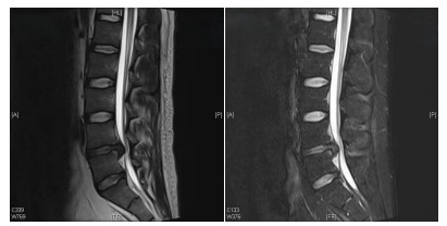
Objective To investigate the efficacy of spinal endoscopic decompression fusion internal fixation in the treatment of lumbar spinal stenosis. Methods Fifty patients with lumbar spinal stenosis in our hospital from September 2019 to September 2021 were selected and randomly divided into the observation group (given total spinal endoscopic decompression fusion internal fixation) and the control group (given traditional open surgery), twenty-five cases/group. The operation time, intraoperative blood loss and hospital stay were compared. The visual analogue scale (VAS) score of incision pain was compared at 24, 48 and 72 hours after operation. The Oswestry disability index (ODI) and Cobb angle were followed up at 3 and 12 months after operation, and the fusion rate was observed at 12 months after operation. Postoperative complications were followed up 12 months after operation. Results The observation group was significantly better than the control group in terms of operative time, intraoperative bleeding and hospital stay (P < 0.05). There was a statistically significant difference in VAS scores at 24, 48 and 72 hours after surgery between the two groups (P < 0.05). The ODI scores at 3 and 12 months postoperative follow-up were significantly improved compared with those before surgery in both groups (P < 0.05). There was no statistically significant difference between the two groups in the change of Cobb angle of the fused segment at 3 or 12 months after surgery and the fusion rate at 12 months after surgery (P > 0.05). The complication rate was 8% (2/25) in the experimental group and 12% (3/25) in the control group, and the difference was statistically significant (P < 0.05). Conclusion The therapeutic effect of spinal endoscopic spinal decompression, lumbar interbody fusion, and percutaneous pedicle nailing for lumbar spinal stenosis are significantly better than that of traditional surgery.
2022, 45(6): 917-920.
doi: 10.12122/j.issn.1674-4500.2022.06.24
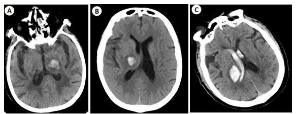
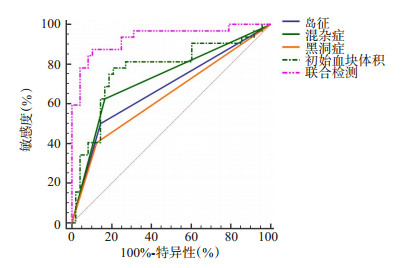
Abstract:
Objective To analyze the clinical value of multi-slice spiral CT (MSCT) pulmonary angiography parameters in the diagnosis of emergency pulmonary embolism. Methods The clinical data of 80 patients with intracerebral hemorrhage who received plain CT imaging examination in our hospital from October 2021 to January 2022 were retrospectively studied. According to whether the hematoma was enlarged, they were divided into the enlarged hematoma group (n=32) and the enlarged non-hematoma group (n=48). The CT imaging signs and hematoma volume of all patients were recorded, and the differences of CT imaging signs and hematoma volume were compared between the two groups. Spearman correlation was used to analyze the correlation between single sign, hematoma volume and hematoma enlargement in patients with intracerebral hemorrhage. ROC curve was used to analyze CT imaging signs and hematoma volume to evaluate the value of hematoma enlargement, and Logistic multifactor regression was used to analyze the related influencing factors of hematoma enlargement. Results The proportion of "island sign", "black hole sign" and "mixed sign" in CT plain scan of hematoma expansion group was significantly higher than that of non-hematoma expansion group (P < 0.05). The volume of hematoma in the enlarged group were significantly higher than that in the non-enlarged group (P < 0.05). Spearman correlation analysis showed that "island sign", "black hole sign", "mixed sign" and hematoma volume were positively correlated with hematoma enlargement in patients with intracerebral hemorrhage (r=0.423, 0.456, 0.427, 0.516, P < 0.05). ROC curve analysis showed that the area under the curve of "island sign", "black hole sign" and "hybrid sign" combined with hematoma volume for evaluation of hematoma enlargement was 0.934, the sensitivity was 87.5%, and the specificity was 89.6%, which were all higher than single sign and hematoma volume. Logistic multifactor regression analysis showed that island sign, black hole sign, mixed sign and newly diagnosed hematoma volume increase were independent risk factors for hematoma enlargement (P < 0.05). Conclusion CT imaging findings and hematoma volume have certain evaluation value for hematoma enlargement in patients with cerebral hemorrhage, and the combined evaluation value is higher.
2022, 45(6): 921-924.
doi: 10.12122/j.issn.1674-4500.2022.06.25
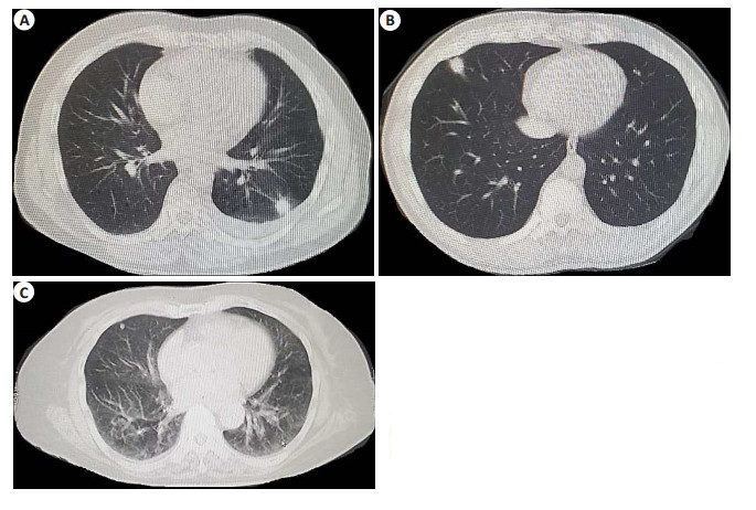
Abstract:
Objective To explore the value of DenseNet network deep learning analysis in differentiating benign and malignant pulmonary nodules. Methods Eighty patients with suspected pulmonary nodules in our hospital from February 2017 to May 2019 were selected. All patients underwent CT scan and artificial intelligence system of DenseNet network deep learning to diagnose benign and malignant, and pathological results were taken as gold standard. We analyzed the diagnostic value of CT images, DenseNet network deep learning analysis combined with CT images in benign and malignant pulmonary nodules. Results CT images showed increased lung density and cloudy shadow, which could clearly show the situation of bronchial vessels. The accuracy rate of benign and malignant nodules was 88.75%, the sensitivity was 76.92%, and the specificity was 94.44%, Kappa value with pathological diagnosis was 0.736 (P < 0.001). The sensitivity and specificity of DenseNet network depth learning combined with CT in evaluating benign and malignant nodules were 96.15% and 88.89%, respectively. The accuracy of densenet network deep learning combined with CT in evaluating benign and malignant nodules was 91.25%, which was higher than that of CT alone (88.75%). It had good consistency with pathological diagnosis (Kappa=0.810, P < 0.001). Conclusion DenseNet network deep learning analysis of CT images in differentiating benign and malignant pulmonary nodules has high accuracy and good consistency with pathological results.
2022, 45(6): 925-928.
doi: 10.12122/j.issn.1674-4500.2022.06.26
Abstract:
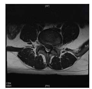
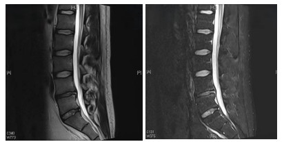
Objective To analyze the correlation between MRI lumbar imaging parameters and lumbar function in patients with lumber disc herniation and evaluate the efficacy of percutaneous foraminoscopy. Methods A total of 92 patients with lumbar disc herniation admitted to our hospital from January 2021 to December 2021 were selected as the observation group, and 76 healthy subjects in the same period were selected as the control group. Percutaneous foraminoscopy was performed on all patients in the observation group, and MRI was performed on all patients. Waist imaging parameters of the two groups were compared. Pearson was used to analyze the correlation between MRI lumbar imaging parameters and lumbar function of lumber disc herniation patients. ROC curve was used to predict the value of MRI lumbar imaging parameters in evaluating the efficacy of percutaneous foraminoscopy. Results The cross sectional area of psoas major muscle, multimultius muscle, sagittal diameter of the middle foramen and the area of the foramen in the observation group were significantly smaller than those in the control group with statistical significance (P < 0.05). Pearson correlation analysis showed that the cross section area of psoas major, the cross section area of multifidus, the sagittal diameter of the middle foramina and the area of the foramina were positively correlated with lumbar function (P < 0.05). ROC curve results showed that MRI lumbar imaging parameters had high sensitivity and specificity in evaluating the postoperative efficacy of percutaneous foraminoscopy. Conclusion MRI lumbar imaging parameters are correlated with lumbar function of lumber disc herniation patients, which can be used as an important basis for evaluating the efficacy of percutaneous foraminoscopy.

2022, 45(6): 929-933.
doi: 10.12122/j.issn.1674-4500.2022.06.27
Abstract:
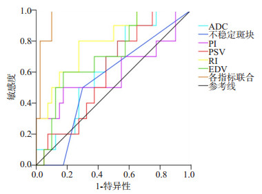
Objective To investigate the clinical value of carotid artery color Doppler ultrasound combined with brain MRI in severity evaluation of anterior circulation atherosclerotic cerebral infarction. Methods A retrospective review was conducted on the data of 50 patients with anterior circulation atherosclerotic cerebral infarction who were admitted to the hospital between January and November. The patients were divided into the mild cerebral infarction group (n=40, NIHSS score ≤7) and the severe cerebral infarction group (n=10, NIHSS score > 7) according to the NIHSS score. All patients were examined with carotid artery color Doppler ultrasound and brain MRI. The value of combination of the two methods in severity evaluation was analyzed. Results The proportion of unstable plaques, stenosis rate, pulsatility index (PI) and resistance index (RI) in the severe cerebral infarction group were higher than those in the mild cerebral infarction group. The peak systolic velocity (PSV) and end-diastolic velocity (EDV) were lower than those in the mild cerebral infarction group (P < 0.05). The apparent diffusion coefficient (ADC) of the severe cerebral infarction group was significantly lower than that of the mild cerebral infarction group (P < 0.05). The detection of T1WI signals and T2WI signals showed no statistically significant difference between the groups (P > 0.05). Spearman correlation analysis found that ADC, unstable plaque, PI, RI, PSV, and EDV were correlated with the severity (P < 0.05). ROC curve analysis found that the AUC values of ADC, unstable plaque, PI, PSV, RI, and EDV to evaluate severe cerebral infarction were 0.638, 0.556, 0.600, 0.608, 0.798 and 0.713, respectively. The AUC of joint evaluation with these indicators was 0.968. Conclusion Carotid artery color Doppler ultrasound and brain MRI both can be used for severity evaluation of patients with anterior circulation atherosclerotic cerebral infarction.
2022, 45(6): 950-956.
doi: 10.12122/j.issn.1674-4500.2022.06.31
Abstract:
Gliomas are the most common primary central nervous system malignancy. O6-methylguanine-DNA methyltransferase (MGMT) promoter methylation is an important molecular feature of glioma, which is closely related to alkylating agent chemotherapy sensitivity, prognosis, risk stratification, and tumor recurrence. Currently, only surgical specimens are used for genetic testing analysis to determine the methylation status of MGMT promoters, but this process has limitations. MRI is currently the most widely used non-invasive examination method for brain tumors. In recent years, a number of advanced magnetic resonance imaging techniques have been used to assess MGMT methylation status non-invasively before surgery, which will help predict treatment response and prognosis. The paper reviewed the research progress of magnetic resonance imaging techniques to predict the methylation status of MGMT including chemical exchange saturation transfer imaging and amide proton transfer imaging, perfusion-weighted imaging (dynamic susceptibility contrast, dynamic contrast enhance, arterial spine labeling, and inflow-based vascular-space-occupancy), diffusion imaging (diffusion tensor imaging, diffusion kurtosis imaging, intravoxel incoherent motion, restriction spectrum imaging), susceptibility weighted imaging, magnetic resonance spectroscopy imaging and the clinical significance of MGMT methylation. In order to provide a preoperative basis for the formulation of individualized treatment plans for patients.
Gliomas are the most common primary central nervous system malignancy. O6-methylguanine-DNA methyltransferase (MGMT) promoter methylation is an important molecular feature of glioma, which is closely related to alkylating agent chemotherapy sensitivity, prognosis, risk stratification, and tumor recurrence. Currently, only surgical specimens are used for genetic testing analysis to determine the methylation status of MGMT promoters, but this process has limitations. MRI is currently the most widely used non-invasive examination method for brain tumors. In recent years, a number of advanced magnetic resonance imaging techniques have been used to assess MGMT methylation status non-invasively before surgery, which will help predict treatment response and prognosis. The paper reviewed the research progress of magnetic resonance imaging techniques to predict the methylation status of MGMT including chemical exchange saturation transfer imaging and amide proton transfer imaging, perfusion-weighted imaging (dynamic susceptibility contrast, dynamic contrast enhance, arterial spine labeling, and inflow-based vascular-space-occupancy), diffusion imaging (diffusion tensor imaging, diffusion kurtosis imaging, intravoxel incoherent motion, restriction spectrum imaging), susceptibility weighted imaging, magnetic resonance spectroscopy imaging and the clinical significance of MGMT methylation. In order to provide a preoperative basis for the formulation of individualized treatment plans for patients.
2022, 45(6): 957-966.
doi: 10.12122/j.issn.1674-4500.2022.06.32
Abstract:
Myocardial work (MW) technology obtains the left ventricular pressure-strain loop by combining two-dimensional speckle tracking echocardiography with noninvasive brachial artery blood pressure, and produces four main parameters to evaluate myocardial systolic function. Compared with the left ventricular ejection fraction, which is widely used in clinic at present, MW has the characteristics of better reducing the impact of load on the evaluation of myocardial function on the basis of strain, providing global and segmental myocardial function information, and early identifying the damage of myocardial function in cardiovascular diseases. This paper first introduces the principle and main parameters of MW, summarizes the research results of the normal reference value range of MW parameters in different populations, then the representative research results of MW applied to the evaluation of myocardial systolic function in clinical cardiovascular diseases are summarized, and finally analyzes the limitations of MW, as well as prospects the good application prospect of MW technology.
Myocardial work (MW) technology obtains the left ventricular pressure-strain loop by combining two-dimensional speckle tracking echocardiography with noninvasive brachial artery blood pressure, and produces four main parameters to evaluate myocardial systolic function. Compared with the left ventricular ejection fraction, which is widely used in clinic at present, MW has the characteristics of better reducing the impact of load on the evaluation of myocardial function on the basis of strain, providing global and segmental myocardial function information, and early identifying the damage of myocardial function in cardiovascular diseases. This paper first introduces the principle and main parameters of MW, summarizes the research results of the normal reference value range of MW parameters in different populations, then the representative research results of MW applied to the evaluation of myocardial systolic function in clinical cardiovascular diseases are summarized, and finally analyzes the limitations of MW, as well as prospects the good application prospect of MW technology.