


Value of MSCT with hypotonic water-filling method in diagnosing TNM staging of gastric cancer
-
摘要:
目的 探究低张水充盈多层螺旋CT(MSCT)在胃癌TNM分期的诊断价值。 方法 选取2017年7月~2020年7月在我院就诊的116例胃癌患者,按照随机数表法将患者分为对照组和观察组,58例/组。对照组进行常规MSCT检查,观察组进行低张水充盈MSCT检查,以病理学检查结果为标准,分析两组诊断胃癌TNM分期的准确性。 结果 两组病理检验TNM分期结果差异无统计学意义(P>0.05),对照组对胃癌患者T1~T4分期的诊断准确性分别为80.00%、70.59%、70.59%和50.00%,总体诊断准确率为70.69%,观察组分别为100%、84.62%、90.48%和80.00%,总体诊断准确率为87.93%;对照组对胃癌患者N0~N2分期的诊断准确性分别为84.00%、73.91%和60.00%,总体诊断准确率为75.86%,观察组分别为90.00%、90.00%和87.50%,总体诊断准确率为89.66%;对照组和观察组对胃癌患者M分期的诊断准确性分别为82.93%、87.93%;观察组T、N分期的诊断准确性高于对照组(P < 0.05),两组M分期诊断准确率差异无统计学意义(P>0.05)。 结论 低张水充盈MSCT诊断胃癌TN分期准确性高于常规MSCT检查,两种方法诊断胃癌M分期效果相近。 Abstract:Objective To explore the diagnostic value of multi-slice spiral CT (MSCT) with hypotonic water-filling method on TNM staging of gastric cancer. Methods A total of 116 patients with gastric cancer who came to the hospital for treatment were selected from July 2017 to July 2020. They were divided into control group and observation group by the random number table method, with 58 cases in each group. The control group underwent routine MSCT examination while the observation group was given MSCT with hypotonic water-filling method. The pathological examination result was used as the standard to analyze the accuracy rates of the two groups in the diagnosis of TNM staging of gastric cancer. Results There were no statistical differences in TNM staging results of pathological examination between the two groups (P > 0.05). The diagnostic accuracy rates of T1-T4 stages of gastric cancer and overall diagnostic accuracy rate were 80.00%, 70.59%, 70.59%, 50.00% and 70.69% in control group, 100%, 84.62%, 90.48%, 80.00% and 87.93% in observation group. The diagnostic accuracy rates of N0- N2 stages of gastric cancer and overall diagnostic accuracy rate were 84.00%, 73.91%, 73.91%, 60.00% and 75.86% in control group, which in observation group were 90.00%, 90.00%, 87.50% and 89.66%, respectively. The diagnostic accuracy rate of M staging was 82.93% in control group and 87.93% in observation group. The diagnostic accuracy rate of TN staging in observation group was higher than that in control group (P < 0.05), but there was no statistical significance in the diagnostic accuracy rate of M staging between the two groups (P > 0.05). Conclusion The accuracy of MSCT with hypotonic water-filling method in diagnosing TN staging of gastric cancer is higher than that of routine MSCT examination. The two methods are similar in diagnosing M staging of gastric cancer. -
Key words:
- gastric cancer /
- TNM staging /
- hypotonic water-filling method /
- multi-slice spiral CT /
- diagnostic value
-

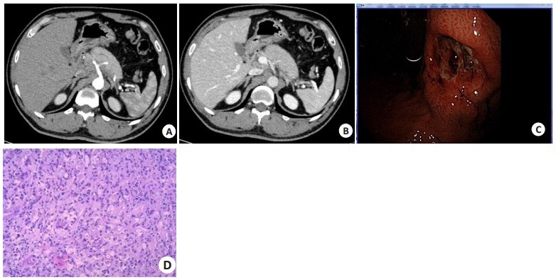
图 1 观察组患者CT、胃镜及病理表现
观察组患者, 男, 67岁, 间断上中腹隐痛不适20 d余. A: 平扫CT; B: 增强CT动脉期; C: 增强CT静脉期; D: 增强CT静脉期冠状位重建. 胃充盈良好, 未见蠕动伪影, 贲门后壁及胃底小弯侧壁明显增厚, 可见结节状、团块状软组织密度影,最厚处约2.6 cm, 增强扫描不均匀强化, 病灶累及浆膜面, 呈结节状改变,周围脂肪间隙模糊; 贲门及胃小弯侧周围可见多发增大淋巴结,增强扫描呈环形强化. E: 电子胃镜示贲门后壁及小弯侧可见隆起型溃疡性病变,周围粘膜明显充血水肿, 触之易出血, 病变累及胃底; F: 病理(HE染色, ×100) 示中分化腺癌.
Figure 1. CT, gastroscopy and pathological findings of patient in observation group.

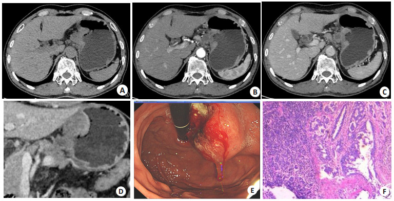
图 2 对照组患者CT、胃镜及病理表现
对照组患者, 男, 39岁, 腹部不适、呕血、黑便2 d. A~B: 常规CT扫描, 增强CT扫描动、静脉期,因胃蠕动较快, 水充盈欠佳, 胃壁挛缩, 结合胃镜检查, CT所示胃角局部壁增厚, 不能明确显示病灶位置、范围大小及侵犯深度. C: 电子胃镜示: 胃角处近后壁可见大小约2.0 cm×2.0 cm深大溃疡型隆起病变,底覆污苔及褐色血迹, 周围黏膜环堤样隆起, 质硬, 出血明显; D: 病理(HE染色,×100)示低分化腺癌, 部分呈印戒细胞.
Figure 2. CT, gastroscopy and pathological findings of patient in control group.
表 1 对照组T分期的诊断准确性比较
Table 1. Comparison of diagnostic accuracy of T staging in control group (n, n=58)
病理T分期 对照组MSCT T分期 合计 T1 T2 T3 T4 T1 4 1 0 0 5 T2 4 24 6 0 34 T3 0 3 12 2 17 T4 0 0 1 1 2 合计 8 28 19 3 58  下载: 导出CSV
表 2 观察组T分期的诊断准确性比较
Table 2. Comparison of diagnostic accuracy of T staging in observation group (n, n=58)
病理T分期 观察组MSCT T分期 合计 T1 T2 T3 T4 T1 6 0 0 0 6 T2 1 22 3 0 26 T3 0 1 19 1 21 T4 0 0 1 4 5 合计 7 23 23 5 58
下载: 导出CSV
表 3 对照组N分期的诊断准确性比较
Table 3. Comparison of diagnostic accuracy of N staging in control group (n, n=58)
病理N分期 对照组MSCT N分期 合计 N0 N1 N2 N0 21 4 0 25 N1 3 17 3 23 N2 0 4 6 10 合计 24 25 9 58
下载: 导出CSV
表 4 观察组N分期的诊断准确性比较
Table 4. Comparison of diagnostic accuracy of N staging in observation group (n, n=58)
病理N分期 观察组MSCT N分期 合计 N0 N1 N2 N0 18 2 0 20 N1 2 27 1 30 N2 0 1 7 8 合计 20 30 8 58
下载: 导出CSV
表 5 对照组M分期的诊断准确性比较
Table 5. Comparison of diagnostic accuracy of M staging in control group (n, n=58)
病理M分期 对照组MSCT M分期 合计 M0 M1 M0 34 7 41 M1 6 11 17 合计 40 18 58
下载: 导出CSV
表 6 观察组M分期的诊断准确性比较
Table 6. Comparison of diagnostic accuracy of M staging in observation group (n, n=58)
病理M分期 观察组MSCT M分期 合计 M0 M1 M0 39 4 43 M1 3 12 15 合计 42 16 58
下载: 导出CSV
-
[1] Rawla P, Barsouk A. Epidemiology of gastric cancer: global trends, risk factors and prevention[J]. Prz Gastroenterol, 2019, 14(1): 26-38. [2] 曹毛毛, 李贺, 孙殿钦, 等. 2000-2019年中国胃癌流行病学趋势分析[J]. 中华消化外科杂志, 2021(1): 102-9. [3] Bray F, Ferlay J, Soerjomataram I, et al. Global cancer statistics 2018: GLOBOCAN estimates of incidence and mortality worldwide for 36 cancers in 185 countries[J]. CA A Cancer J Clin, 2018, 68(6): 394-424. doi: 10.3322/caac.21492 [4] 顾鹏飞, 邓靖宇, 梁寒. 胃癌术前分期研究进展[J]. 中国肿瘤临床, 2019, 46(1): 6-11. doi: 10.3969/j.issn.1000-8179.2019.01.629 [5] 中华医学会肿瘤学分会, 中华医学会杂志社. 中华医学会胃癌临床诊疗指南(2021版)[J]. 中华医学杂志, 2022, 102(16): 1169-89. doi: 10.3760/cma.j.cn112137-20220127-00197 [6] 封俊, 高德培, 廖承德, 等. MSCT胃低张充气成像对胃癌进行术前分期诊断的临床应用[J]. 中国CT和MRI杂志, 2018, 16(3): 97-9, 106. https://www.cnki.com.cn/Article/CJFDTOTAL-CTMR201803031.htm [7] Edge SB. AJCC cancer staging manual. 7th ed. [M]. New York: Springer, 2010. [8] 沈朝军, 姜研, 沈艳, 等. MSCT三期动态增强扫描在胃癌术前分期以及淋巴结转移诊断中的应用价值[J]. 蚌埠医学院学报, 2021, 46 (11): 1595-9. https://www.cnki.com.cn/Article/CJFDTOTAL-BANG202111026.htm [9] 熊焕煜, 万玲, 杨爽. MSCT在胃癌术前分期诊断中的应用价值[J]. 中国现代普通外科进展, 2022, 25(5): 384-6. https://www.cnki.com.cn/Article/CJFDTOTAL-PWJZ202205011.htm [10] 秦书敏, 刘亚良, 黄光建, 等. 3.0T MRI与MSCT对胃癌术前T分期的诊断价值[J]. 解放军医药杂志, 2018, 30(12): 19-22. https://www.cnki.com.cn/Article/CJFDTOTAL-HBGF201812006.htm [11] 吕晓波, 樊鹏飞, 景斐华, 等. 低张气充盈法多层螺旋CT增强扫描在胃部病变内镜治疗术前的评估价值[J]. 中华消化病与影像杂志: 电子版, 2022, 12(3): 150-3. https://www.cnki.com.cn/Article/CJFDTOTAL-ZHYE202203005.htm [12] 顾茵茵, 殷海燕. 低张水充盈与普通水充盈对胃癌MSCT诊断的对照分析[J]. 影像研究与医学应用, 2019, 3(5): 105-6. https://www.cnki.com.cn/Article/CJFDTOTAL-YXYY201905069.htm [13] 张春梅, 杨晓, 熊鸣. 胃窗超声造影、MSCT及两者联合诊断胃癌术前TNM分期与手术后病理一致性研究[J]. 中国CT和MRI杂志, 2021, 19(10): 155-7. https://www.cnki.com.cn/Article/CJFDTOTAL-CTMR202110049.htm [14] 秦佳敏, 刘广国, 王宏. MSCT在胃癌分期诊断及化疗疗效预测中的应用[J]. 影像科学与光化学, 2022, 40(3): 651-4. https://www.cnki.com.cn/Article/CJFDTOTAL-GKGH202203042.htm [15] 王山, 云昊, 陈国. MSCT三维重建、MRI联合经腹超声胃充盈造影在胃癌TNM分期诊断中的应用价值[J]. 中国临床医学影像杂志, 2020, 31(7): 486-9, 494. https://www.cnki.com.cn/Article/CJFDTOTAL-LYYX202007010.htm [16] 许欣, 全志成, 陈亮. MRI与MSCT对术前胃癌淋巴结分期诊断的临床价值[J]. 临床误诊误治, 2019, 32(10): 94-8. https://www.cnki.com.cn/Article/CJFDTOTAL-LCWZ201910023.htm [17] 韩超, 陈新晖, 赵宝琼, 等. MSCT用于胃癌淋巴结转移早期诊断的临床价值[J]. 海南医学, 2021, 32(6): 746-9. https://www.cnki.com.cn/Article/CJFDTOTAL-HAIN202106018.htm [18] 于华隆, 姚增武, 张翼飞, 等. 多层螺旋CT在晚期胃癌转化治疗后N分期及淋巴结转移判断中的临床应用分析[J]. 中国普外基础与临床杂志, 2020, 27(2): 158-62. https://www.cnki.com.cn/Article/CJFDTOTAL-ZPWL202002007.htm [19] 骆栋梁, 张维春, 赵宝安, 等. 多排螺旋CT在胃癌患者术前临床分期评估中的应用价值[J]. 中国肿瘤临床与康复, 2018, 25(2): 164-7. https://www.cnki.com.cn/Article/CJFDTOTAL-ZGZK201802011.htm [20] 江小云, 赵铭, 陈淑君, 等. 螺旋CT诊断老年胃癌患者术前分期的价值[J]. 分子影像学杂志, 2021, 44(1): 151-4. doi: 10.12122/j.issn.1674-4500.2021.01.30 [21] 王会丰, 刘鑫, 韩文, 等. 超声内镜、MSCT扫描在胃癌术前诊断和分期中的应用分析[J]. 中国CT和MRI杂志, 2019, 17(11): 120-2. https://www.cnki.com.cn/Article/CJFDTOTAL-CTMR201911038.htm -

 点击查看大图
计量
- 文章访问数: 164
- HTML全文浏览量: 65
- PDF下载量: 9
- 被引次数: 0