

 Find Duplicates
 Check Document
 Submission(new)
 Experts Office
 Editorial Office
2021 Vol. 44, No. 5
Display Method:
2021, 44(5): 733-738.
doi: 10.12122/j.issn.1674-4500.2021.05.01
Abstract:
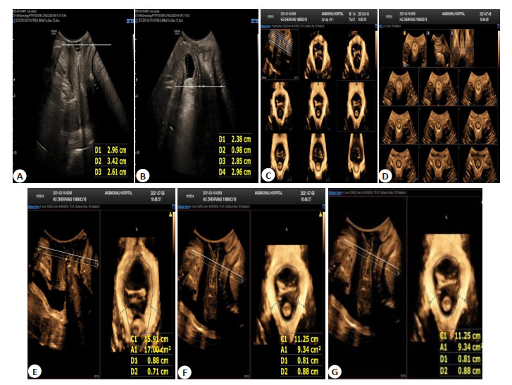
Objective To analyze the influence of different delivery modes on pelvic floor structure and function in early postpartum period. Methods Sixty-seven primiparas who received reexamination in obstetrics clinic of the hospital at 6-8 weeks after delivery were selected between December 2019 and June 2020. According to the delivery mode, the primiparas enrolled were divided into cesarean section group (n=23) and natural delivery group (n=44). The bladder neck position, urethral tilt angel, posterior urethravesical angle and cervical mouth position were observed using transperineal three-dimensional pelvic floor ultrasound under resting state and on maximum Valsalva, respectively. The bladder neck descent, urethral rotation angle and cervix down distance under maximum Valsalva action state were calculated. The formation rate of urethral funnel, formation rate and type of vesicocele, and formation rate of uterine prolapse were compared. The areas of levator ani muscle hiatus and thicknesses of left and right levator ani muscle under maximum Valsalva action state, anal sphincter contraction state and resting state were compared. The presence or absence of internal and external anal sphincter and levator ani muscle tear were observed. Results The bladder neck position under resting state was lower than that in the cesarean section group (t=3.43, P < 0.05). The bladder neck descent and urethral rotation angle on Valsalva were larger than those in the cesarean section group (t=3.53, 3.82, P < 0.05). The incidence of vesicocele was higher than that in the cesarean section group (t=11.075, P < 0.05). APLAM under resting state, anal sphincter contraction state and Valsalva were larger than those in the cesarean delivery group (t=4.17, 2.95, 2.80, P < 0.05). The incidence of type II vesicocele was higher than that of the other two types in the two groups, and the incidence of type II vesicocele in natural delivery group was higher than that in the cesarean section group (χ2 =13.39, P < 0.05). The differences in cervical mouth position and posterior urethravesical angle under resting state, cervix down distance on Valsalva, the incidences of urethral funnel formation, uterine prolapse, rectocele and excessive perineal movement were not significant (P>0.05). There was no significant difference in the thicknesses of left and right levator ani muscle under the three states. No internal and external anal sphincter injury and laceration of levator ani muscle were found in the two groups. Conclusion The above results indicate that transperineal three-dimensional pelvic floor ultrasound can be used to dynamically observe the indicators of anterior, middle and posterior pelvic organs. All delivery modes can cause different degrees of damage to pelvic floor. Compared with natural delivery, cesarean section is better in terms of anterior pelvic cavity and areas of levator ani muscle hiatus.
2021, 44(5): 739-743.
doi: 10.12122/j.issn.1674-4500.2021.05.02
Abstract:
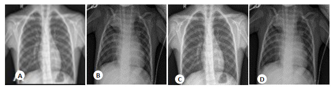
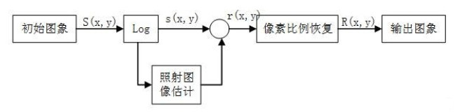
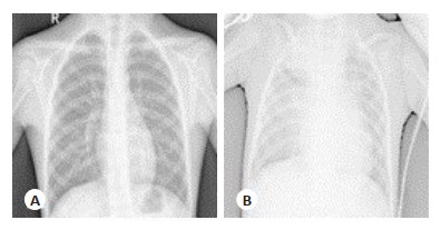
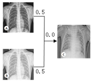
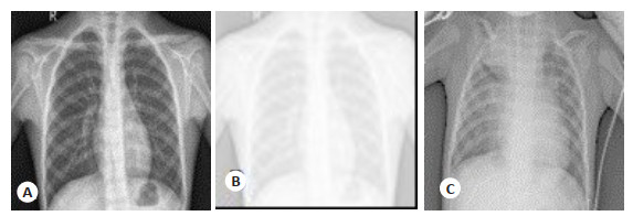
Objective To enhance Retinex optimization method and pneumonia X- ray image features. Methods A Retinex optimization method for enhancement of pneumonia X-ray image features was proposed. The edge of the X-ray image was centralized for information reconstruction. Retinex was used for feature enhancement. The image was weighted and combined with the original image to retain features to the maximum extent. Results Compared with the initial image training, the accuracy was increased by 2.57 percentage points, the sensitivity accuracy was lost by 0.17 percentage points, and the specificity accuracy was increased by 7.15 percentage points. Conclusion The improved algorithm can enable the machine to quickly and automatically identify pneumonia and non-pneumonia, which greatly improves the diagnostic efficiency during the period of high incidence of pneumonia.
2021, 44(5): 744-748.
doi: 10.12122/j.issn.1674-4500.2021.05.03
Abstract:
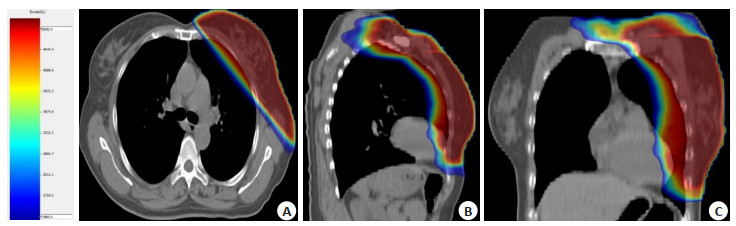
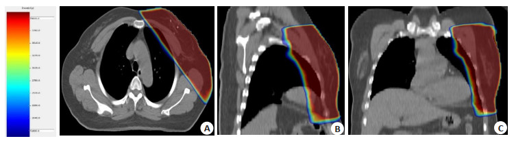
Objective To compare the side effection of skin and cosmetic scores between postoperative hypofraction radiotherapy and convention fraction radiotherapy for early breast cancer patiens with breast-conserving surgery, through balancing the covariates of the two groups with propensity score matching method. Methods The patients (n=319) who underwent breast-conserving surgery at Shenzhen People's Hospital from Jan. 2015 to Jul. 2018 were retrospectively analyzed. There were 104 patients in the hypofraction radiotherapy group and the other 215 patients in the convention fraction radiotherapy group, which matched by propensity scores to obtain two new groups with balanced covariates.We analyzed the samples of the two groups. Results There were 99 patients in the convention fraction radiotherapy group and 100 patients in the hypofraction radiotherapy group after propensity score matched. There were 86.9% and 90% patients presented early skin side effection, respectively. The 14.1% and 10% of patients in the two groups occured wet peeling, which had no significant difference (P=0.111). The incidence of grade I skin side effection in the two groups at 6 months, 1 year, and 2 years were 86.9%, 90.0%; 96.0%, 92.0%; 99.0%, 96.0%(P=0.471). There were no late grade III or worse skin side effection in the two groups. The good rates of cosmetic scores between the convention fraction radiotherapy group and hypofraction radiotherapy group were 78.8% and 84.0% (P=0.361) after 2 years. Conclusion There is no significant difference in side effection of skin or cosmetic scores between postoperative hypofraction radiotherapy and convention radiotherapy for early breast cancer patiens with breast-conserving surgery.
2021, 44(5): 749-753.
doi: 10.12122/j.issn.1674-4500.2021.05.04
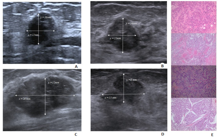
Abstract:
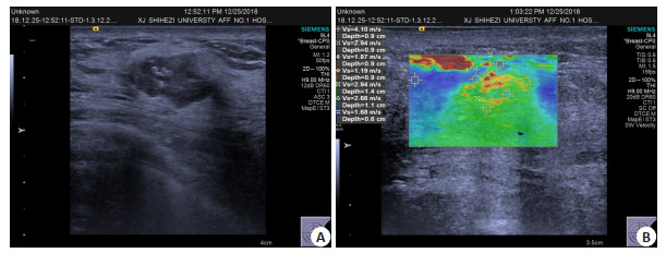
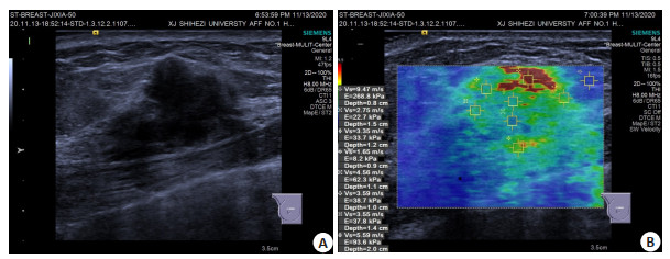
Objective To analyze the ultrasonographic features and immunohistochemical indexes of mucinous carcinoma of the breast, and improve the accuracy in the diagnosis of mucinous carcinoma. Methods The ultrasonographic features (maximum diameter, aspect ratio, high echo halo, metastasis, elastic score), shear wave velocity (SWVmax, SWVratio, SWVmean) and immunohistochemical detection (ER, PR, HER-2, Ki-67) of 56 cases were analyzed retrospectively. 24 cases with mucinous carcinoma were used as experimental group and 32 cases with invasive ductal carcinoma were used as control group.The differences of ultrasonic features and immunohistochemical indexes between the two groups were compared. The correlation between ultrasonic features and immunohistochemical indexes was analyzed. Results Compared with invasive ductal carcinoma, mucinous carcinoma showed clear boundary, high echo halo, posterior echo enhancement, less than 4 score and BI-RADS classification < 4b (χ2=4.419, 5.171, 6.162, 4.743, 14.000, P < 0.05). The positive rates of SWVmax, ER and HER-2 were significantly different between the two groups (t=-2.069, χ2=4.371, 4.051, P < 0.05). Conclusion Compared with invasive ductal carcinoma, the expression of ultrasound features and immunohistochemical markers in mucinous carcinoma of breast are different, which can provide reference for the diagnosis and treatment of mucinous Carcinoma of breast.
2021, 44(5): 754-758.
doi: 10.12122/j.issn.1674-4500.2021.05.05
Abstract:
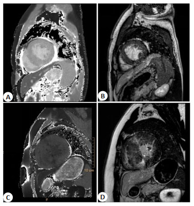
Objective To explore the application of gadolinium delayed enhancement combined with cardiac magnetic resonance T1 mapping in the clinical examination of patients with cardic amyloidosis (CA) and hypertrophic cardiomyopathy (HCM). Methods In this prospective study, 40 CA patients and 55 HCM patients diagnosed in our hospital from December 2018 to December 2020 were selected as the research objects, and 54 healthy patients were selected as the control group. The cardiac function, cardiac morphology, and MRI related indicators in three groups were compared. Results The difference between the levels of LVESV and LVEDV of three groups was significant (P < 0.05). By pairwise comparison, the levels of LVESV and LVEDV were the HCM group, the control group and the CA group in descending order. There was no significant difference in LVEF between the three groups of patients (P > 0.05). The left ventricular wall thickness and left ventricular mass of the three groups were the HCM group, the CA group and the control group in descending order (P < 0.05). The T1 values of the three groups were the CA Group, the HCM group and the control group in descending order (P < 0.05). After the enhancement, the T1 value was the HCM group, the control group and the CA group in descending order. The extracellular volume was the CA group, the control group and the HCM group in descending order (P < 0.05). Conclusion Gadolinium delayed enhancement combined with cardiac magnetic resonance T1 mapping has a good differential diagnosis ability in the clinical examination of CA and HCM patients.
2021, 44(5): 759-763.
doi: 10.12122/j.issn.1674-4500.2021.05.06
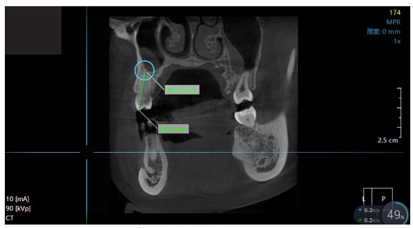
Abstract:
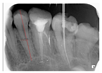
Objective To compare the value of digital periapical film and cone- beam CT (CBCT) in measuring tooth length. Methods Clinical data of 103 cases of tooth extraction orthodontic patients (a total of 200 premolars) admitted to our hospital from June 2017 to June 2020 were retrospectively analyzed. All patients underwent digital periapical film and CBCT examination. The actual measurement with a vernier caliper was taken as the gold standard. InVivoDental software and parallel long-focus parallel projection technology were used to measure the tooth lengths of different examination methods. The image lengths of teeth in axially, sagittally and coronally examined by CBCT and those by digital periapical detection were compared in different dental positions, and the difference between the actual length and image lengths was calculated. Results There was no significant difference between the tooth length measured by digital periapical method and the actual tooth length in different positions (P > 0.05). The differences between the tooth length of mandibular molar area, canine area, anterior area axially measured by the CBCT and the actual tooth length were significant (P < 0.05). There was no significant difference in the tooth length between the maxillary molar area, maxillary premolar area, maxillary canine area, maxillary anterior area and mandibular premolar area and the actual tooth length (P > 0.05). The differences between the tooth length, sagittally measured by CBCT, of the maxillary molar area, the mandibular molar area, the mandibular premolar area, the mandibular canine area and the mandibular anterior area with the actual tooth length were significant (P < 0.05). There was no significant difference between the tooth length of the maxillary premolar area, the maxillary canine area and the maxillary anterior area with the actual tooth length (P > 0.05). The differences between the tooth length, coronally measured by CBCT, of the maxillary molar area, the maxillary canine area, the maxillary anterior area, the mandibular premolar area, the mandibular canine area and the mandibular anterior area, and the actual tooth length were significant (P < 0.05). There was no significant difference between the tooth length of the maxillary premolar area and the mandibular molar area and the actual tooth length (P > 0.05). Conclusion Compared with CBCT, digital periapical film under the parallel long- focus parallel projection technology can measure the tooth length more accurately.
2021, 44(5): 764-770.
doi: 10.12122/j.issn.1674-4500.2021.05.07
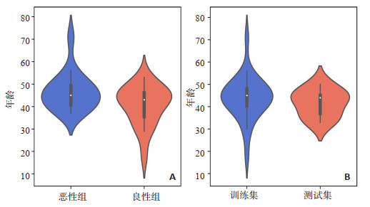
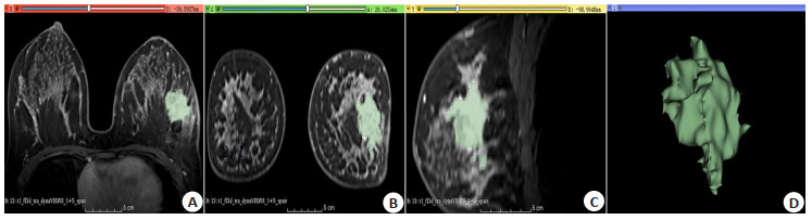
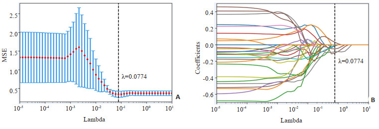
Abstract:
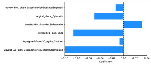
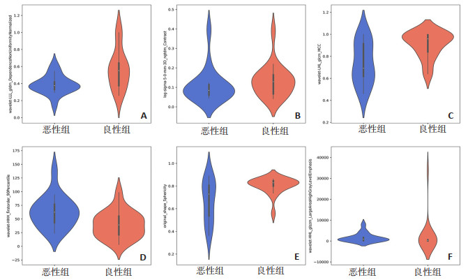
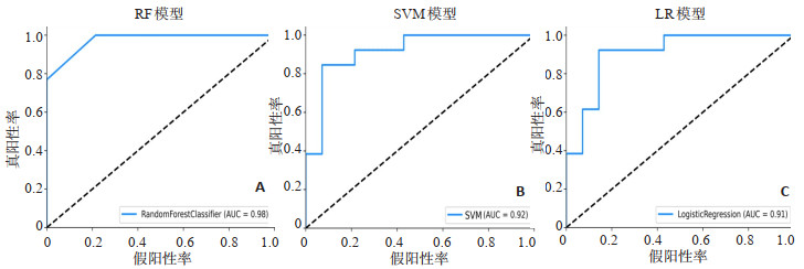
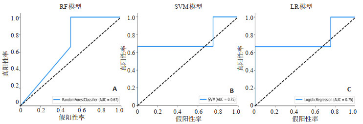
Objective To construct MRI radiomic prediction models for the differentiation of benign and malignant lesions of breast based on random forest, support vector machine and logistic regression classifiers, and evaluate the diagnostic value of the above models. Methods A Retrospective analysis of dynamic contrast-enhanced MRI images of 34 patients with breast lesions, who underwent MRI imaging examinations and obtained surgical pathology, was conducted in Southern University of Science and Technology Yantian Hospital. The cases were divided into training set (n=27) and test set (n=7) according to the ratio of 0.8∶0.2. The 3D Slicer software was used to delineate the target area of breast lesions and generate 3D volume of interest. Then 1037 radiomic features was extracted for each volume of interest. The LASSO was performed to reduce dimensionality. Then three classifiers, including random forest, support vector machine and logistic regression, were used to construct prediction models for differentiating benign and malignant breast lesions in the training set, and evaluated in the test set. Results Six radiomics signatures were selected after dimensional reduction by LASSO. The classification efficiency of the three models were very good (AUC>0.90) in the training set, and the logistic regression radiomic prediction model was the most stable one in differentiating benign and malignant breast lesions. Conclusion The MRI radiomic prediction models based on random forest, support vector machine and logistic regression has good diagnostic efficiency in differentiating benign and malignant breast lesions. The logistic regression model is more stable.The radiomics can provide a new method for the prediction of benign and malignant breast lesions.
2021, 44(5): 771-775.
doi: 10.12122/j.issn.1674-4500.2021.05.08
Abstract:
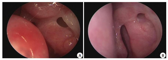
Objective To explore the effect of endoscopic endoscopic internal capsule nasal anastomosis (EES-DCR) combined with lacrimal duct dilation and drainage tube in the treatment of patients with chronic dacryocystitis and lacrimal duct obstruction. Methods We selected 120 patients with chronic dacryocystitis combined with lacrimal duct obstruction in our hospital from January 2018 to January 2020. A prospective randomized trial study was used to divide the patients into an observation group and a control group, 60 cases/group. The observation group was treated with EES-DCR combined with lacrimal duct dilation and drainage tube. The control group was treated with EES-DCR combined with melamine. The clinical effect evaluation, operation time, and tear arc height above the lower eyelid margin were compared between the two groups at different times after surgery. The degree of tearing, tear film rupture time, tear secretion experiment, pain degree of the affected eye and complications were compared. Results Evaluation 3 months after the operation, the observation group had a cure rate of 91.67%, an improvement rate of 8.33%, and a control group had a cure rate of 86.67%, an improvement rate of 11.67%, and an inefficiency of 1.67%. There was no significant difference in clinical efficacy between the observation group and the control group (P>0.05). 6 months after the operation, the observation group had a cure rate of 88.33%, an improvement rate of 11.67%, a control group had a cure rate of 73.33%, an improvement rate of 25.00%, and an inefficiency of 1.67%. The observation group's clinical efficacy was better than the control (P < 0.05). 3 months after the operation, the observation group had a normal rate of 88.33%, a high rate of 8.33%, and a higher rate of 3.33% of the tear arc above the lower eyelid margin. The normal rate of the tear arc above the eyelid margin was 76.67%, the high rate was 15.00%, and the higher rate was 8.33%. There was no significant difference in the height of the tear arc above the lower eyelid margin between the two groups (P>0.05). There was no significant difference in the VAS score of the control group (P>0.05). One week after the operation, the VAS score of the observation group was lower than that of the control group and the difference was significant (P < 0.05). There were no significant differences in the degree score, tear film rupture time, and tear secretion experiment (P>0.05). One month after surgery, the degree of tearing and tear secretion experiment in the observation group were lower than those in the control group, and the tear film rupture time was longer than the control group (P < 0.05). Conclusion The postoperative effect of EES-DCR combined with lacrimal duct dilation and drainage tube in the treatment of patients with chronic dacryocystitis and lacrimal duct obstruction is better than that of EES-DCR combined with melody
2021, 44(5): 776-781.
doi: 10.12122/j.issn.1674-4500.2021.05.09
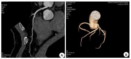
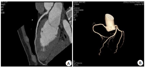
Abstract:
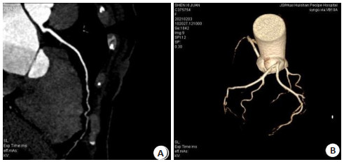
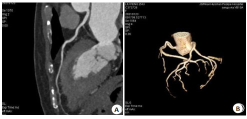
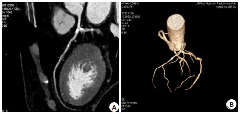
Objective To explore the correlation between blood circulation velocity and the enhancement effect of coronary CT angiography (CTA), and to analyze the optimization scheme of contrast medium. Methods A total of 161 patients who underwent coronary CT angiography in our hospital from January 2020 to April 2021 were selected and divided into fast group (n=32), medium group (n=35) and slow group (n=94) according to blood circulation velocity. The relationship between blood circulation speed and enhancement effect was analyzed. The slow group was divided into three subgroups: group A, group B and group C. The contrast agent concentration and injection rate of group A, fast group and medium group were the same (iodine concentration was 320 mgI/mL and injection rate was 5 mL/s). The iodine concentration of group B was 350 mgI/mL and injection rate was 5.5 mL/s, while the iodine concentration of group C was 370 mgI/mL and injection rate was 5.5 mL/s. The occurrence of artifacts, ventricular septal display, coronary CT value and image quality were compared. Methods The artifact probability of fast group, medium group, B group and C group was lower than that of a group, and fast group was lower than that of medium group, and C group was lower than that of B group (P < 0.05). The CT value of aortic root, image quality score and ventricular septal display score of fast group, medium group, B group and C group were higher than those of A group, and the fast group was higher than that of medium group, and the C group was higher than that of B group (P < 0.05). Conclusions The velocity of blood circulation can increase the enhancement effect of coronary CTA and clearly show the interventricular septum. For the patients with slow blood circulation, the contrast medium concentration and injection rate can be increased appropriately to ensure the image quality.
2021, 44(5): 782-786.
doi: 10.12122/j.issn.1674-4500.2021.05.10
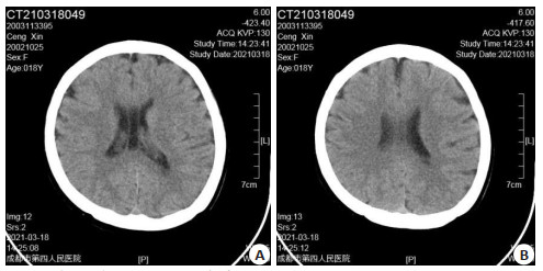
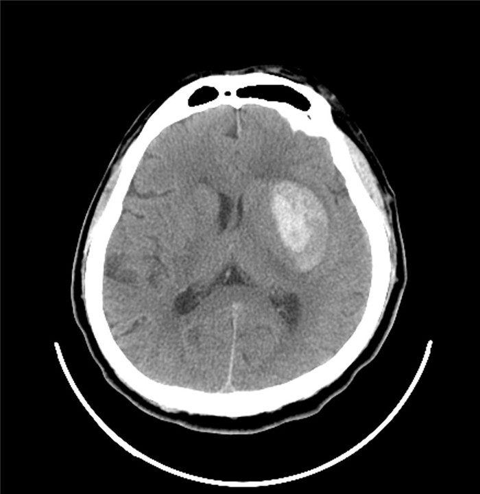
Abstract:
Objective To explore the relationship between the changes of head CT scan parameters and cognitive function abnormalities in patients with early-onset schizophrenia. Methods We selected 80 first-onset schizophrenia patients (case group), 80 physical examination volunteers (control group) with basically matched factors such as age and gender. The diagnosis time of the patients was February 2017 to March 2021. The patients acceptted head CT scans. We compared the brain structure abnormalities, brain lobe parameters, and cognitive function scores found in the CT examination of the two groups. The correlation analysis was used to explore the correlation between brain structure abnormalities, brain lobe parameters and patients' cognitive functions. Methods The overall incidence of cortical, medullary, mixed cortical and medullary, and brain atrophy in the case group were significantly higher than those in the control group (P < 0.05). The average CT value of the frontal lobe of the case group was lower than the control group (P < 0.05). Two groups had no significant difference in CT values in the temporal lobe, parietal lobe, and occipital lobe (P>0.05). The scores of attention/alertness, working memory, social cognition, and overall cognition were lower than those of the control group (P < 0.05). Two groups had scores on word memory, visual learning, reasoning and problem solving(P>0.05). The information processing speed, attention/alertness, working memory, social cognition, and overall cognition scores of patients with brain atrophy in the case group were lower than those without brain atrophy(P < 0.05). The two groups had no significant differences in the scores of word memory, visual learning, reasoning and problem solving (P>0.05). The frontal lobe CT value of the case group and the speed of information processing, working memory, social Cognition and overall cognition scores were positively correlated (P < 0.05). Conclusions CT scan of the brain of patients with early-onset schizophrenia can find that the detection rate of brain atrophy is significantly increased. CT value of the frontal lobe is decreased. These changes are closely related to the impaired cognitive function of the patient.
2021, 44(5): 787-791.
doi: 10.12122/j.issn.1674-4500.2021.05.11
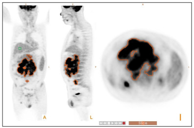
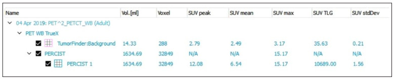
Abstract:
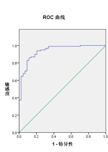
Objective To evaluate the predictive significance of F-18 FDG PET/CT quantization parameters SUVpeak, MTV and TLG in patients with diffuse large B cell lymphoma (DLBCL) before chemotherapy. Methods Seventy-six patients with DLBCL were involved between August 2011 and November 2018 who had undergone F-18 FDG PET/CT scan prior to treatment. The patients were divided into progress group (n=40) and non-progressive group (n=36), death group (n=6) and survival group (n= 70). Maximum standardized uptake value (SUVmax), peak standardized uptake value (SUVpeak), metabolic tumor volume (MTV) and total lesion glycolysis (TLG) were measured by a automatic VOI software. The differences in PET parameters between the progression group and the non-progressive group, and between the death group and the survival group were compared. The best cut-off value of PET parameters through the ROC curve was obtained. Kaplan-Meier survival analysis was used to compare survival rates. The clinical variables Data and PET parameters were used for univariate and multivariate analysis of survival and progression. Methods The median SUVmax, SUVpeak, MTV and TLG in the progression group were 30.82(12.64-71.16), 19.89(9.35-52.23), 786.49(135.06-8795.16) cm3 and 4618.76(653.88-37361.18), and 34.53(3.82-52.41), 15.76(3.18-32.37), 116.05 (54.14-2642.96) cm3 and 420.18(276.97-9409.09) in the non-progressive group. The differences in SUVpeak, MTV and TLG between progressive group and non progressive group were significant(p < 0.05). The median SUVmax, SUVpeak, MTV and TLG were 50.47 (19.95-130.14), 37.40(16.95-82.63), 2195.11(231.85-8361.82) cm3 and 14712.77(3371.5-28302.65) in the death group; and 30.82 (3.82-52.41), 19.43(3.18-38.62), 252.10(54.14-8795.16) cm3 and 1219.53(276.97-37361.18) in the survival group.The differences in the SUVmax, SUVpeak, MTV, and TLG between the dead group and the survival group were significant (P < 0.05). The survival rate of patients when SUVpeak, MTV and TLG are less than the cut-off value was significantly higher than when they are greater than the cut-off value. SUVpeak, MTV, TLG and international prognostic index scores were positively correlated with progression-free survival. Conclusions SUVpeak, MTV and TLG can all provide important predictive value in the prognosis of DLBCL patients.
2021, 44(5): 792-789.
doi: 10.12122/j.issn.1674-4500.2021.05.12
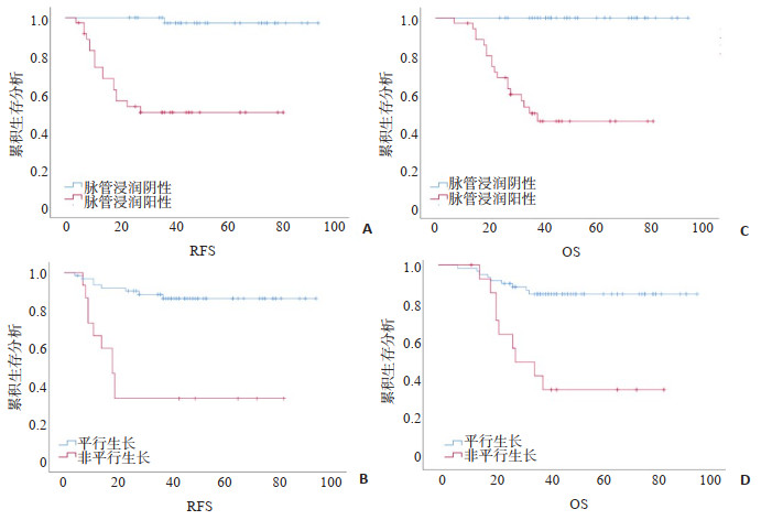
Abstract:
Objective To investigate the value of clinic-pathological characteristics and ultrasound features in predicting the prognosis of triple negative breast cancer (TNBC). Methods Patients with TNBC treated between January 2012 and December 2017 were retrospectively included. Patients' clinic-pathological, ultrasound features, recurrence-free survival, and overall survival were collected. Cox regression and Kaplan-Meier analysis were used to determine the prognostic factors of TNBC. Methods A total of 78 TNBC patients were included with a median follow-up of 40 months. Eighteen patients (23.1%) experienced recurrence and 18 patients (23.1%) occurred deaths. Univariate Cox analysis showed that pre/peri-menopausal patients, maximum tumor diameter >2 cm, positive axillary lymph node, Ki-67 expression >30%, fibroglandua breast, non-parallel oriented, and lymphatic vascular invasion are associated with poorer prognosis (P < 0.05). Multivariable Cox analysis found that only non-parallel oriented and lymphatic vascular invasion were two independent predictors of recurrence-free survival and overall survival (P < 0.05). Conclusions Peripheral lymphatic vascular invasion and tumor non-parallel growth are associated with poor prognosis in TNBC patients.
2021, 44(5): 799-803.
doi: 10.12122/j.issn.1674-4500.2021.05.13
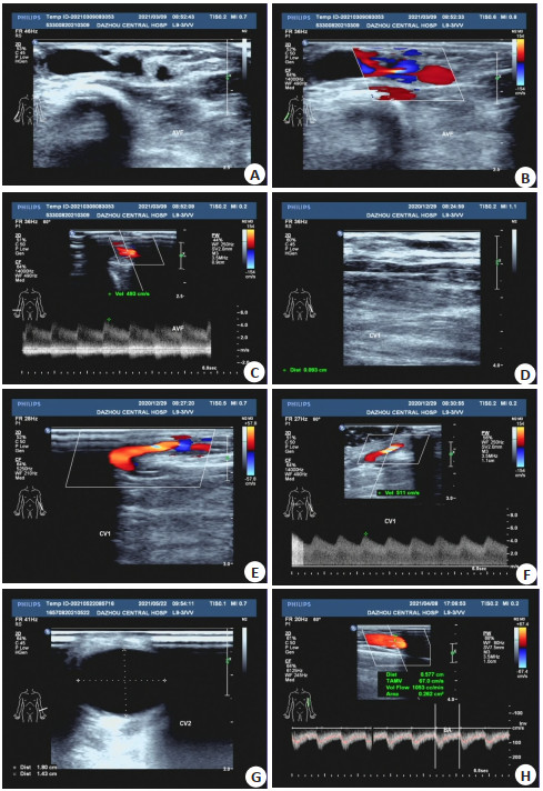
Abstract:
Objective To analyze the value of ultrasound in evaluating hemodynamics and maturity of internal arteriovenous fistula (AVF). Methods The clinical data of 143 hemodialysis patients who underwent the first operation of venous fistula in our hospital from February 2019 to April 2020 were selected retrospectively. All patients were examined by color Doppler ultrasound before and after operation, and the related hemodynamic indexes, blood flow and diameter of radial artery and cephalic vein were observed before and after operation. Six weeks after operation, the maturity of the patients was observed and the relationship between Internal fistula maturity and Cephalic vein hemodynamics was analyzed. Results At 1 week, 3 weeks and 6 weeks after operation, the peak systolic velocity and diastolic peak velocity were significantly higher than those before operation, while the RI was significantly lower than that before operation(P < 0.05). The blood flow and diameter of radial artery and cephalic vein were significantly larger than those before operation(P < 0.05). There was no significant difference in peak systolic velocity, diastolic peak velocity, blood flow and diameter of radial artery and cephalic vein at 1 week, 3 weeks and 6 weeks after operation. Six weeks after operation, the maturity of AVF was 69.23%, peak systolic velocity, diastolic peak velocity, diameter and blood flow of cephalic vein in mature group were significantly higher than those in immature group(P < 0.05). RI was significantly lower than that in immature group(P < 0.05). ROC curve for predicting AVF maturity at 6 weeks after operation showed that the area under ROC curve was 0.938, and the critical value for predicting AVF maturity was 387.98 mL/min. Conclusion Color Doppler ultrasound can be used to evaluate the maturity of internal arteriovenous fistula in patients with arteriovenous fistula.
2021, 44(5): 809-813.
doi: 10.12122/j.issn.1674-4500.2021.05.15
Abstract:
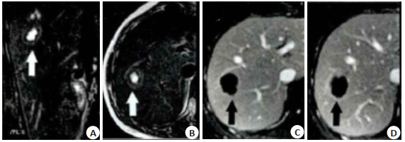
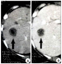
Objective To investigate the MRI and CT findings and diagnostic value of solitary necrotic nodule (SNN) of the liver. Methods Forty cases of liver SNN patients in our hospital from January 2014 to June 2020 were selected, including 22 cases of MRI examination (MRI group) and 18 cases of CT examination (CT group). The value of MRI and CT in the diagnosis of liver SNN were analyzed. The differences of MRI and CT images of simple coagulative necrosis type, liquefying necrosis type and multi nodule fusion type were analyzed. Results The accuracy of MRI in diagnosing liver SNN was 95.45%, which was significantly higher than that in CT group (P < 0.05). In the CT group, there was no significant difference in the plain and enhanced CT findings of the simple coagulation necrosis type, liquefaction necrosis type and multi nodule fusion type (P> 0.05). The lesions of simple coagulation necrosis, liquefaction necrosis and multi nodule fusion showed low density on plain CT scan, accounting for 83.33%, 42.86% and 40.00%. There was no enhancement in the three types of lesions, including 83.33%, 57.14% and 80.00% of the capsule of lesions had delayed enhancement. In MRI group, the low signal ratio of T2WI in liquefying necrosis was significantly higher than that of coagulation necrosis and multi nodule fusion (P < 0.05). The proportion of slightly high signal in T2WI of multi nodule fusion type was significantly higher than that of liquefaction necrosis type (P < 0.05). The low signal ratio of T1WI sequence in the single coagulable necrosis type, liquefaction necrosis type and multi-nodule fusion type lesions was 100.00%. The sensitivity, specificity, accuracy, positive predictive value and negative predictive value of T2WI sequence low signal for the identification of SNN with liquefaction necrosis were 100.00%, 78.57%, 86.36%, 72.73% and 100.00% respectively. The sensitivity, specificity, accuracy, positive predictive value and negative predictive value of slightly higher signal of T2WI sequence identifies SNNs with multi-nodular fusion were 100.00%, 68.75%, 77.27%, 54.55% and 100.00%, respectively for the identification of SNN with liquefaction necrosis. The proportion of lesions of the necrotic type, liquefactive necrosis type and multi-tuberculous fusion without enhancement at the arterial, portal and delayed stages was 100.00%, but there was enhancement in the edge of all lesions at the delayed stage. Conclusion MRI and CT have certain application value in the diagnosis of liver SNN, among which MRI has better diagnostic value.
2021, 44(5): 814-819.
doi: 10.12122/j.issn.1674-4500.2021.05.16
Abstract:
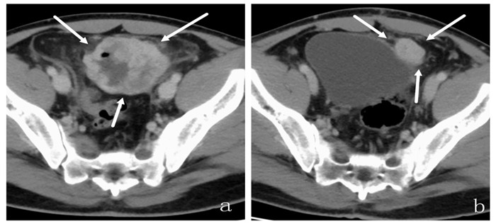
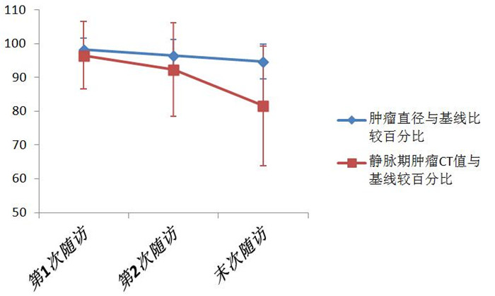
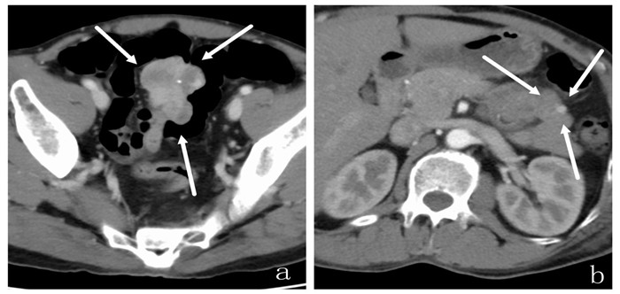
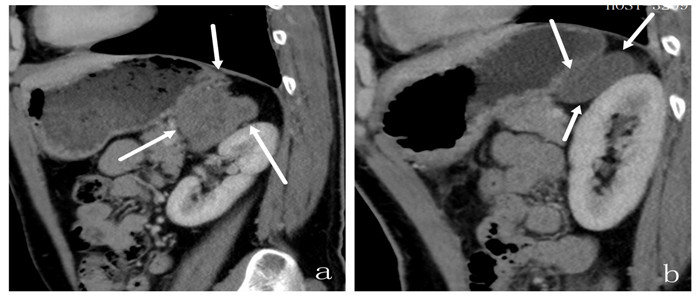
Objective To explore the value of multi-slice spiral CT (MSCT) in evaluation of the therapeutic effect of gastrointestinal stromal tumor (GIST). Methods We retrospectivly analyzed the MSCT data of 42 patients with GIST who received treatment in our hospital from January 2018 to December 2019. Thirty-four cases received surgical treatment, 8 cases received molecular targeted therapy for inoperable or postoperative recurrence or metastasis. Surgical treatment was followed up by MSCT for recurrence and metastasis. After the follow-up of MSCT for molecular targeted therapy, Choi criteria were used to evaluate the curative effect and evaluate the characteristics of the lesion. Results GIST patients who underwent surgery were followed up for 6 to 24 months. Twelve cases recurred with an average tumor diameter of 1.64±0.49 cm.Seven cases had clear borders and 5 cases infiltrated growth. Eight cases had uniform internal density and 4 case had liquefaction necrosis area. Eight cases had a round shape and 4 cases were oval. Seven cases had intracavitary growth and 5 cases had extracavity growth. Eight cases had uniform strengthening, 4 case had inhomogeneous strengthen and 2 cases Calcification. The 8 patients that received molecular targeted therapy were followed up for 6 to 24 months. Among them, 2 cases had clear boundaries, 3 cases had smooth edges, 3 cases had blurred edges. Six cases had uniform enhancement after enhancement, 2 cases had uneven enhancement after enhancement. Four cases had mild enhancement, 2 cases had moderate enhancement, and 2 cases had severe enhancement. Five cases with peripheral infiltration, 3 cases without peripheral infiltration. Five cases showed cystic degeneration and necrosis. The disease control rate of the first MSCT followed-up were 75.0%, the second disease control rate were 75.0%, and the last disease control rate were 62.5%. With the followed-up of MSCT, the CT values of the longest diameter of the tumor and the venous stage tumors of GIST patients decreased. The average decrease of the CT value compared with the baseline was higher than the decrease of the long diameter of the tumor. After treatment, 9 cases had metastatic lesions, including 4 cases of liver metastasis, 2 cases of abdominal metastasis, 1 case of multiple metastases and 2 cases of pelvic metastasis. Conclusion MSCT has a good application value in the evaluation of the therapeutic effect of gastrointestinal stromal tumors. It can provide important imaging data for the evaluation of the efficacy and treatment guidance of GIST patients after surgery or molecular targeted therapy.
2021, 44(5): 820-823.
doi: 10.12122/j.issn.1674-4500.2021.05.17
Abstract:
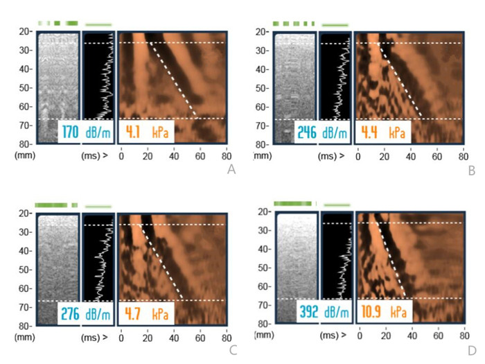
Objective To explore the application value of liver transient elastography and ultrasound diagnostic parameters in fatty liver diagnosis and disease severity evaluation. Methods A total of 120 patients with fatty liver in our hospital between January 1, 2019 and March 1, 2021 were enrolled as observation group. Fifty healthy subjects with physical examination during the same period were treated as control group. All patients underwent liver color Doppler ultrasound and transient elastography. According to the results of liver routine examination (liver size, liver parenchymal echo), the patients in observation group were given disease severity evaluation, and then divided into mild group (n=54), moderate group (n=44) and severe group (n=24). BMI, subcutaneous fat layer thickness, liver tissue sound velocity and liver stiffness value were compared among control group, mild fatty liver group, moderate fatty liver and severe fatty liver group, respectively. Kappa consistency test was used to analyze the consistency of liver transient elastography and color Doppler ultrasound in diagnosing fatty liver. Linear trend chi-square test was applied to analyze the correlation between liver transient elastography and color Doppler ultrasound in the diagnosis of fatty liver. Results The BMI, subcutaneous fat layer thickness and liver stiffness value of control group, mild group, moderate group and severe group and the liver tissue sound velocity of moderate group and severe group were increased (P < 0.05). There was no significant difference in liver tissue sound velocity among control group, mild group and moderate group (P>0.05). Liver transient elastography and color Doppler ultrasound showed good consistency in the diagnosis of fatty liver (Kappa=0.67, P < 0.05). Chi-square test showed that the liver transient elastography and color Doppler ultrasound were significantly relevant in diagnosing fatty liver. The liver transient elastography was more discriminating than color Doppler ultrasound for mild-to-moderate fatty liver (total variation χ2= 298.25, linear regression component χ2=203.47, deviation from linear regression component χ2=91.43, P < 0.05). Conclusion Liver transient elastography and ultrasound are consistent in the diagnosis of fatty liver, but liver transient elastography can distinguish the severity of the disease more finely.
2021, 44(5): 824-829.
doi: 10.12122/j.issn.1674-4500.2021.05.18
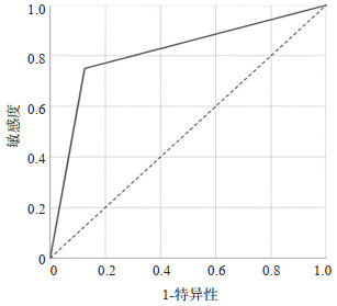
Abstract:
Objective To explore differences of heart rate between hypertensive emergency and hypertensive urgency. Methods The data of patients with hypertensive crisis presenting to emergency department of Shantou Central Hospital between January 2019 and December 2019 was documented. Based on the presence or absence of acute target organ involvement, clustering of hypertensive crisis into emergency or urgency. The relationship between clinical characteristics, types and quantities of medications taken, medication compliance, blood pressure parameters and heart rate were analyzed. The differences in blood pressure parameters and heart rate among different types of hypertensive emergency patients were analyzed. The different predictors between hypertensive emergency and hypertensive urgency were used to analyze the predictive value for hypertensive emergency patients in hypertensive crisis. The value of heart rate in diagnosing hypertensive emergency was analyzed. Results A total of 186 patients of hypertensive crisis in sinus rhythm presenting to emergency department were enrolled. Hypertensive emergency accounted for 44.1%, and hypertensive urgency accounted for 55.9%. The average heart rate of women was higher than men (P=0.002). The average heart rate of the hypertensive emergency was higher than those with urgency (P < 0.001). Compared with urgency, hypertensive emergency has a higher proportion of diabetes and poor medication compliance; and hemodynamic parameters including systolic blood pressure, diastolic blood pressure, mean arterial pressure, and pulse pressure relates neither to urgency nor to emergency. Finally, hypertensive emergency in hypertensive crisis patients may be predicted by diabetes mellitus and heart rate. The area under the curve and optimal cut-off value of heart rate to diagnose hypertensive emergency were 0.813 and 86 b/min. Conclusion Hemodynamic parameters relate neither to hypertensive emergency nor to urgency. The mean heart rate of hypertensive emergencies, especially those with acute heart failure, is higher than that of hypertension urgencies. The heart rate threshold of 86 b/min may be a reliable clinical indicator for distinguishing hypertensive emergency or urgency in hypertensive crisis. It is especially suitable for the clinical thinking of diagnosis and treatment in emergency department. In addition, hypertensive crisis patients with diabetes are more prone to emergency.
2021, 44(5): 830-834.
doi: 10.12122/j.issn.1674-4500.2021.05.19
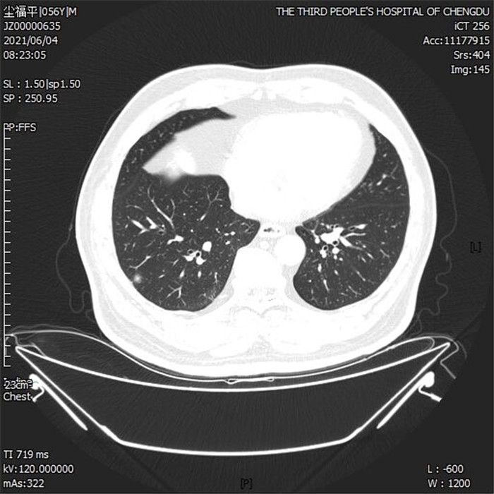
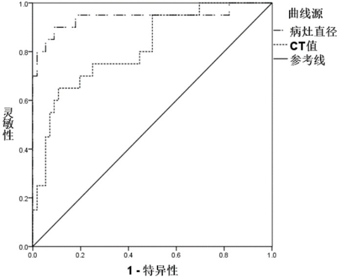
Abstract:
Objective To investigate the value of low-dose chest CT scan, carcinoembryonic antigen (CEA) and cytokeratin 19 fragment antigen (Cyfra21-1) in the diagnosis of early lung cancer. Methods A total of 108 patients with pulmonary sub solid nodules from January 2019 to January 2021 in our hospital were selected. All patients were given low-dose CT scanning. The serum CEA and CYFRA21-1 in patients with lung cancer and non lung cancer were compared, and the value of CT combined with serum CEA and CYFRA21-1 in the diagnosis of early lung cancer was analyzed. Results Seventy-six patients were pathologically diagnosed as early lung cancer (including 34 cases of carcinoma in situ, 22 cases of micro invasive carcinoma and 20 cases of invasive carcinoma), and 32 patients were non lung cancer. The serum CEA and Cyfra21-1 in lung cancer patients were significantly higher than those in non-lung cancer patients (P < 0.05). The sensitivity and negative predictive value of CT combined with serum CEA and Cyfra21-1 in the diagnosis of early lung cancer were significantly higher than that of CT (P < 0.05). The diameter and CT values of invasive carcinoma were significantly higher than those of carcinoma in situ and microinvasive carcinoma (P < 0.05). There was no significant difference in CEA and Cyfra21-1 among patients with carcinoma in situ, microinvasive carcinoma and invasive carcinoma (P>0.05). The area under the ROC curve of lesion diameter and CT value for the diagnosis of invasive carcinoma were 0.941 and 0.816, respectively (P < 0.05), the cut-off value was 15.86 mm and -422.52 Hu respectively. The sensitivity were 90.00% and 65.00%, and the specificity were 91.10% and 89.30% respectively. Conclusion Low dose chest CT, CEA and Cyfra21-1 level have good value in the diagnosis of early lung cancer, and lesion diameter and CT value have certain application value in the differentiation of invasive cancer.
2021, 44(5): 835-839.
doi: 10.12122/j.issn.1674-4500.2021.05.20
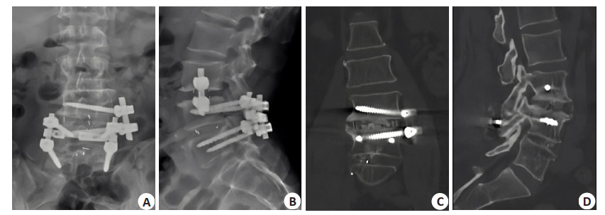
Abstract:
Objective To investigate the effect of rivaroxaban on hemorheology, inflammatory factors and coagulation function in patients with spinal trauma after spinal fusion. Methods A total of 102 patients with spinal trauma who underwent spinal fusion in our hospital from January 2019 to January 2020 were selected. The patients were randomly divided into observation group (n=51) and control group (n=51) according to the random number table arrangement method. The control group was given routine postoperative treatment, and the observation group was given rivaroxaban treatment, once a day, one tablet once a day for 14 days. The hemorheology (whole blood low shear viscosity, whole blood medium shear viscosity, whole blood high shear viscosity, hematocrit, plasma viscosity and platelet adhesion rate), inflammatory factors (IL-8, IL-10, TNF-α and CRP), coagulation function indexes (TT, Pt, APTT, FIB and D-D) were compared between the two groups at 1 and 14 days after operation. Results After treatment, the hemorheology of whole blood low shear viscosity, whole blood medium shear viscosity, whole blood high shear viscosity, hematocrit, plasma viscosity and platelet adhesion rate of the two groups were reduced to a certain extent(P < 0.05). The inflammatory factors IL-8 and IL-10 in the observation group were significantly lower than those in the control group (P < 0.05). The levels of TNF-α and CRP in the observation group were significantly lower than those in the control group (P < 0.05). The coagulation indexes such as TT, Pt, APTT, FIB and D-D in the observation group were lower than those in the control group (P < 0.05). Conclusion Rivaroxaban can effectively improve the hemorheology of patients with spinal trauma after spinal fusion, reduce the level of related inflammatory factors, and do not significantly change the coagulation function, which has a certain safety and clinical effect.
2021, 44(5): 840-844.
doi: 10.12122/j.issn.1674-4500.2021.05.21
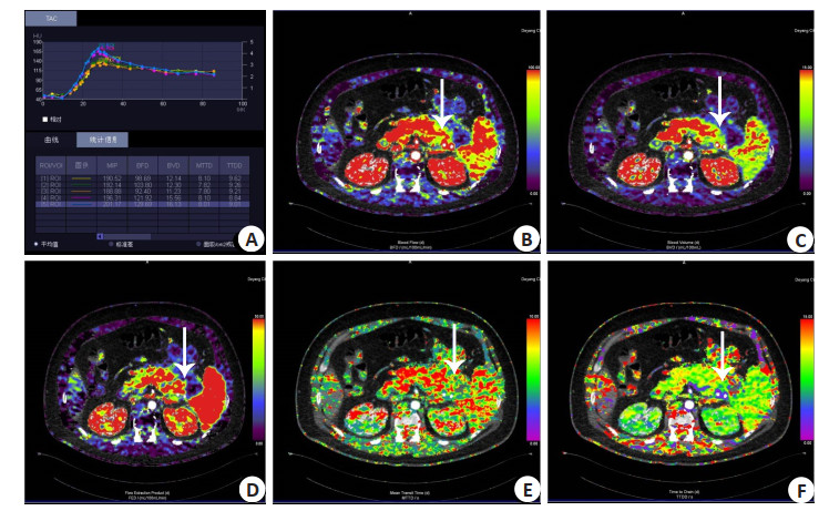
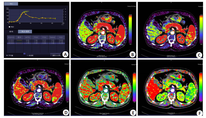
Abstract:
Objective To investigate the value of low-dose CT perfusion in the assessment of patients with moderately severe hyperglycemic acute pancreatitis. Methods Retrospective study was performed in 76 patients with hyperglycemic acute pancreatitis. Patients were dichotomized into two groups. Group A: 54 patients with moderately severe hyperglycemic acute pancreatitis.Group B: 22 patients with mild hyperglycemic acute pancreatitis. The perfusion images were acquired with the third-generation dual-source CT (DSCT) and analyzed with the pancreatic perfusion software package. We analyzed the laboratory data and perfusion parameters, including serum amylasemia (AMY), Lipase (LPS), total cholesterol (TC), triglyceride (TG), high density lipoprotein (HDL), low density lipoprotein(LDL), apoprotein A1(Apo-A1), apoprotein B (Apo-B) blood glucose (BG) and blood flow (BF), blood volume (BV), flow extract product (FEP), mean transit time (MTT), time to peak (TTP) and maximal intensity projection (MIP). Result The laboratory indicators and perfusion parameters of TC, TG, HDL, MTT, TTP were higher in group A, while BV, BF, MIP, FED were lower respectively (P < 0.05). There was a significant inverse correlation between the clinical grades and perfusion parameters(BV, BF, MIP) in hyperglycemic acute pancreatitis (r=-0.835、-0.753、-0.543、P < 0.05). Conclusion The third-generation dual-source low-dose CT perfusion is useful for evaluating severity of the microcirculation of the pancreas in patients with hyperglycemic acute pancreatitis, which is conducive to early clinical treatment and intervention.
2021, 44(5): 845-848.
doi: 10.12122/j.issn.1674-4500.2021.05.22
Abstract:
Objective To explore the value of the CT angiography examination in the left atrium, pulmonary vein and esophagus in guiding the anatomical position during radiofrequency ablation of atrial fibrillation. Methods Sixty-one cases of atrial fibrillation and 31 cases of non-atrial fibrillation underwent CT angiography examination of the left atrium, pulmonary veins and esophagus in our hospital from July 2018 to October 2020 were enrolled. The patients were divided into set experimental group and control group. The diameters of pulmonary vein ostia, diameter of the esophagus, left atrium anteroposterior diameter and the left atrium volume as well as the anatomical types of easophagus were compared between two groups. Results The superoinferior diameters of right inferior pulmonary vein ostia of experimental group were larger than those of control group(P < 0.05). The anteroposterior diameters of right superior pulmonary vein ostia of experimental group were smaller than those of control group (P < 0.05). The mean length of left atrium posterior wall segment, median least distance between esophageal anterior wall and left atrium, mean thickness of left atrium posterior wall, and mean thickness of esophagus segment showed no significant difference between groups (P >0.05). The left atrium anteroposterior diameter and the left atrium volume were larger in experimental group than in control group (P < 0.05). The experimental group included 39 cases of type Ia, 12 cases of type Ib, 11 cases of type Ic, 5 cases of type IIa, 3 cases of type IIb and 2 cases of type IIc; while control group included 13 cases of type Ia, 6 cases of type Ib, 6 cases of type Ic, 3 cases of type IIa, 2 cases of type IIb and 1 case of type IIc. Conclusion Anatomical guidance by CT angiography of left atrium and pulmonary veins combined with esophagus has great value in the treatment of atrial fibrillation by radiofrequency ablation, which can effectively reveal the changes of pulmonary vein and atrium.
2021, 44(5): 849-852.
doi: 10.12122/j.issn.1674-4500.2021.05.23
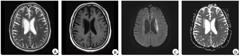
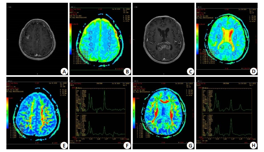
Abstract:
Objective To explore the value of diffusion tensor imaging (DTI) and 1H MRS in the differential diagnosis of brain gliomas and in the prediction of peritumoral invasion of high-grade gliomas. Methods In this prospective study, 69 patients with brain glioma who were treated in our hospital from February 2017 to January 2020 were selected. According to the pathological staging of central nervous system tumor of who in 2007, there were 36 patients with low-grade glioma and 33 patients with high-grade glioma. The DTI and 1H of the two groups were compared to analyze the diagnostic value of DTI and 1H MRS in the diagnosis of peritumoral infiltration of high-grade gliomas. Result ADC of low-grade gliomas was significantly higher than that of high-grade gliomas (t=4.480, P < 0.001). NAA/Cr (t=8.477, P < 0.001), CHO/Cr (t=5.851, P < 0.001) and Lip + Lac/ Cr (t=19.141, P < 0.001) of low-grade gliomas were significantly lower than those of high-grade gliomas. CHO/NAA (t=2.493, P= 0.015) of low-grade gliomas were significantly higher than those of high-grade gliomas In tissues. NAA/Cr (t=2.503, P=0.015) of low-grade glioma patients was significantly lower than that of high-grade glioma patients, CHO/NAA (t=6.937, P < 0.001), CHO/ Cr (t=14.267, P < 0.001) and lip + Lac/Cr (t=2.322, P=0.023) of low-grade glioma patients were significantly higher than that of high-grade glioma patients; the diagnostic sensitivity of DTI and 1H MRS combined diagnosis was significantly higher than that of single detection. Conclusion DTI, 1H MRS imaging performance of brain glioma in the differential diagnosis and high-grade glioma peritumoral infiltration of high-grade predictive efficiency, it is recommended that clinical promotion.
2021, 44(5): 853-855.
doi: 10.12122/j.issn.1674-4500.2021.05.24
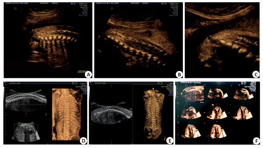
Abstract:
Objective To explore the clinical value of multimodal ultrasound in screening for fetal non severe closed spina bifida. Methods We summarized 19 cases of fetal spinal deformity diagnosed by indirect multi-mode ultrasound prenatal screening from January 2017 to January 2020. The value of multi-mode ultrasound in prenatal screening of fetal spinal deformity was analyzed. Results Among the 38 564 pregnant women, 19 cases were diagnosed as fetal spina bifida, of which 18 cases were detected by prenatal ultrasound. The detection rate of ultrasonic spina bifida was 0.047% (18/38 564). The remaining one case was missed diagnosis because the fetus was breech position and no available imaging data was obtained. The missed diagnosis rate was 5.26% (1/19). Eighteen cases of fetal spina bifida were voluntarily terminated pregnancy, and confirmed by autopsy. Among 19 cases of spina bifida, 15 cases were open spina bifida and 4 cases were closed spina bifida. Among 15 cases of open spina bifida, 6 cases were complicated with hydrocephalus, 4 cases were varus. Prenatal ultrasound showed ventricular dilatation, disappearance of posterior cranial fossa, lemon head sign, ventricular septal defect and other imaging signs. Four cases of closed spina bifida were combined with low conus medullaris, and one case was combined with intraspinal lipoma. Conclusion Multimodal ultrasound is safety and repeatability, which can effectively screen fetal spina bifida.
2021, 44(5): 856-862.
doi: 10.12122/j.issn.1674-4500.2021.05.25
Abstract:
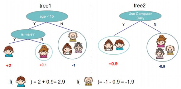
The XGBoost algorithm was first proposed in 2014. Based on the boosting algorithm, it has shown its excellent performance and usability in many data science competitions. At present, the classification and regression models for prediction based on XGBoost algorithm have been widely used for data analysis in health care, finance, education, manufacturing, and other fields. In the medical field, XGBoost has been used for disease diagnosis, prediction of risk, outcomes, prognosis, rational and safe drug use, drug research and development, etc. It provides solutions with great possibilities to improve the efficiency and quality of decision making and reduce the false positive rate. At the same time, XGBoost can automatically learn the splitting direction when processing missing values. It can also simulate nonlinear effect when dealing with large data sets with a high efficiency and accuracy.
The XGBoost algorithm was first proposed in 2014. Based on the boosting algorithm, it has shown its excellent performance and usability in many data science competitions. At present, the classification and regression models for prediction based on XGBoost algorithm have been widely used for data analysis in health care, finance, education, manufacturing, and other fields. In the medical field, XGBoost has been used for disease diagnosis, prediction of risk, outcomes, prognosis, rational and safe drug use, drug research and development, etc. It provides solutions with great possibilities to improve the efficiency and quality of decision making and reduce the false positive rate. At the same time, XGBoost can automatically learn the splitting direction when processing missing values. It can also simulate nonlinear effect when dealing with large data sets with a high efficiency and accuracy.
2021, 44(5): 863-867.
doi: 10.12122/j.issn.1674-4500.2021.05.26
Abstract:
Cervical cancer has been the second leading cause of cancer- related deaths in women all over the world. The incidence rate of cervical cancer is higher in current years, and the main age of cervical cancer has been younger. Pap smears are more often used for the screening of cervical cancer, so more cervical intraepithelial neoplasia (CIN) have been found at earlier stage. Despite the high cure rate, conventional treatments of CIN may damage cervical stroma, thus can lead to reduce fertility such as infertility, abortion or preterm delivery. Therefore, a novel treatment for CIN that can leave fertility virtually intact, with similar cure rate, is urgently needed. Photodynamic therapy (PDT) is an effective therapy for CIN and it could maintain correct tissue structures and functions. PDT is applied for the cases with CIN especially for the younger patients who have the desire for future fertility. This review provides an overview of CIN and PDT. Furthermore, comparison of conventional treatments of CIN and PDT are comprehensively presented, especially investigating the therapeutic effects and clinical efficacy of these treatments in preserving fertility. Finally, we also discuss the limitations and future of PDT. This review aims to introduce the latest research progress of PDT for the treatment of cervical intraepithelial neoplasia, and would indicate new directions for our clinical work about CIN.
Cervical cancer has been the second leading cause of cancer- related deaths in women all over the world. The incidence rate of cervical cancer is higher in current years, and the main age of cervical cancer has been younger. Pap smears are more often used for the screening of cervical cancer, so more cervical intraepithelial neoplasia (CIN) have been found at earlier stage. Despite the high cure rate, conventional treatments of CIN may damage cervical stroma, thus can lead to reduce fertility such as infertility, abortion or preterm delivery. Therefore, a novel treatment for CIN that can leave fertility virtually intact, with similar cure rate, is urgently needed. Photodynamic therapy (PDT) is an effective therapy for CIN and it could maintain correct tissue structures and functions. PDT is applied for the cases with CIN especially for the younger patients who have the desire for future fertility. This review provides an overview of CIN and PDT. Furthermore, comparison of conventional treatments of CIN and PDT are comprehensively presented, especially investigating the therapeutic effects and clinical efficacy of these treatments in preserving fertility. Finally, we also discuss the limitations and future of PDT. This review aims to introduce the latest research progress of PDT for the treatment of cervical intraepithelial neoplasia, and would indicate new directions for our clinical work about CIN.
2021, 44(5): 868-872.
doi: 10.12122/j.issn.1674-4500.2021.05.27
Abstract:
In recent years, the incidence of lung cancer has been increasing year by year, and it is also the most fatal disease among malignant tumors. The survival rate of advanced patients is extremely low. Angiogenesis and lymph node metastasis are two very important characteristics of malignant tumors. It is generally believed that the expression of integrin αvβ3 is related to both of these characteristics. The radionuclide-labeled arginine-glycine-aspartic acid peptide can specifically bind to integrins, which can then reflect the changes in tumor blood vessels. To achieve the early monitoring of lung cancer primary tumors, metastases and efficacy, this article reviews the research progress of 99mTc-3PRGD2 SPECT/CT in lung tumor diagnosis at home and abroad in recent years.
In recent years, the incidence of lung cancer has been increasing year by year, and it is also the most fatal disease among malignant tumors. The survival rate of advanced patients is extremely low. Angiogenesis and lymph node metastasis are two very important characteristics of malignant tumors. It is generally believed that the expression of integrin αvβ3 is related to both of these characteristics. The radionuclide-labeled arginine-glycine-aspartic acid peptide can specifically bind to integrins, which can then reflect the changes in tumor blood vessels. To achieve the early monitoring of lung cancer primary tumors, metastases and efficacy, this article reviews the research progress of 99mTc-3PRGD2 SPECT/CT in lung tumor diagnosis at home and abroad in recent years.
2021, 44(5): 873-877.
doi: 10.12122/j.issn.1674-4500.2021.05.28
Abstract:
18F- FDG PET/CT has been widely used in the diagnosis, staging, efficacy monitoring and prognosis evaluation of malignant tumors. SUVmax is the most commonly used semi-quantitative index, but it is only a single point estimation, ignoring the change of 18F- FDG uptake distribution, and it is susceptible to many factors. Dynamic 18F- FDG PET/CT is a continuous acquisition process that allows quantitative analysis of the net uptake rate of FDG in tumor by using a kinetic model. Compared with traditional SUVs, dynamic scanning can provide early assessment of small metabolic changes in tumors, reflecting tracer uptake more directly and effectively. However, due to its long scanning time and complicated parameter analysis, the clinical application is limited. Nowadays, with the development of a variety of simplified scanning protocols, dynamic 18F-FDG scanning has achieved preliminary research results in a variety of malignant tumors.
18F- FDG PET/CT has been widely used in the diagnosis, staging, efficacy monitoring and prognosis evaluation of malignant tumors. SUVmax is the most commonly used semi-quantitative index, but it is only a single point estimation, ignoring the change of 18F- FDG uptake distribution, and it is susceptible to many factors. Dynamic 18F- FDG PET/CT is a continuous acquisition process that allows quantitative analysis of the net uptake rate of FDG in tumor by using a kinetic model. Compared with traditional SUVs, dynamic scanning can provide early assessment of small metabolic changes in tumors, reflecting tracer uptake more directly and effectively. However, due to its long scanning time and complicated parameter analysis, the clinical application is limited. Nowadays, with the development of a variety of simplified scanning protocols, dynamic 18F-FDG scanning has achieved preliminary research results in a variety of malignant tumors.
2021, 44(5): 878-881.
doi: 10.12122/j.issn.1674-4500.2021.05.29
Abstract:
Chronic low back pain (CLBP) is a common symptom in modern society, more and more evidence shows that the central nervous system plays a crucial role in the occurrence and development of non-specific CLBP. Functional magnetic resonance imaging can reveal detailed neurological activity in the brain of patients with CLBP and track changes in cerebral function. Arterial spin-labeled perfusion imaging evaluates pain-related cerebral perfusion changes by labeling water hydrogen protons in arterial blood to obtain perfusion information such as cerebral blood flow. This article reviews the basic and clinical research of functional magnetic resonance imaging and Arterial spin-labeled perfusion imaging in CLBP, and discusses the application value and research prospect of functional magnetic resonance imaging and arterial spin-labeled in CLBP.
Chronic low back pain (CLBP) is a common symptom in modern society, more and more evidence shows that the central nervous system plays a crucial role in the occurrence and development of non-specific CLBP. Functional magnetic resonance imaging can reveal detailed neurological activity in the brain of patients with CLBP and track changes in cerebral function. Arterial spin-labeled perfusion imaging evaluates pain-related cerebral perfusion changes by labeling water hydrogen protons in arterial blood to obtain perfusion information such as cerebral blood flow. This article reviews the basic and clinical research of functional magnetic resonance imaging and Arterial spin-labeled perfusion imaging in CLBP, and discusses the application value and research prospect of functional magnetic resonance imaging and arterial spin-labeled in CLBP.
2021, 44(5): 882-884.
doi: 10.12122/j.issn.1674-4500.2021.05.30
Abstract:
Dysphagia is just an independent risk factor for adverse outcomes in stroke patients. Imaging examination is the main inspection and evaluation method for dysphagia, which can identify lesions that cause dysphagia and guide treatment. It is required to have an in-depth understanding of imaging evaluation methods for dysphagia in order to incorporate the characteristics of rehabilitation nursing into the multidisciplinary collaborative diagnosis and treatment model for dysphagia, which conduce to reduce the complications and improve the nursing effect of stroke patients. Video fluoroscopic swallowing examination is the most commonly used method to check swallowing function, and it is the "gold standard" for clinical evaluation of dysphagia; Ultrasound examination is used as an auxiliary examination method to assess the swallowing function of patients with dysphagia; CT has good spatiotemporal resolution to display the movement of food boluses and swallowing organsthree- dimensionally and dynamically; MRI can show the lesions of dysphagia and the related brain function networks. Such imaging techniques provide a basis for the rehabilitation and nursing of dysphagia.
Dysphagia is just an independent risk factor for adverse outcomes in stroke patients. Imaging examination is the main inspection and evaluation method for dysphagia, which can identify lesions that cause dysphagia and guide treatment. It is required to have an in-depth understanding of imaging evaluation methods for dysphagia in order to incorporate the characteristics of rehabilitation nursing into the multidisciplinary collaborative diagnosis and treatment model for dysphagia, which conduce to reduce the complications and improve the nursing effect of stroke patients. Video fluoroscopic swallowing examination is the most commonly used method to check swallowing function, and it is the "gold standard" for clinical evaluation of dysphagia; Ultrasound examination is used as an auxiliary examination method to assess the swallowing function of patients with dysphagia; CT has good spatiotemporal resolution to display the movement of food boluses and swallowing organsthree- dimensionally and dynamically; MRI can show the lesions of dysphagia and the related brain function networks. Such imaging techniques provide a basis for the rehabilitation and nursing of dysphagia.