

 Find Duplicates
 Check Document
 Submission(new)
 Experts Office
 Editorial Office
2021 Vol. 44, No. 6
column
Display Method:
2021, 44(6): 885-891.
doi: 10.12122/j.issn.1674-4500.2021.06.01
Abstract:
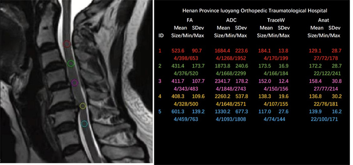
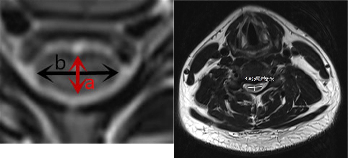
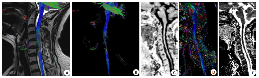
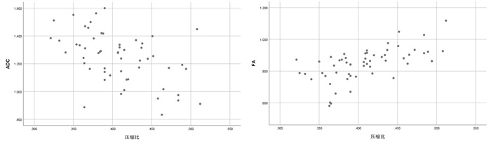
Objective To investigate the diagnostic value of diffusion tensor imaging (DTI) parameters after self- contrast in cervical spondylotic myelopathy. Methods Fifty-six patients with cervical spondylotic myelopathy admitted to our hospital from October 2020 to May 2021 were selected as the case group, while 62 healthy volunteers were selected as the healthy group. DTI imaging was performed in all included subjects, and the ratios of cervical spinal cord parameters to cervical 2/3 segments were taken to compare the differences in fractional anisotropy (FA) and apparent diffusion coefficient (ADC) before and after taking the ratios in healthy groups, and to explore whether taking the ratio method can reduce individual differences in DTI imaging parameters. The parameters of the healthy case group after taking the ratio were compared. The spinal cord aspect ratio was measured in combination with conventional magnetic resonance. The correlation between FA and ADC and spinal cord aspect ratio was analyzed. Finally, the diagnostic efficacy of FA and ADC for cervical spondylotic myelopathy after taking the ratio was judged and the diagnostic cut-off value was given by plotting the receiver operating characteristic curve. Results There were individual differences in FA and ADC values in the healthy group, and the individual differences in FA and ADC in the healthy group disappeared after taking the ratio (P > 0.05). Compared with the healthy group, FA decreased and ADC increased after the ratio in the case group, and there was a moderate positive correlation between FA and spinal cord aspect ratio after the ratio in the case group (r=0.617, P < 0.05), and a low negative correlation between ADC and spinal cord aspect ratio after the ratio (r=-0.478, P < 0.05). The diagnostic efficacy of FA for cervical spondylotic myelopathy was 0.821 and the cut-off value was 0.917 after the ratio. The diagnostic efficacy of ADC for cervical spondylotic myelopathy was 0.791 and the cut-off value was 1.140. The diagnostic efficacy of the combination of the two was 0.896. Conclusion The ratio of DTI parameters can reduce the influence of individual difference, and has more clinical practical significance in the diagnosis of cervical spondylotic myelopathy. The ratio of FA and ADC has certain correlation with the compression aspect ratio of spinal cord.
2021, 44(6): 892-899.
doi: 10.12122/j.issn.1674-4500.2021.06.02
Abstract:
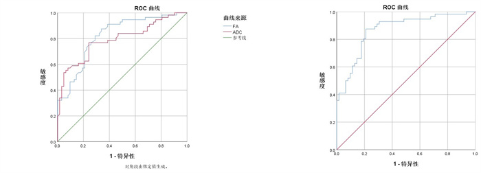
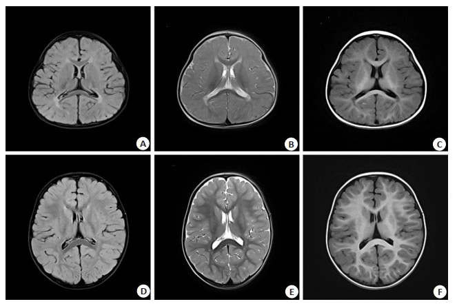
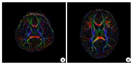
Objective To explore the value of diffusion tensor imaging (DTI) in the diagnosis of cerebral white matter injury and prognosis of neonates with hypoxic-ischemic encephalopathy. Methods Sixty neonates undergoing MRI examination admitted to our hospital from August 2018 to June 2021 were selected as the research subjects. All patients were younger than 2 years old and had a history of hypoxic-ischemic encephalopathy. The patients were divided into two groups, the signs and scores of conventional craniocerebral scans were recorded. The regions of interest were identified on DTI images, the values of FA and ADC were measured. The measured areas of white matter including forelimbs of the internal capsule, craniocerebrum of the midbrain, hind limbs of the internal capsule, knee of the internal capsule, corticospinal tract, center of half ovale, retrothalamic radiation, inferior longitudinal tract, auditory radiation, visual radiation, corpus callosum body, knee of the corpus callosum, frontal white matter, pressure of the corpus callosum, and white matter of the parietal lobe. Three-dimensional images of the fibrous tracts of the corpus callosum and the corticospinal tracts were produceded. The plain scan signs, FA values and ADC values of the two groups were compared and analyzed. The independent influencing factors of white matter injury was screened by multivariate logistic regression analysis, the fitting degree and effectiveness were evaluated. Results Brain MRI plain scan showed significant differences in the signs of left thalamus, bilateral center of semi- ovale and corpus callosum between the two groups (P < 0.05). The FA value of each part of the group of children with cerebral palsy were smaller than the cerebral palsy group, sac and FA values of two groups of children, hind legs, bilateral middle cerebral cerebral peduncle, corticospinal tract, half egg circle center, thalamus after radiation, the longitudinal fasciculus, listen to the radiation, radiation, corpus callosum knee, parietal white matter, corpus callosum, the body and the left side of the corpus callosum lentiform nucleus have significant difference (P < 0.05). There were significant differences in ADC values between the two groups in bilateral semi-ovale center, left posterior limb of internal capsule, left posterior thalamic radiation, bilateral corticospinal tract, left parietal lobe, corpus callosum body part and compression part, and left thalamus (P < 0.05). Multivariate regression showed that the FA values of the left posterior limb of the internal capsule, the left cortical cord and the corpus callosum were independent predictors of white matter injury. The ROC AUC of the three factors combined in the treatment of white matter injury in children with hypoxic- ischemic encephalopathy was 0.887, the specificity and sensitivity were 90.38% and 85.12% respectively, and the accuracy was 0.761. Conclusion Hypoxic-ischemic encephalopathy can damage the white matter fiber bundles, decrease the FA value and increase the ADC value. DTI can clearly reflect the development and damage degrees of the nerve fiber bundles in children, which can be used as the diagnosis and prognosis basis of hypoxic-ischemic encephalopathy.
2021, 44(6): 900-904.
doi: 10.12122/j.issn.1674-4500.2021.06.03
Abstract:
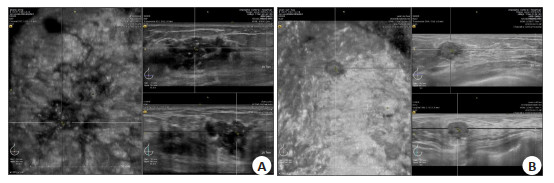
Objective To analyze the relationship between breast cancer automatic breast volume (ABVS) coronal image features, serum Ki67 antigen and C-erbB-2. Methods A total of 78 breast cancer patients in our hospital from January 2016 to December 2020, 81 lesions were collected. ABVS coronal features, including size, boundary, worm erosion sign, convergent sign, microcalcification, hyperechoic ring and immunization were obtained. The immunohistochemical method was used for detection. The difference between Ki67 antigen and C- erbB- 2 in breast cancer ABVS coronal ultrasound features and age of patients was compared. Multivariate Logisitc regression analysis was conducted for indicators with statistically significant differences and for clinically considered indicators that might be significant. Results Size (≥2 cm, OR=4.400) was a risk factor for the high expression of Ki67. Microcalcification (OR=2.741) was a risk factor for C-erbB-2 positive. Boundary, worm-eaten sign, convergence sign, hyperechoic ring and age had no correlation with Ki67 and C-erbB-2 (P > 0.05). Conclusion The characteristic signs of the ABVS coronal surface of breast cancer indirectly reflect the molecular biological behavior of tumor cells, which can be used to evaluate the treatment and prognosis of breast cancer.
2021, 44(6): 905-910.
doi: 10.12122/j.issn.1674-4500.2021.06.04
Abstract:
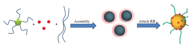
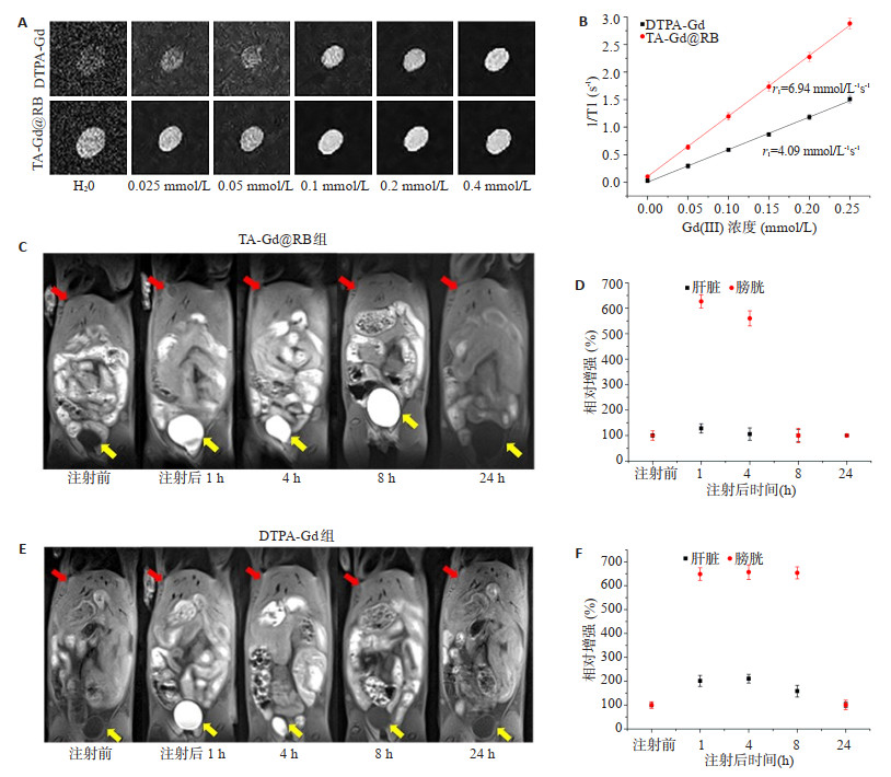
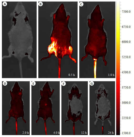
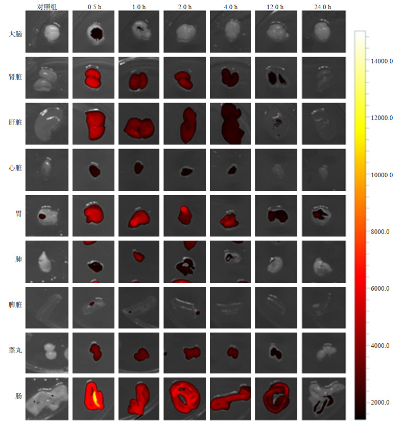
Objective To explore the magnetic resonance imaging/fluorescence imaging (MRI/FI) dual-modality imaging of normal mice organs by a nano-imaging probe (TA-Gd@RB) constructed based on metal gadolinium-catechol. Methods Firstly, the relaxation performance of TA-Gd@RB and DTPA-Gd in vitro was compared. Secondly, 20 normal KM mice were selected and randomly divided into four groups, with 5 in each group: one group was injected with TA-Gd@RB, and one group was injected with clinical DTPA-Gd, the liver and bladder of the mice were MRI scanned respectively. In addition, the other group was injected with TA-Gd@RB again, last group was injected with PBS, fluorescence imaging of mice in vivo and in vitro were performed respectively. Results The results of in vitro relaxation efficacy showed that the relaxation rate of TA-Gd@RB in the experimental group was 6.94 mmol-1s-1, which was 1.7 times that of the clinical DTPA-Gd relaxation rate of 4.09 mmol-1s-1. By analyzing the enhancement effect, amplitude and duration of liver and bladder in mice, TA-Gd@RB had better enhancement effect, higher enhancement amplitude and longer duration of enhancement than DTPA-Gd. Fluorescence imaging results showed that TA-Gd@RB had better fluorescence imaging results in major organs in the body, which were consistent with MRI results. Conclusion TA-Gd@RB have a high relaxation effect in vitro. TA-Gd@RB performs both MRI and FI in vivo and in vitro imaging, and has excellent FI/MRI dual-modal imaging performance.
2021, 44(6): 911-916.
doi: 10.12122/j.issn.1674-4500.2021.06.05
Abstract:
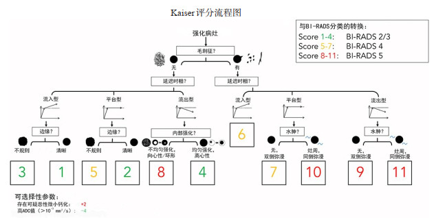
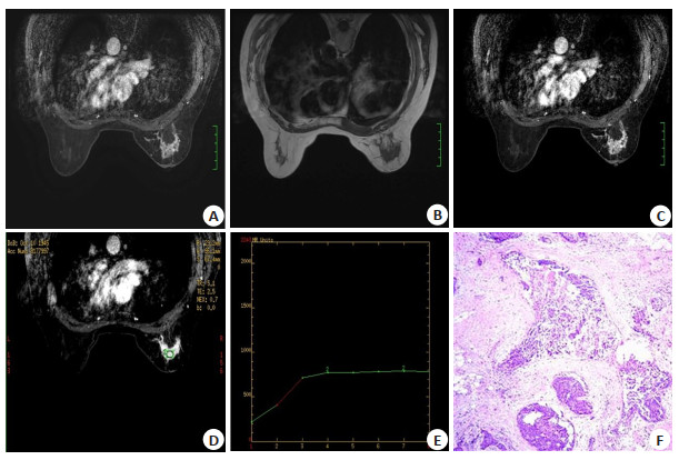
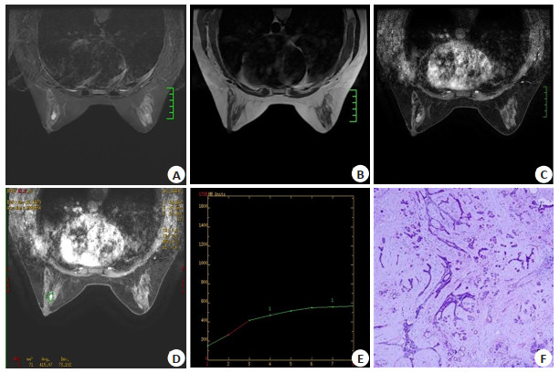
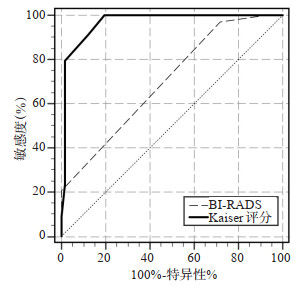
Objective To explore the efficacy of different cut-off values in Kaiser scoring system as diagnostic decision support tool in MRI diagnosis of breast lesions, and explore the best reference cut-off value of benign and malignant lesions so as to avoid excessive breast biopsy, and compare the diagnostic efficiency with the traditional BI-RADS classification diagnostic criteria. Methods The imaging data of 101 breast lesions from 92 patients examined by 1.5 T MRI before operation and confirmed by operation or puncture pathology were reviewed and analyzed. Two intermediate radiologist independently scored all cases according to Kaiser score system, to explore the diagnostic efficacy of Kaiser score system in MRI of breast lesions and compare it with traditional BI-RADS classification diagnosis. The sensitivity, specificity and likelihood ratio of Kaiser score under different critical values were calculated, and the consistency of Kaiser score between the two readers was analyzed. Results There were 101 lesions from 92 patients, including 34 malignant lesions (33.66%) and 67 benign lesions (66.34%). The AUC value of Kaiser score system was 0.969 and that of BI-RADS classification was 0.702. There was a significant difference between two diagnostic methods (P < 0.01). Using the critical value ≥4, ≥5 and ≥6 as the cut-off value of benign and malignant lesions, we could accurately identify 100.00%, 100.00% and 91.98% malignant lesions, and the specificity is 79.10%, 80.60% and 88.06%. Taking the high cut-off value of 7 as the reference cut-off value for judging benign and malignant lesions, we could correctly identify 79.41% of malignant lesions, with a specificity of 98.51% and a positive likelihood ratio of 53.21. The Kaiser scores of the two readers were consistent (Kappa=0.89, P < 0.01). Conclusion The application of Kaiser score in MRI diagnosis of breast lesions is beneficial to improve the diagnostic accuracy, taking ≥5 as the diagnostic threshold for judging benign and malignant lesions. The best comprehensive diagnostic efficiency could be obtained, and for specific diagnostic objectives, the reasonable use of high diagnostic threshold in Kaiser score could enhance diagnostic confidence and avoid excessive breast biopsy to the greatest extent.
2021, 44(6): 917-921.
doi: 10.12122/j.issn.1674-4500.2021.06.06
Abstract:
Objective To evaluate the impact of the timing of percutaneous catheterization and drainage in the treatment of moderately severe acute pancreatitis (MSAP) on patient outcomes and complications. Methods A total of 113 patients with MSAP who were hospitalized in the Second People's Hospital of Guangdong Province from July 2017 to April 2021 were enrolled. According to whether they underwent ultrasound-guided percutaneous catheter drainage (PCD), they were divided into early PCD group, late PCD group and control group, and the basic data of the patients were recorded (age, gender, BMI, etiology), compared the three groups of clinical outcomes (number of patients converted to severe acute pancreatitis, number of patients converted to surgery, number of patients who died), laboratory test indicators (white blood cell count, serum amylase, CRP and blood calcium), clinical efficacy time indicators (systemic inflammatory response time, abdominal pain time, bowel sound recovery time, diet recovery time and total hospital stay), adverse events (abdominal infection, intra-abdominal hemorrhage, catheter blockage) and complications Symptoms (pancreatic pseudocyst, pancreatic abscess, abdominal compartment syndrome, and multiple organ tissue failure). Results The treatment success rate of the early PCD group and the late PCD group was significantly higher than that of the control group, while the mortality, the rate of severe acute pancreatitis patients, and the surgical operation rate were significantly lower than those of the control group, and the treatment success rate of the early PCD group was significantly higher than that of the late PCD group (P < 0.05); The systemic inflammatory response time, bowel sound recovery time and hospital stay in the PCD group were less than those in the control group, and the systemic inflammatory response time and hospital stay in the early PCD group were significantly less than those in the late PCD group (P < 0.05); The improvement of white blood cell count, serum amylase, CRP and blood calcium in the PCD group was better than that of the control group, and the improvement of serum amylase and CRP in the early PCD group was better than that in the late PCD group (P < 0.05); PCD The complications of pancreatic pseudocyst, pancreatic abscess, abdominal compartment syndrome and multiple organ tissue failure in the control group were significantly less than those in the control group (P < 0.05), and the early PCD group had significantly less abdominal compartment syndrome and multiple organ tissue failure In the advanced PCD group, there was no significant difference in abdominal infection and bleeding among the three groups. Conclusion For patients with MSAP, waiting for the buildup to form a package at a later stage before proceeding with PCD does not produce any additional benefits. Early PCD treatment can effectively improve the success rate of MSAP patients, reduce hospitalization time and complications.
2021, 44(6): 922-926.
doi: 10.12122/j.issn.1674-4500.2021.06.07
Abstract:
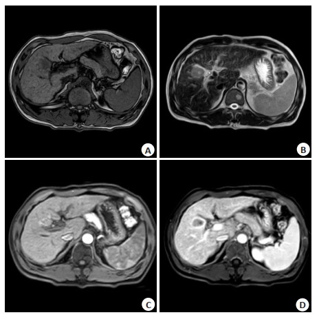
Objective To analyze the MRI findings of hepatobiliary phase (HBP) doughnut-like nodules in patients with liver cirrhosis and the relationship between HBP nodule signal with cholestasis. Methods The imaging and clinical data of 551 patients with liver cirrhosis who underwent MRI with Gd-EOB-DTPA from January 1st, 2019 to July 31st, 2021 were analyzed retrospectively. Sixty patients with HBP doughnut nodules were analyzed. The frequency of HBP doughnut-like nodules in different causes of liver cirrhosis, the signal characteristics on MR plain scan, diffusion weighted imaging (DWI) and enhanced sequence and follow-up of nodules were analyzed. The relationship between HBP nodule signal and cholestasis was analyzed. Of the 60 patients with HBP doughnut nodules, 31 cases had liver cancer in liver background and some underwent surgical treatment. In order to avoid the interference of this factor, they were excluded, and the other 3 cases were excluded due to incomplete data. A total of 112 patients were finally included, 26 patients with HBP doughnut-like nodules, 27 patients with high signal nodules and 59 patients with isosignal nodules were included. Whether there were differences in cholestasis markers such as total bilirubin, direct bilirubin, indirect bilirubin, alkaline phosphatase, glutamyltransferase, total bile acid between HBP doughnut-like nodules, high signal nodules and iso-signal nodules were analyzed. Results Among 551 patients with liver cirrhosis, HBP doughnut-like nodules accounted for 10.9% (60/551). There were significant differences in the frequency of doughnut-like nodules in different causes of liver cirrhosis (P=0.032). There were 688 doughnut-like nodules in 60 patients, single in 18.3% and multiple in 81.7%. On fs-T2WI sequence, 77% of the nodules showed isointensity, 22.5% showed hyperintensity and 0.4% showed hypointensity. On in/out phase T1WI, 89.1% of the nodules showed isointensity, 7% showed hyperintensity and3.9% showed hypointensity. On DWI, 4.5% nodules showed hyperintensity, but there was no limited diffusion. The remaining 95.5% of nodules showed isointensity. All nodules had no enhancement in arterial phase, 48.5% of nodules began to enhancement in portal phase, 42.8% began to enhancement in delayed phase, and 8.7% nodules had no enhancement in multiple phases. 25 patients were followed up without malignant transformation. There was no significant difference (P > 0.05) in age, sex, direct bilirubin, indirect bilirubin, alkaline phosphatase and liver function between HBP doughnut- like nodules, high signal nodules and iso-signal nodules group. The differences in total bilirubin, glutamyltransferase and total bile acid between the groups were significant (P < 0.05). Multiple comparisons showed that there were significant differences in total bilirubin between HBP doughnut-like nodules group and iso-signal group, in glutamyltransferase between HBP doughnut-like nodules and high signal nodules group (P < 0.05). Conclusion HBP doughnut-like nodules in are not uncommon. They have characteristic imaging manifestations of no arterial phase enhancement, portal blood supply and no limited diffusion, and have no malignant tendency during follow-up. The formation of HBP doughnut-like nodules and high signal nodules are not related to cholestasis.
2021, 44(6): 927-931.
doi: 10.12122/j.issn.1674-4500.2021.06.08
Abstract:
Objective To evaluate the value of diffusion- weighted magnetic resonance imaging (DWI) combined with three- dimensional digital mammography (DBT) in the diagnosis of benign and malignant breast lesions. Methods A total of 118 cases of breast DWI and DBT in our hospital were retrospectively analyzed. The signs of calcification were found by DBT. There were 51 cases with breast cancer. The pathological types included invasive ductal carcinoma, invasive ductal carcinoma, ductal carcinoma in situ, medullary carcinoma, invasive lobular carcinoma, mucinous adenocarcinoma. Benign breast lesions were found in 67 cases, including fibroadenoma, adenosis and intraductal papilloma. All patients were scored for calcification on DBT images, and the apparent diffusion coefficient (ADC) value of the corresponding lesion area was measured. The diagnostic efficacy of ADC value, calcification score and their combination in breast calcified lesions was analyzed. Results ADC value of breast cancer group was lower than benign breast lesions group (P < 0.05). Calcification score of breast cancer group was higher than benign lesion group (P < 0.05). The area under the curve (AUC) of ADC value for the diagnosis of benign and malignant breast lesions was 0.853, and the AUC of calcification score for the diagnosis of benign and malignant breast lesions was 0.855. Delong test showed that the two methods had the same diagnostic efficiency for benign and malignant breast lesions with calcification, and the difference was not significant (P=0.625). Logistic regression analysis of the two methods (calcification score+ADC value) showed that the AUC value of the combination of the two methods was 0.903, which had high diagnostic efficiency. Calcification score, ADC value and the combination of the two methods was used to carry out Delong test. The results showed that the difference of receiver operating characteristic curve (ROC) combined with ROC and ADC ROC of the two methods were statistically significant (P=0.0409, 0.0216). Conclusion There is no difference in the differential diagnosis of calcification between diffusion- weighted imaging and three- dimensional digital mammography. The diagnostic efficiency of the combination of the two methods is significantly higher than that of a single method, which provides a more reliable basis for the diagnosis and differential diagnosis of breast lesions with calcification.
2021, 44(6): 932-936.
doi: 10.12122/j.issn.1674-4500.2021.06.09
Abstract:
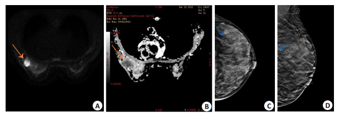
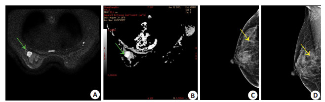
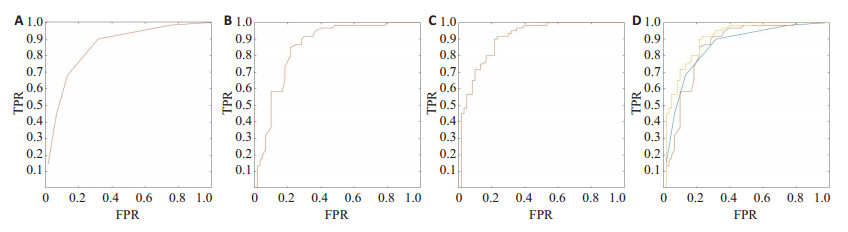
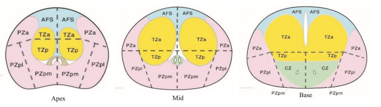
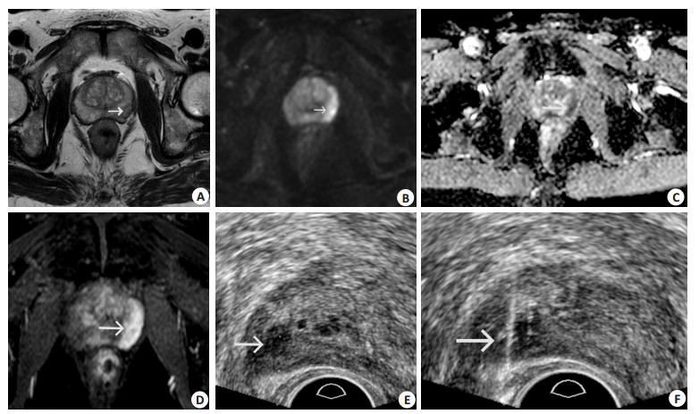
Objective To evaluate the diagnostic value of MRI/transrectal ultrasound (TRUS) cognitive fusion targeted prostate biopsy (CFTB) combined with Transrectal Ultrasound-guided systematic prostate biopsy (SB) for patients with prostate specific antigen level 4-20 ng/mL. Methods A total of 337 patients with serum PSA level 4-20 ng/mL who underwent TRUS- guided prostate biopsy for the first time in our hospital were included. They were randomly divided into CFTB+SB group (n=177) and SB group (n=160) according to whether MRI examination was performed before the biopsy. The positive rate of puncture, positive rate of single needle, detection rate of clinically significant prostate cancer (CSPCa) and clinically insignificant prostate cancer were compared between two groups. Results The positive rate of puncture, positive rate of single needle and detection rate of CSPCa in CFTB + SB group were significantly higher than those in SB (40.1% vs 26.3%, 17.5% vs 10.3%, 38.4% vs 20.6%, P < 0.05). While the detection rate of clinically insignificant prostate cancer in CFTB + SB group was significantly lower than those in SB (1.7% vs 5.6%, P < 0.05). The consistency comparison of CSPCa detection rates in CFTB+SB group showed that CSPCa detection rates had high consistency betwen CFTB + SB and CFTB only (Kappa=0.860). The CSPCa detection rate of of CFTB + SB was higher than that of CFTB (40.1% vs 36.1%, P < 0.05). Conclusion TB guided by MRI/TRUS cognitive fusion combined with SB have high clinical diagnostic value for patients with serum prostate specific antigen level 4- 20 ng/mL, who were suspected prostate cancer prostate.
2021, 44(6): 937-940.
doi: 10.12122/j.issn.1674-4500.2021.06.10
Abstract:
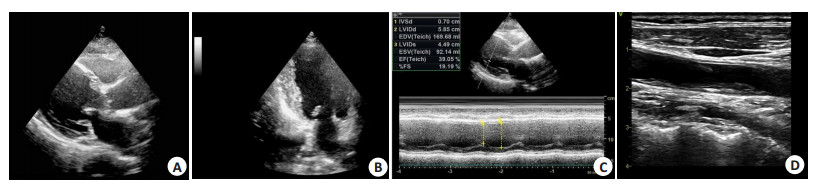
Objective To investigate the clinical value of two-dimensional echocardiography (2DE) in evaluating the severity of non-ST-segment elevation myocardial infarction (NSTEMI) in the elderly. Methods The image data of 126 elderly patients with NSTEMI in our hospital from June 2016 to June 2020 were respectively analyzed. All patients received 2DE and coronary arteriography (CAG). CAG examination showed that 124 patients had at least one main coronary artery and/or branch with diameter stenosis > 50%, then 2DE was used to detect the regional wall motion abnormality (RWMA) among those patients. The characteristics of arterial lesions was compared between RWMA and non-RWMA patients. The 2DE examination indicated that 86 of 126 patients had RWMA, meantime, the value of 2DE in identifying infarct related artery was analyzed using CAG as "gold standard". Killip grading was used as a criterion for the classification of severity of acute myocardial infarction patients, and the intima media thickness of middle artery wall in 2DE examination of patients with different grades was compared. Results Among the 124 patients, 2DE examination showed that 85 patients had RWMA and 39 patients had no RWMA. The proportions of single-vessel lesions, double-vessel lesions and multivessel lesions showed significant difference between RWMA and non-RWMA patients (P < 0.05). CAG accurately identified the infarct- related arteries of 86 patients, and the lesions were found at left anterior descending artery in 26 cases, right coronary artery in 40 cases, and left circumflex in 20 cases. CAG results indicated that the accuracy of RWMA in predicting infarct-associated arteries was 95.35%. The intima media thickness of patients of Killip grade Ⅰ-Ⅱ was significantly lower than that of Ⅲ-Ⅳ patients (P < 0.05). Conclusion Application of 2DE can effectively judge the severity of elderly patients with NSTEMI and locate the infarct-related arteries.
2021, 44(6): 941-944.
doi: 10.12122/j.issn.1674-4500.2021.06.11
Abstract:
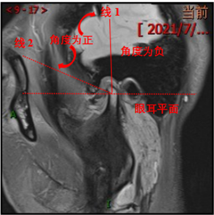
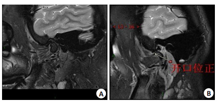
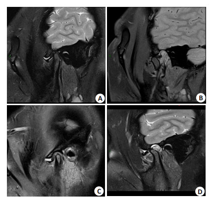
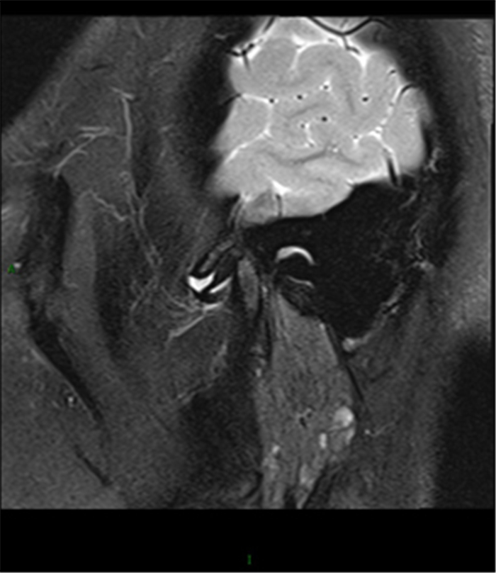
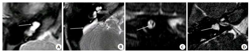
Objective To investigate the clinical features and MRI findings of 182 patients with temporomandibular joint disorders. Methods A total of 364 temporomandibular joints of 182 patients with temporomandibular joint disorder in our hospital from September 2018 to June 2021 were retrospectively selected. The clinical data, including general data, clinical features and MRI images were collected. Results There were 364 joints in 182 patients with temporomandibular joint disorders, including 162 joints with reversible disc displacement, 114 joints with irreversible disc displacement, 27 joints with lateral displacement, 60 joints with disc injury, 32 joints with disc perforation, 47 cases with condyle mutation and destruction, and 31 joints with capsule and ligament injury. The clinical characteristics of patients were complex and changeable, but most patients generally presented with joint pain, popping, mouth opening limitation, mandibular deviation and other symptoms. MRI images showed clear articular disc position, shape, thickness change and joint effusion. Conclusion Patients with temporomandibular joint disorder have various clinical characteristics. MRI can accurately reflect the changes of the position, shape and thickness of the joint disc during the progression of temporomandibular joint disorder, and improve the accuracy of clinical diagnosis.
2021, 44(6): 945-949.
doi: 10.12122/j.issn.1674-4500.2021.06.12
Abstract:
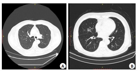
Objective To explore the application value of preoperative enhanced multi-slice spiral CT scan in the surgical treatment for elderly patients with non-small cell lung cancer (NSCLC). Methods We retrospectively analyzed the clinical data of 120 elderly patients with NSCLC treated in the hospital between January 2020 and March 2021. All patients were subjected to enhanced multi- slice spiral CT scan. The consistency between multi- slice spiral CT and pathological results in diagnosing TNM stage and lymph node metastasis of NSCLC were analyzed. The operative conditions and postoperative complications of patients treated by different surgical methods were compared. Results The accuracy rates of preoperative enhanced multi-slice spiral CT scan in the diagnosis of T stage, N stage, lymph node metastasis and clinical stage were 89.17%, 90.83%, 87.10%, and 88.33%, which were consistent with pathological results (Kappa=0.789, 0.792, 0.787, 0.776). In the 120 patients with NSCLC, 98 received surgical treatment, including 57 patients undergoing pulmonary wedge resection and lymph node dissection, and 41 patients undergoing total lobectomy and lymph node dissection. The operation time of patients undergoing pulmonary wedge resection were significantly shorter than those of patients undergoing total lobectomy. The intraoperative blood loss and number of lymph nodes removed were less or smaller than those of patients undergoing total lobectomy, and the incidence of complications was significantly lower than that in patients undergoing total lobectomy (P < 0.05). Conclusion Enhanced multi- slice spiral CT scan has high accuracy in the diagnosis of preoperative stage and lymph node metastasis of NSCLC, thereby providing guidance for the selection of surgical methods.
2021, 44(6): 950-953.
doi: 10.12122/j.issn.1674-4500.2021.06.13
Abstract:
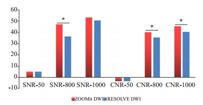
Objective To compare the nasopharyngeal image quality with RESOLVE DWI and ZOOMit DWI in 3.0 T MR. Methods Forty patients who underwent 3.0 T MR nasopharyngeal scan in hospital from January to October 2021 were retrospectively analyzed. Two different diffusion imaging sequences, ZOOMit DWI and RESOLVE DWI, were scanned simultaneously, and the two diffusion weighted images were quantitatively analyzed and subjectively scored. Results The subjective scores of image sharpness, artifact and distortion were significantly different between two different diffusion imaging sequences (P < 0.05). ZOOMit DWI sequences had a better sharpness and less distortion and artifacts. SNR and CNR (b=50 s/mm2) of the two diffusion-weighted sequences showed no significant difference. The SNR and CNR (b=800 and b=1000 s/mm2) of ZOOMit DWI were better than that of RESOLVE DWI. Conclusion ZOOMit DWI sequence has better image quality than RESOLVE DWI sequence and it is suitable for MRI diffusion imaging of nasopharynx.
2021, 44(6): 954-960.
doi: 10.12122/j.issn.1674-4500.2021.06.14
Abstract:
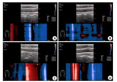
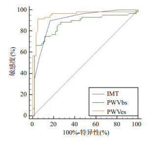
Objective To investigate the correlation between the degree of carotid arteriosclerosis by the ultrafast pulse wave velocity (UFPWV) technology and the degree of essential hypertensive retinopathy. Methods The UFPWV technology was used to measure carotid artery intima-media thickness (IMT), the carotid PWV at the beginning of systole (PWV-BS), and the carotid PWV at the end of systole (PWV-ES) in 120 patients with essential hypertension and to collect the images of the patient' s binocular fundus for clinical grading. We collected data on patients' age, gender, course of hypertension, blood pressure, smoking history, BMI, HDL-C, LDL-C, TC, TG, UA, Cr and urea. The correlation between the above factors and fundus changes and compared the predictive effects of IMT and UFPWV values on the severity of fundus grading were analyzed. Results PWV-ES, PWV-BS, and IMT in hypertensive patients were found to be positively correlated with the degree of retinopathy (P < 0.001). In multivariate logistic regression analysis, PWV-ES, PWV-BS, and IMT were identified as important independent predictors of hypertensive retinopathy. The prediction effect of PWV-ES was better than that of PWV-BS and IMT, and the difference was significant (all P < 0.05). When the value of PWV-ES was greater than the cut-off value of 8.97, PWV-ES indicated that hypertensive retinopathy lesions were serious (P < 0.05). Conclusion The degree of carotid arteriosclerosis in patients with essential hypertension is correlated with the degree of retinopathy. The UFPWV technology may be used as a simple, safe, efficient, and accurate diagnostic index for clinical auxiliary prediction of hypertensive retinopathy.
2021, 44(6): 961-964.
doi: 10.12122/j.issn.1674-4500.2021.06.15
Abstract:
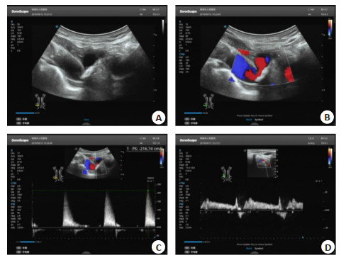
Objective To analyze the diagnostic value of ultrasound probe combined with CT angiography in subclavian steal syndrome. Methods A total of 102 patients with subclavian steal syndrome in our hospital from November 2015 to November 2020 were selected as the observation group, and 100 healthy people were selected as the control group. Toshiba color Doppler ultrasound diagnostic instrument was used to examine the patients. High frequency linear array probe, phased array probe and convex array probe were used to examine the patients. The subjects were examined by GE Revolution 64 slice spiral CT angiography. Logistic regression model was used to analyze the combined application model of multiple ultrasound probes combined with CT angiography in the diagnosis of subclavian steal syndrome. The ROC curve was drawn to analyze the value of each index in predicting subclavian steal syndrome. Results The diagnostic model of subclavian steal syndrome using multiple ultrasound probes combined with CT angiography was log(P)=0.732×ultrasound exploration+0.811×CT angiography+ 0.473. The number of positive patients detected by ultrasound exploration and CT angiography was 92 and 92, respectively, and the number of positive patients detected by combined application was 101. The sensitivity, specificity and area under curve of combined application of multiple ultrasound probes and CT angiography in the diagnosis of subclavian steal syndrome were significantly higher than those of single application of each index (P < 0.05). Conclusion The combined application of multiple ultrasound probes and CT angiography has high value in the evaluation and diagnosis of subclavian steal syndrome.
2021, 44(6): 965-968.
doi: 10.12122/j.issn.1674-4500.2021.06.16
Abstract:
Objective To analyze the acetabular index before and after MRI measurement, evaluate the development of acetabulum before and after hip joint in children, and analyze the influencing factors of developmental dysplasia of the hip (DDH). Methods A total of 174 children with suspected DDH admitted to our hospital from December 2018 to October 2020 were selected. The clinical data, birth weight, pregnant hip position and other data of all the children were recorded, and MRI scanning was performed. The acetabular index was analyzed by different schemes. Scheme 1: The line from the center of femoral head to the weakest part of acetabulum and the line from the weakest point of acetabulum to the outer edge of anterior/posterior wall of bony acetabulum were used. The angle between the weakest point of acetabulum and the center of femoral head and the weakest point of acetabulum to the outer edge of anterior/posterior wall of cartilaginous acetabulum was recorded as A/PBAI. Scheme 2: The angle between the middle point of bilateral Y-shaped cartilage and the outer edge of anterior/posterior wall of cartilaginous acetabulum was recorded as A/PBAI, and the angle between the middle point of bilateral Y-shaped cartilage and the outer edge of anterior/posterior wall of cartilaginous acetabulum was recorded as A/PCAI. Intra class correlation coefficient (ICC) was used to evaluate the consistency of acetabular index, and Forward Logistics regression model was used to explore the independent factors influencing DDH. Results The consistency of ABAI, ACAI, PBAI and PCAI measured by Scheme 1 was significantly higher than that measured by Scheme 2, and the ICC of each index detected by Scheme 1 was >0.75. There were significant differences between DDH group and non DDH group in pregnant breech position, unequal length of lower limbs, congenital muscular torticollis, swaddling position, joint bounce, and the differences were significant (P < 0.05). Sex torticollis, swaddling posture and joint bounce were independent risk factors for DDH (P < 0.05). Conclusion The DDH imaging examination of MRI, the method of measuring acetabular index with the line from the thinnest weakness of the acetabulum to the central point of the femoral head as the baseline has a high consistency. It can accurately evaluate the acetabular development before and after the hip joint in children. The pregnant breech position, unequal length of lower limbs, congenital muscular torticollis, swaddling position and joint bounce are independent risk factors for DDH.
2021, 44(6): 969-973.
doi: 10.12122/j.issn.1674-4500.2021.06.17
Abstract:
Objective To investigate the relationship between quantitative parameters of dynamic contrast-enhanced magnetic resonance (DCE-MRI) scanning and pathological features and HIF-1α levels in patients with primary hepatocellular carcinoma (HCC). Methods A total of 94 patients with primary liver cancer admitted from January 2019 to December 2020 were selected as the study subjects, and 40 patients with benign liver nodules admitted during the same period were selected as the control group. All subjects underwent DCE-MRI to assess microvascular density (MVD), transport coefficient from intracellular space to extracellular space (Ktrans), rate constant of extracellular space to intracellular space (Kep), percentage of extracellular space volume (Ve), and HIF-1α in both groups. The quantitative parameters of DCE-MRI were analyzed among patients with different tumor stages, different degrees of differentiation, whether there was metastasis or not, and different levels of HIF-1α. Results The MVD, Ktrans, Kep, Ve and HIF-1α in HCC patients were higher than those in control group (P < 0.05). The MVD, Ktrans, Kep and Ve in stage Ⅲ/Ⅳ were higher than those in stage Ⅰ/Ⅱ (P < 0.05). The MVD, Ktrans, Kep and Ve in patients with low differentiation were higher than those in patients with high differentiation (P < 0.05). The MVD, Ktrans, Kep and Ve in HCC patients with lymph node metastasis were higher than those without lymph node metastasis (P < 0.05). The MVD, Ktrans, Kep and Ve in patients with HIF-1α≥162.96 ng/L were higher than those in patients with HIF-1α < 162.96 ng/L (P < 0.05). The clinical stage of HCC was positively correlated with MVD and Ve (P < 0.05), and lymph node metastasis of HCC was positively correlated with Ktrans and Ve (P < 0.05). The HIF-1α was positively correlated with MVD, Ktrans, Kep and Ve (P < 0.05). Conclusion DCE-MRI quantitative parameters are closely related to the pathological characteristics and HIF-1α of HCC patients, and they can be used to evaluate the disease progression of HCC patients.
2021, 44(6): 974-977.
doi: 10.12122/j.issn.1674-4500.2021.06.18
Abstract:
Objective To investigate the application of ultrasound guided percutaneous transluminal angioplasty on arteriovenous fistula stenosis in maintenance hemodialysis patients. Methods The clinical data of 40 maintenance hemodialysis patients with arteriovenous fistula stenosis in our hospital from January 2018 to December 2019 were retrospectively analyzed. All patients accepted blood vessels assessment by ultrasound before operation, and underwent percutaneous transluminal angioplasty under ultrasound intervention guidance during operation. The technical success rate, clinical success rate and occurrence of complications were recorded, and the blood flow parameters before and after operation were compared. Results Ultrasound could clearly display the stenosis location, and guide the process of balloon dilatation for arteriovenous fistula stenosis. After percutaneous transluminal angioplasty, the color ultrasound showed that the stenosis and thrombosis disappeared in 40 patients, and both the technical success rate and clinical success rate were 100%. Only 2 cases had mild subcutaneous hematoma at the puncture site, and no serious complications occurred in all patients. The vessel diamete, hemodialysis blood flow and internal fistula blood flow of patients after operation were significantly higher than those before operation (P < 0.05). Conclusion Ultrasound guided percutaneous transluminal angioplasty has high success rate and vascular patency rate for arteriovenous fistula stenosis. Ultrasound can be used as a safe and effective guidance method, and is of certain clinical application value.
2021, 44(6): 978-982.
doi: 10.12122/j.issn.1674-4500.2021.06.19
Abstract:
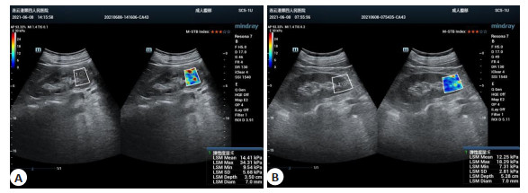
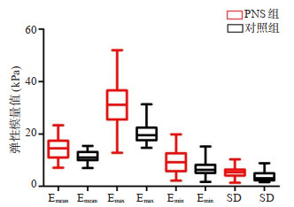
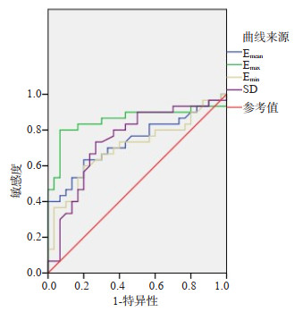
Objective To investigate the application value of shear wave elastography elastic modulus value in the comparative study between patients with early primary nephrotic syndrome (PNS) and healthy subjects and the correlation of renal function indicators. Methods Patients with early PNS CKD stage1 [Patients with normal renal function (blood urea nitrogen and serum creatinine) in normal stage] confirmed by clinical and pathological examination were involved. The characteristics of renal elastic modulus values in 30 PNS patients were analyzed, and the bilateral kidneys of 30 healthy subjects were selected as the control group. The difference of renal elastic modulus values between the two groups and the correlation between the mean renal elastic modulus (Emean) in the PNS group and the 24-hour urinary protein volume and serum β2-microglobulin volume (β2-MG) were analyzed. Results There was no significant difference of renal length, thickness and renal cortex of bilateral kidneys between PNS group and control group (P > 0.05). The mean elastic modulus (Emean), maximum value (Emax), minimum value (Emin) and dispersion of PNS group were higher than those of control group (P < 0.05). When the maximum elastic modulus was 22.45 kPa, the sensitivity, specificity and accuracy of the two groups were 80.0%, 93.3% and 86.7%, respectively. The area under the curve was the largest (0.857), the diagnostic efficiency was the highest. In PNS group, Emean was positively correlated with 24-hour urinary protein volume and serum β2-microglobulin volume, but there was no correlation with blood urea nitrogen and serum creatinine. Conclusion The quantitative analysis technique of shear wave elastography elastic modulus value can provide pathological basis and molecular imaging basis for noninvasive determination of the changes of renal hardness and the degree of injury in early PNS, and provide reliable basis for diagnosis and differentiation of early PNS. It can provide valuable assessment of renal function injury in early PNS by combining 24-hour urinary protein volume and serum β2-microglobulin volume.

2021, 44(6): 983-987.
doi: 10.12122/j.issn.1674-4500.2021.06.20
Abstract:
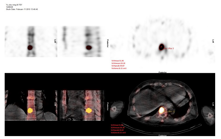
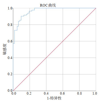
Objective To investigate the differential diagnosis value of SPECT/CT bone quantitative analysis in bone metastases of elderly prostate cancer patients. Methods From January to December in 2019, 158 patients with elderly prostate cancer who underwent whole body bone scan were enrolled. SPECT/CT bone quantitative analysis were preformed on abnormal radioactive concentration lesions and 182 normal vertebral. The differences of SUVmax among the benign, malignant, and normal vertebral were compared. A total of 144 lesions of 46 patients from January to April in 2019 are plotted ROC curve to obtain cut-off values of SUVmax. A total of 293 lesions from May to December 2019 were selected to compare the diagnostic efficacy between SPECT/CT bone quantitative analysis and conventional SPECT/CT. Results A total of 437 lesions (254 malignants/183 benigns) were determined with pathology or follow-up results. The SUVmax of malignant lesions was significantly higher than benign lesions (43.93±19.09 vs 15.26±6.81, P < 0.01) and normal vertebral (6.54±1.19, P < 0.01). Using the best cut-off value of SUVmax≥19.2 as the diagnostic criterion, the accuracy, sensitivity, specificity, positive prediction rate, and negative prediction rate of conventional SPECT/CT and SPECT/CT bone quantitative analysis were 81.6% and 94.5%, 93.0% and 96.5%, 65.2% and 91.7%, 79.2% and 94.3%, 86.8% and 94.9%, respectively. The accuracy, specificity, positive prediction rate and negative prediction rate of SPECT/CT bone quantitative diagnosis were significantly higher than conventional SPECT/CT. Conclusion SPECT/CT bone quantitative analysis has important differential diagnosis value in bone metastases of elderly prostate cancer, and it has the potential for clinical application.
2021, 46(6): 988-991.
doi: 10.12122/j.issn.1674-4500.2021.06.21
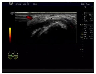
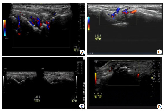
Abstract:
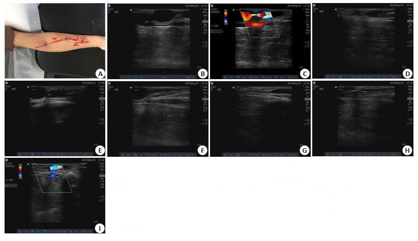
Objective To analyze the diagnostic value of high frequency ultrasound for wrist and knuckle lesions in patients with rheumatoid arthritis (RA). Methods Clinical data of 60 patients with RA admitted to Beijing Haidian Hospital from January 2019 to December 2020 were retrospectively collected as the RA group.The physical data of 60 healthy patients who underwent physical examination in Beijing Haidian Hospital were selected as the healthy control group. All patients underwent high- frequency ultrasound examination. The general data, synovial thickness, joint effusion, tenosynovitis, synovial thickening, bone erosion and pannus were compared between the two groups, and the distribution of synovial thickening and pannus in the joints of RA patients statistically analyzed. Results Synovial thickness of carpal joint, metacarpophalangeal joint and proximal interphalangeal joint in the RA group were higher than those in the healthy control group (P < 0.05). The detection rates of joint effusion, tenosynovitis, synovial thickening, bone erosion and pannus of each joint in the RA group (20.53%, 1.89%, 36.59%, 3.56%, 5.83%) were higher than those in the healthy control group (7.95%, 0.15%, 2.58%, 0.38%, 0.53%, P < 0.05). The synovial thickening and pannus distribution of various joints in RA patients were higher in carpal joint (64.17%, 18.33%) than in metacarpophalangeal joint (52.00%, 7.17%, 31.50%, 5.83%, P < 0.05), and which in metacarpophalangeal joint was higher than in proximal interphalangeal joint (P < 0.05). Conclusion High-frequency ultrasound could better display the pathological conditions of wrist and knuckle joints in RA patients, and had become an effective imaging examination method for RA diagnosis.
2021, 44(6): 992-996.
doi: 10.12122/j.issn.1674-4500.2021.06.22
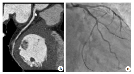
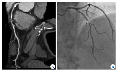
Abstract:
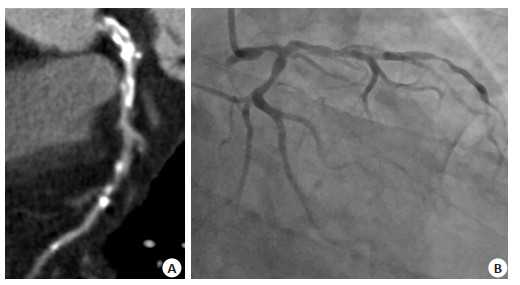
Objective To compare the diagnostic value of low-dose computeirzed tomography angiography and digital subtraction angiography in severe coronary artery stenosis. Methods Eighty-eight patients with clinically suspected or confirmed coronary atherosclerotic heart disease were selected as the research objects. Vascular analysis software was applied to reconstruct images to analyze the nature of coronary plaques detected by computed tomography angiography and evaluate them for stenosis. The result were compared with the stenosis of the lesion detected by digital subtraction angiography of the corresponding segment. Results Digital subtraction angiography was taken as the gold standard, computed tomography angiography detected moderate to severe stenosis at 855 segments caused by coronary artery diseased plaques, there were 396 segments of soft plaques and 459 segments of hard plaques among them. The test results of hard plaques leading to moderate stenosis, hard plaques leading to severe stenosis, moderate stenosis caused by soft plaque and severe stenosis caused by soft plaque had no significant difference (P>0.05). Conclusion Both low-dose computeirzed tomography angiography and digital subtraction angiography have high accuracy in the diagnosis of coronary heart disease. Low-dose computer tomography angiography is economical, convenient and practical to be used as the first choice for clinical screening of coronary heart disease.
2021, 44(6): 997-1001.
doi: 10.12122/j.issn.1674-4500.2021.06.23
Abstract:
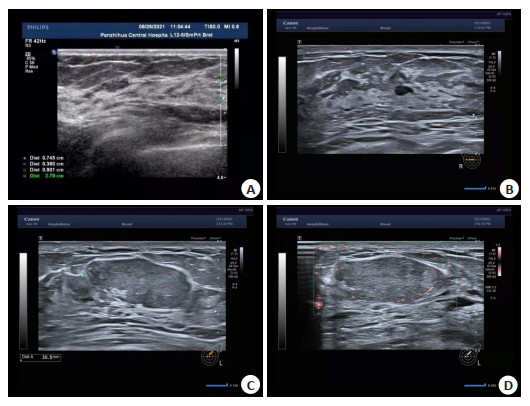
Objective To observe the effect of ultrasound-guided minimally invasive surgery on pain and breast appearance in patients with breast fibroma. Methods A total of 127 patients with single benign breast fibroma who received treatment in our hospital from November 2020 to April 2021 were retrospectively selected. They were divided into open resection group (n=44) and minimally invasive resection group (n=83) according to surgical methods. In the open resection group, conventional open resection was performed, while in the minimally invasive resection group, ultrasound-guided minimally invasive resection was performed. Perioperative related indicators, postoperative breast cosmetic effect, patient satisfaction, postoperative complications and visual analog scale (VAS) score before and after surgery were observed and compared between the two groups. Results Under ultrasonography of both mammary glands, the breast skin echo was clear, and the gland echo was scattered in light spots, with uneven distribution and irregular sparse areas. The operative time, intraoperative blood loss, incision length, scar length, incision healing time and hospital stay in minimally invasive rotation group were significantly shorter than those in open resection group (P < 0.05). VAS scores in minimally invasive rotation group were significantly lower than those in open resection group at 6, 12 and 24 h after surgery (P < 0.05). The rate of excellent breast cosmetic effect and satisfaction score on breast shape, size, softness and symmetry were significantly higher than those in open resection group (P < 0.05). The incidence of postoperative complications in minimally invasive rotatectomy group was significantly lower than that in open resection group (P < 0.05). Conclusion Ultrason-guided minimally invasive rotatectomy for breast fibroma patients has significant advantages such as less trauma, quick recovery and less postoperative complications. It can significantly relieve postoperative pain of patients, and has good postoperative aesthetics and high patient satisfaction.
2021, 44(6): 1002-1006.
doi: 10.12122/j.issn.1674-4500.2021.06.24
Abstract:
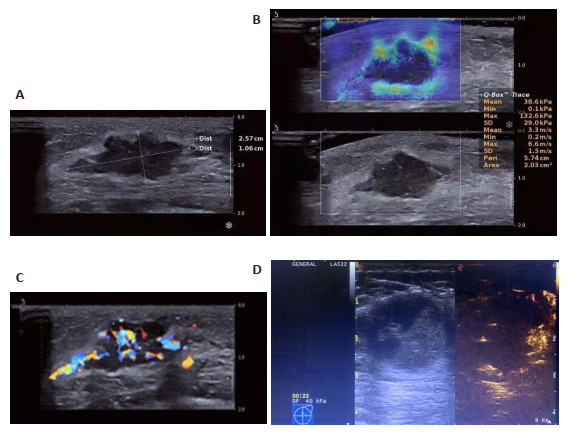
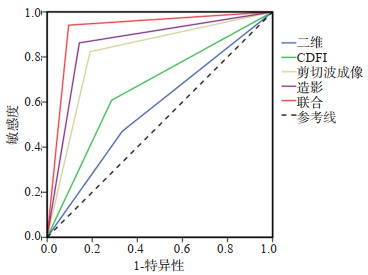
Objective To compare the multimodal ultrasonographic parameters (two-dimensional high frequency ultrasound, color Doppler flow imaging (CDFI), shear wave elasticity imaging, contrast-enhanced ultrasound) and clinicopathological appearances of breast cancer. Methods Sixty-one patients (72 lesions) with breast space occupying lesions from April 2018 to September 2020 in our hospital were enrolled. Two-dimensional high frequency ultrasound, color Doppler flow imaging, shear wave elasticity imaging, contrast-enhanced ultrasound and pathological examinations were performed on all patients. Using pathological results as gold standard, the sensitivity and accuracy of each examination in the diagnosis of breast cancer were analyzed. Results Pathological results showed that among 61 patients (72 lesions), 43 were malignant (51 lesions) and 18 were benign (21 lesions). In the diagnosis of breast cancer, the sensitivity, specificity and accuracy were 47.06%, 66.67% and 52.78% through two-dimensional high frequency ultrasound, those of color Doppler flow imaging were 60.78%, 71.43% and 63.89%, and those of shear wave elasticity imaging were 82.35%, 80.95% and 81.94%, and those of contrast-enhanced ultrasound were 86.27%, 85.71% and 86.11%. The sensitivity, specificity and accuracy of combined examinations in the diagnosis of breast cancer were 94.12%, 90.48% and 93.06%, respectively. The accuracy of combined examination in the diagnosis of breast cancer was higher than that of single examination (P < 0.05). Conclusion Combined examinations can significantly improve accuracy of diagnosis of breast cancer, which is highly consist with pathological results.
2021, 44(6): 1007-1012.
doi: 10.12122/j.issn.1674-4500.2021.06.25
Abstract:
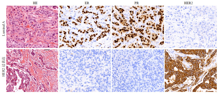
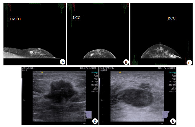
Objective To investigate the correlation between molecular subtypes and imaging features of breast cancer in women aged 18-30 years. Methods The images and pathological data of 195 patients with primary breast cancer diagnosed by postoperative pathology were retrospectively analyzed. According to the expression levels of ER, PR HER2 and Ki-67 they were divided into Luminal A group (n=55), Luminal B group (n=79), HER2 overexpression group (n=41) and triple negative group (n=20). The clinical and pathological data of each subtype were analyzed retrospectively. Results The molecular typing proportion of 195 cases of young breast cancer was Luminal B type >Luminal A>HER2 overexpression >triple negative type. More patients with Luminal A breast cancer were in clinical T1 stage and histological grade Ⅰ(P < 0.05). More patients with Luminal B stage were in clinical T2 stage and biopsy grade (P < 0.05). More patients with TNBC were in stage T3 stage and histological grade (P < 0.05). TNBC had more clinical T3 and histological grade Ⅲ (P < 0.05). X-ray results showed that Luminal type A was characterized by Luminal mass with calcification, parvopleomorphism, and marginal burrs (P < 0.05). Luminal B tumor, parvopleomorphism & line-like, with blurred margins were more common (P < 0.05). HER2 overexpression tumor, with calcification, line-like or line-like branching, with blurred margins were more common (P < 0.05). Triple negative tumor, parvopleomorphism, with blurred margins were more common (P < 0.05). Ultrasonographic results showed that most of the three negative internal echoes were uniform, while the other three types were inhomogeneous (P < 0.05). Conclusion The immunohistochemical subtypes of breast cancer in young women under 18 to 30 years old are closely related to some imaging features. Therefore, preoperative molecular typing could be preliminarily determined according to the comprehensive findings of X-ray and ultrasound.
2021, 44(6): 1013-1016.
doi: 10.12122/j.issn.1674-4500.2021.06.26
Abstract:
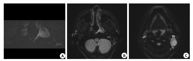
Objective To analyze the clinical characteristics and inner ear MR features in children with absent auditory brainstem response (ABR). Methods Children in the otolaryngology department with extremely severe sensorineural deafness between July 2018 to May 2021 were included. Each of these children had shown no recordable ABR to presented at maximum levels (100 dB). There were 58 males and 41 females, a total of 99 cases with 190 ears. All patients had been evaluated with magnetic resonance imaging of the inner ear, and a perinatal history of some of the children were collected: prematurity, hyperbilirubinemia, low birth weight, history of birth asphyxia, external ear malformation, preterm abortion, family history of deafness, and viral infection during pregnancy. Results A total of the 67 children with perinatal history were included. There were 24 children with a history of hyperbilirubinemia (35.8%); 12 children with a history of low birth weight (17.9%), 11 children with a history of preterm birth(16.4%), 10 children with a history of preterm abortion(14.9%), 10 children with a history of viral infection during pregnancy (14.9%), 5 children with a history of birth asphyxia (7.5%), 4 children with a family history of deafness(6%), and 1 child with a history of external ear malformation(1.5%). The association between gender, age, perinatal risk factors, and inner ear malformations were not significant. Inner ear malformations were found in 43 of 192 ears (22.6%), and Only 17 (8.9%) ears had single anomaly; others (13.7%) had multiple anomalies. In the 43 deformed ears, 81 deformities were found: 35 ears with cochlear nerve dysplasia (18.4%), 20 ears with semicircular canal dysplasia(10.5%), 12 ears with cochlear anomalies(6.3%), 11 ears with large vestibular aqueducts(5.8%), and 3 ears with vestibular malformations (1.5%). A significant correlation was foud between semicircular canal anomalies and cochlear dysplasia (P < 0.05) and cochlear malformations (P < 0.01). Conclusion Children with hearing loss may have various etiologies and should be actively sought out for possible causes to select the best treatment option and improve prognosis.
2021, 44(6): 1017-1023.
doi: 10.12122/j.issn.1674-4500.2021.06.27
Abstract:
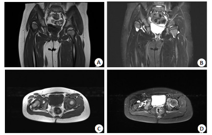
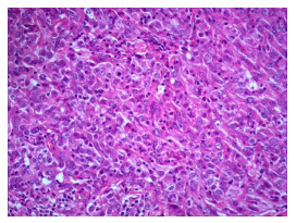
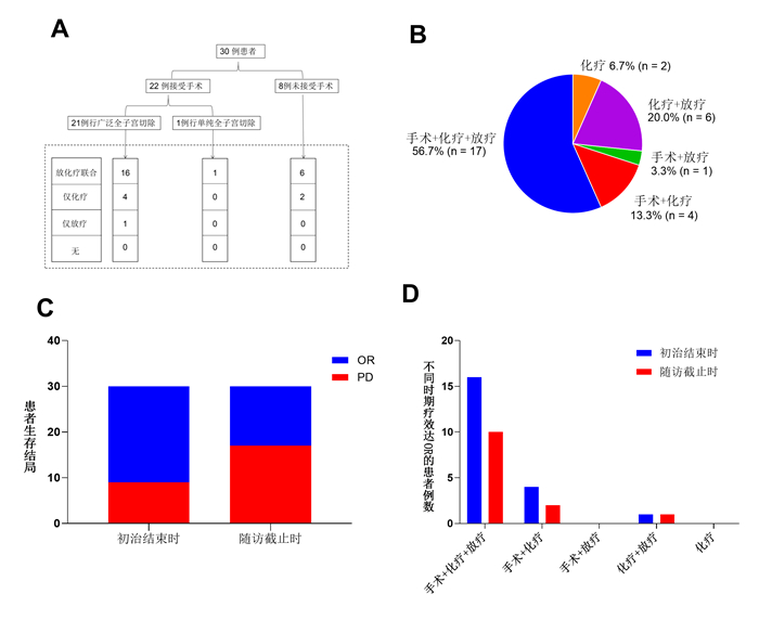
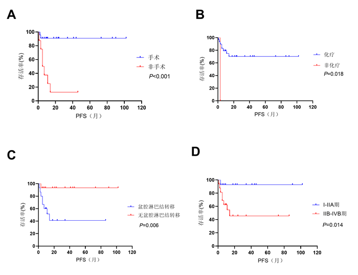
Objective To analyze prognostic factors of patients with neuroendocrine carcinoma of the cervix (NECC). Methods Thirty patients with NECC between December 2009 and July 2020 in Affiliated Cancer Hospital & Institute of Guangzhou Medical University were retrospectively reviewed. Clinicopathologic characteristics and primary treatments were reviewed. The end of the follow-up time was March 31st, 2021.The factors were analyzed using Kaplan- Meier and Cox regression methods. Results With a median follow-up of 43 months (9-135 months), 13 (43.3%) patients were alive. The median PFS was 16 months (1-102 months), median OS was 19 months (3-102 months). The median age was 44.5 years (21-65 years). Univariate analysis showed that surgery (P < 0.001), chemotherapy (P=0.018), pelvic lymph node metastasis (P=0.006) and stage (P=0.014) were significant prognostic factors for PFS. For OS, only surgery and pelvic lymph node metastasis were significant prognostic factors(P < 0.001, P=0.033). In multivariate analyses, surgeryand chemotherapy were independent prognostic factors for PFS (P= 0.002, P=0.005). Surgery was the sole independent prognostic factor for OS (P=0.002). Age, stage, tumor size, histology, pelvic lymph node metastasis, cycles of chemotherapy and radiotherapy were not independent prognostic factors. Conclusion The prognosis of NECC is closely related with surgery and chemotherapy. Patients with indication for surgery should be treated with it. Chemotherapy was advised as adjuvant therapy even in early stage.

2021, 44(6): 1024-1028.
doi: 10.12122/j.issn.1674-4500.2021.06.28
Abstract:
Ovarian tumors and endometrial cancer are seriously threaten women's health and even life-threatening.With the continuous improvement of medical technology and health awareness, the incidence of ovarian tumors and endometrial cancer are also increasing.Early detection, early diagnosis and early treatment have become the principles to standardize the response to both.Ultrasonography and curettage are the main screening methods for ovarian tumors and endometrial cancer, respectively.The rate of misdiagnosis and missed diagnosis are high.Magnetic resonance diffusion-weighted imaging and its derivative techniques are increasingly used in the diagnosis of ovarian tumors and endometrial cancer because of their advantages of both high-resolution, seminal, and multi-directional imaging.This article reviews the application of different diffusion models in ovarian tumors and endometrial cancer, in order to obtain the diagnostic value of different diffusion models for ovarian and endometrial tumors.
Ovarian tumors and endometrial cancer are seriously threaten women's health and even life-threatening.With the continuous improvement of medical technology and health awareness, the incidence of ovarian tumors and endometrial cancer are also increasing.Early detection, early diagnosis and early treatment have become the principles to standardize the response to both.Ultrasonography and curettage are the main screening methods for ovarian tumors and endometrial cancer, respectively.The rate of misdiagnosis and missed diagnosis are high.Magnetic resonance diffusion-weighted imaging and its derivative techniques are increasingly used in the diagnosis of ovarian tumors and endometrial cancer because of their advantages of both high-resolution, seminal, and multi-directional imaging.This article reviews the application of different diffusion models in ovarian tumors and endometrial cancer, in order to obtain the diagnostic value of different diffusion models for ovarian and endometrial tumors.
2021, 44(6): 1029-1033.
doi: 10.12122/j.issn.1674-4500.2021.06.29
Abstract:
Stroke is a serious disease endangering people's life and safety, ranking first in the cause of disability and mortality in China. Post-stroke depression is the most common brain dysfunction after stroke, which aggravates the cognitive dysfunction after stroke, delays the rehabilitation process after stroke, and increases the disability, mortality and recurrence rate of stroke. Diffusion tensor imaging, which can display the structure of white matter in vivo, has been widely used in the study of the pathogenesis of brain diseases, and has gradually become an indicator of curative effect evaluation. This paper reviews the application of diffusion tensor imaging in post-stroke depression, in order to explore its research status and application progress.
Stroke is a serious disease endangering people's life and safety, ranking first in the cause of disability and mortality in China. Post-stroke depression is the most common brain dysfunction after stroke, which aggravates the cognitive dysfunction after stroke, delays the rehabilitation process after stroke, and increases the disability, mortality and recurrence rate of stroke. Diffusion tensor imaging, which can display the structure of white matter in vivo, has been widely used in the study of the pathogenesis of brain diseases, and has gradually become an indicator of curative effect evaluation. This paper reviews the application of diffusion tensor imaging in post-stroke depression, in order to explore its research status and application progress.
2021, 44(6): 1034-1040.
doi: 10.12122/j.issn.1674-4500.2021.06.30
Abstract:
In recent years, with the speedy evolvement of medical imaging diagnosis, a growing number of ultrasound imaging technologies have been used in the diagnosis and treatment of breast cancer. Neoadjuvant chemotherapy is the standard treatment plan for breast cancer patients, which can effectively reduce the clinical stage of tumors and improve the breastconserving rate of patients, thereby improving the prognosis of patients. At present, the main ultrasound diagnostic methods include conventional ultrasound, contrast-enhanced ultrasound, ultrasound elastography, three-dimensional ultrasound, et cetera, which combines with neural network deep learning technology. It can be more efficiently and accurately evaluate the curative effect of neoadjuvant chemotherapy and the evaluation of breast cancer invasiveness. This article reviews the research progress of multi-modal ultrasound combined with deep learning in the evaluation of the efficacy of neoadjuvant chemotherapy and invasiveness for breast cancer.
In recent years, with the speedy evolvement of medical imaging diagnosis, a growing number of ultrasound imaging technologies have been used in the diagnosis and treatment of breast cancer. Neoadjuvant chemotherapy is the standard treatment plan for breast cancer patients, which can effectively reduce the clinical stage of tumors and improve the breastconserving rate of patients, thereby improving the prognosis of patients. At present, the main ultrasound diagnostic methods include conventional ultrasound, contrast-enhanced ultrasound, ultrasound elastography, three-dimensional ultrasound, et cetera, which combines with neural network deep learning technology. It can be more efficiently and accurately evaluate the curative effect of neoadjuvant chemotherapy and the evaluation of breast cancer invasiveness. This article reviews the research progress of multi-modal ultrasound combined with deep learning in the evaluation of the efficacy of neoadjuvant chemotherapy and invasiveness for breast cancer.
2021, 44(6): 1041-1046.
doi: 10.12122/j.issn.1674-4500.2021.06.31
Abstract:
Visual fluorescence imaging is a new surgical assistant technology, which has been widely used in many surgical fields in recent years. Fluorescent dyes have real-time visual imaging characteristics, which can display different anatomical structures such as tumor, blood supply, lymph and nerve, so as to improve the accuracy of operation and postoperative recovery, and may change the operation method. For chest diseases, visual fluorescence imaging technology has good clinical guiding significance in the detection and positioning of pulmonary nodules, the identification of tumor cutting edge and residue, the identification of lung segment plane in thoracoscopic surgery, the prevention of anastomotic leakage after esophagectomy, the identification of intraoperative thoracic duct and the treatment of postoperative chylous leakage, and the mapping of sentinel lymph nodes. In order to evaluate the different needs for fluorescence guidance in different surgical scenes, we described the preclinical research and clinical application progress of imaging technology and fluorescent agent respectively. So far, it marks the increasing maturity of visual fluorescence imaging technology and its wide application prospect in thoracic surgery.
Visual fluorescence imaging is a new surgical assistant technology, which has been widely used in many surgical fields in recent years. Fluorescent dyes have real-time visual imaging characteristics, which can display different anatomical structures such as tumor, blood supply, lymph and nerve, so as to improve the accuracy of operation and postoperative recovery, and may change the operation method. For chest diseases, visual fluorescence imaging technology has good clinical guiding significance in the detection and positioning of pulmonary nodules, the identification of tumor cutting edge and residue, the identification of lung segment plane in thoracoscopic surgery, the prevention of anastomotic leakage after esophagectomy, the identification of intraoperative thoracic duct and the treatment of postoperative chylous leakage, and the mapping of sentinel lymph nodes. In order to evaluate the different needs for fluorescence guidance in different surgical scenes, we described the preclinical research and clinical application progress of imaging technology and fluorescent agent respectively. So far, it marks the increasing maturity of visual fluorescence imaging technology and its wide application prospect in thoracic surgery.