


Correlation between blood circulation velocity and enhancement effect of coronary CT angiography and optimization of contrast medium
-
摘要:
目的 探究血液循环速度与冠状动脉CT血管造影(CTA)增强效果的相关性,并分析对比剂优化方案。 方法 选取2020年1月~2021年4月于我院行冠状动脉CT血管造影患者161例,按照血液循环速度分为快速组(n=32)、中等组(n=35)和缓慢组(n= 94),分析血液循环速度与增强效果关系,并将缓慢组分为A、B、C 3个亚组,A组与快速组、中等组使用的对比剂浓度和注射速率相同(均为碘浓度320 mgI/mL,速率5 mL/s);B组采用对比剂的碘浓度为350 mgI/mL,速率5.5 mL/s;C组采用对比剂的碘浓度为370 mgI/mL,速率5.5 mL/s,比较各组伪影发生、室间隔显示、冠脉CT值及图像质量情况。 结果 快速组、中等组、B组、C组产生伪影概率均低于A组,且快速组低于中等组,C组低于B组(P < 0.05);快速组、中等组、B组、C组主动脉根部CT值、图像质量评分、室间隔显示评分均高于A组,且快速组高于中等组,C组高于B组(P < 0.05)。 结论 血液循环速度能增加冠脉CTA增强效果,清晰显示室间隔,对于血液循环较慢者可适当增加对比剂浓度和注射速率以保证图像质量。 -
关键词:
- 血液循环速度 /
- 冠状动脉CT血管造影 /
- 对比剂 /
- 冠心病
Abstract:Objective To explore the correlation between blood circulation velocity and the enhancement effect of coronary CT angiography (CTA), and to analyze the optimization scheme of contrast medium. Methods A total of 161 patients who underwent coronary CT angiography in our hospital from January 2020 to April 2021 were selected and divided into fast group (n=32), medium group (n=35) and slow group (n=94) according to blood circulation velocity. The relationship between blood circulation speed and enhancement effect was analyzed. The slow group was divided into three subgroups: group A, group B and group C. The contrast agent concentration and injection rate of group A, fast group and medium group were the same (iodine concentration was 320 mgI/mL and injection rate was 5 mL/s). The iodine concentration of group B was 350 mgI/mL and injection rate was 5.5 mL/s, while the iodine concentration of group C was 370 mgI/mL and injection rate was 5.5 mL/s. The occurrence of artifacts, ventricular septal display, coronary CT value and image quality were compared. Methods The artifact probability of fast group, medium group, B group and C group was lower than that of a group, and fast group was lower than that of medium group, and C group was lower than that of B group (P < 0.05). The CT value of aortic root, image quality score and ventricular septal display score of fast group, medium group, B group and C group were higher than those of A group, and the fast group was higher than that of medium group, and the C group was higher than that of B group (P < 0.05). Conclusions The velocity of blood circulation can increase the enhancement effect of coronary CTA and clearly show the interventricular septum. For the patients with slow blood circulation, the contrast medium concentration and injection rate can be increased appropriately to ensure the image quality. -

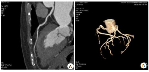
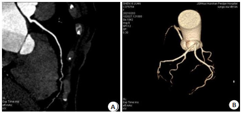
图 1 女,58岁,心功能1级,注射造影剂速率5 mL/s,二尖瓣无反流,主动脉根部对比剂达峰值时间25 s,主动脉根部CT值598 Hu,对比剂碘浓度320 mgI/mL
A: 左前降支曲面重建显示主动脉及冠脉增强效果、图像质量、对比度均较好,左前降支斑块和血管边缘显示清晰; B: 冠脉树容积重建显示冠脉主干、主要分支及细小分支均显示清晰.
Figure 1. Female, 58 years old, cardiac function grade 1, injection rate 5 mL/s. There was no mitral regurgitation, the peak time of contrast medium in aortic root was 25 s, the CT value of aortic root was 598 Hu, and the iodine concentration of contrast medium was 320 mgI/mL.

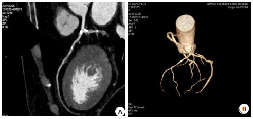
图 2 女,60岁,心功能2级,二尖瓣轻度反流,造影剂注射速率5 mL/s,主动脉根部对比剂达峰值时间28 s,主动脉根部CT值513 Hu,对比剂碘浓度320 mgI/mL
A: 左前降支曲面重建显示主动脉及冠脉增强效果中等,图像质量及对比度一般,左前降支斑块、血管边缘显示尚清晰; B: 冠脉树容积重建显示冠脉主干、主要分支尚清晰,细小分支稍模糊.
Figure 2. Female, 60 years old, cardiac function grade 2, mild mitral regurgitation, injection rate of contrast agent 5 mL/s, peak time of aortic root contrast agent 28 s, CT value of aortic root 513 Hu, iodine concentration of contrast agent 320 mgI/mL.

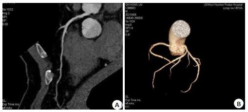
图 3 女,83岁,心功能2级,二尖瓣轻度反流,造影剂注射速率5 mL/s主动脉根部对比剂达峰值时间30 s,主动脉根部CT值300 Hu,对比剂碘浓度320 mgI/mL
A: 左前降支曲面重建显示主动脉及冠脉增强效果、图像质量、对比度均一般左前降支斑块、血管边缘尚清晰; B: 冠脉树容积重建显示冠脉主干、主要分支尚清晰,细小分支模糊。近中段见钙化影非钙化斑块影官腔轻-中度狭窄
Figure 3. Female, 83 years old, cardiac function grade 2, mild mitral regurgitation, injection rate of contrast agent 5 mL/s, peak time of aortic root contrast agent 30 s, CT value of aortic root 300 Hu, iodine concentration of contrast agent 320 mgI/mL.

图 4 男,51岁,心功能1级,二尖瓣轻度反流,造影剂注射速率5.5 mL/s主动脉根部对比剂达峰值时间24 s,主动脉根部CT值402 Hu,对比剂碘浓度350 mgI/mL
A: 左前降支曲面重建显示主动脉及冠脉增强效果不佳,图像质量一般,对比度差,左前降支斑块、血管边缘尚清晰,局部心肌内走形; B: 冠脉树容积重建显示冠脉主干、主要分支尚清晰,细小分支模糊.
Figure 4. Male, 51 years old, cardiac function grade 1, mild mitral regurgitation, injection rate of contrast agent 5.5 mL/s, peak time of aortic root contrast agent 24 s, CT value of aortic root 402 Hu, iodine concentration of contrast agent 350 mgI/mL.

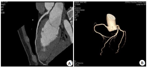
图 5 男,52岁,心功能1级,二尖瓣轻度反流,造影剂注射速率5.5 mL/s,主动脉根部对比剂达峰值时间24 s,主动脉根部CT值445 Hu,对比剂碘浓度370 mgI/mL
A: 左前降支曲面重建显示主动脉及冠脉增强效果中等,图像质量及对比度一般,左前降支斑块、血管边缘尚清晰; B: 冠脉树容积重建显示冠脉主干、主要分支尚清晰,细小分支显示稍模糊.
Figure 5. Male, 52 years old, cardiac function grade 1, mild mitral regurgitation, injection rate of contrast medium 5.5 mL/s, peak time of aortic root contrast medium 24 s, aortic root CT value 445 Hu, contrast medium iodine concentration 370 mgI/mL.
表 1 一般资料比较
Table 1. Comparison of general data(Mean±SD)
组別 男/女(n) 年龄(岁) BM(kg/m2) 心率(次/min) 快速组(n=32) 20/12 58.72±9.32 24.85±1.92 66.32±2.57 中等组(n=35) 23/12 59.24±8.27 25.03±1.38 65.17±3.15 A组(n=30) 19/11 60.35±7.29 24.96±1.63 64.93±4.72 B组(n=33) 24/9 60.26±7.54 25.10±1.34 65.04±3.89 C组(n=31) 21/10 61.25±8.23 24.24±1.62 64.98±4.26  下载: 导出CSV
表 2 各组对比剂注射方案
Table 2. Contrast agent injection scheme of each group
组別 对比剂量(mL) 注射速度(mL/s) 碘浓度(mgI/mL) 快速组(n=32) 60 5.0 320 中等组(n=35) 60 5.0 320 A组(n=30) 60 5.0 320 B组(n=33) 66 5.5 350 C组(n=31) 66 5.5 370
下载: 导出CSV
表 3 各组伪影发生情况
Table 3. Artifacts in each group (n)
组別 0级 1级 2级 快速组(n=32) 31 1 0 中等组(n=35) 28 7 0 A组(n=30) 4 21 5 B组(n=33) 16 16 1 C组(n=31) 29 2 0 x2 15.240 P < 0.001
下载: 导出CSV
表 4 各组室间隔显示评分
Table 4. Ventricular septal display score of each group (n)
组別 1分 2分 3分 快速组(n=32) 0 2 30 中等组(n=35) 1 6 28 A组(n=30) 23 6 1 B组(n=33) 18 7 8 C组(n=31) 9 16 6 x2 7.225 P < 0.001
下载: 导出CSV
表 5 各组冠脉CT值、图像质量比较
Table 5. Comparison of coronary CT value and image quality in
组別 主动脉根部CT值(Hu) 图像质量评分(分) 快速组(n=32) 336.72±23.14* 2.75±0.21* 中等组(n=35) 322.41±20.23* 2.34±0.54* A组(n=30) 291.11±23.60 1.32±0.21 B组(n=33) 309.24±15.43* 1.95±0.42* C组(n=31) 317.61±11.82* 2.30±0.53* F 164.258 265.422* P < 0.001 < 0.001 *P < 0.05 vs A组
下载: 导出CSV
-
[1] 吴舒窈, 刘艳, 宋倩. 冠心病发病及预后的影响因素研究[J]. 中国全科医学, 2018, 21(29): 3562-70. doi: 10.3969/j.issn.1007-9572.2018.00.249 [2] 姚福会. 64排螺旋CT冠状动脉CTA图像质量影响因素分析[J]. 国际心血管病杂志, 2017, 16(44): 224-5. https://www.cnki.com.cn/Article/CJFDTOTAL-CTMR201302040.htm [3] Collet C, Onuma Y, Andreini D, et al. Coronary computed tomography angiography for heart team decision- making in multivessel coronary artery disease[J]. Eur Heart J, 2018, 39(41): 3689-98. http://www.onacademic.com/detail/journal_1000040880488810_ce35.html [4] 樊刚, 李波, 董莉, 等. 血管内超声与64排螺旋CT冠状动脉CTA对冠脉钙化病变定性、定量检测价值比较[J]. 中国CT和MRI杂志, 2021, 19(1): 90-2. doi: 10.3969/j.issn.1672-5131.2021.01.031 [5] 中国心血管病预防指南写作组中华心血管病杂志编辑委员会. 中国心血管病预防指南(2017[) J]. 中华心血管病杂志, 2018, 46(1): 10-25. doi: 10.3760/cma.j.issn.0253-3758.2018.01.004 [6] SCOT- HEART Investigators, Newby DE, Adamson PD, et al. Coronary CT angiography and 5-year risk of myocardial infarction [J]. N Engl J Med, 2018, 379(10): 924-33. doi: 10.1056/NEJMoa1805971 [7] 黄超, 万维佳, 姚宇环, 等. 320排容积CT冠状动脉减影技术在临床应用中的影响因素分析[J]. 中国医疗设备, 2020, 35(10): 126-30. doi: 10.3969/j.issn.1674-1633.2020.10.028 [8] 于易通, 尹卫华, 马伟, 等. 冠状动脉CTA个体化对比剂注射方案实现图像质量均一化的可行性研究[J]. 放射学实践, 2019, 34(4): 427- 31. https://www.cnki.com.cn/Article/CJFDTOTAL-FSXS201904014.htm [9] Lee JM, Choi KH, Koo BK, et al. Prognostic implications of plaque characteristics and Stenosis severity in patients with coronary artery disease[J]. J Am Coll Cardiol, 2019, 73(19): 2413-24. doi: 10.1016/j.jacc.2019.02.060 [10] 陈伟彬, 冯莉, 张伟杰, 等. 优化对比剂注射方案对冠状动脉CTA图像的影响[J]. 临床放射学杂志, 2017, 36(11): 1712-5. https://www.cnki.com.cn/Article/CJFDTOTAL-LCFS201711044.htm [11] 付永波, 朱冬梅, 胡绍波, 等. 冠脉CTA与血管超声评估T2DM合并CHD患者颈动脉与冠脉粥样硬化的关系[J]. 中国CT和MRI杂志, 2020, 18(1): 48-50, 68. doi: 10.3969/j.issn.1672-5131.2020.01.016 [12] Chang HJ, Lin FY, Lee SE, et al. Coronary atherosclerotic precursors of acute coronary syndromes[J]. J Am Coll Cardiol, 2018, 71(22): 2511-22. doi: 10.1016/j.jacc.2018.02.079 [13] 陈伟彬, 冯莉, 张伟杰, 等. 对比剂不同注射方案对冠状动脉CTA图像质量的影响[J]. 医学研究杂志, 2017, 46(5): 171-4, 3. https://www.cnki.com.cn/Article/CJFDTOTAL-YXYZ201705044.htm [14] 张栋青, 史朴军, 蔡显圣, 等. 自动选择最佳期相在高心率患者冠状动脉CTA成像中的应用[J]. 中国医疗设备, 2018, 33(6): 67-70. doi: 10.3969/j.issn.1674-1633.2018.06.017 [15] Poon M, Lesser JR, Biga C, et al. Current evidence and recommendations for coronary CTA first in evaluation of stable coronary artery disease[J]. J Am Coll Cardiol, 2020, 76(11): 1358-62. doi: 10.1016/j.jacc.2020.06.078 [16] 赵月玲, 王莉. 冠状动脉CTA检查过程中的图像质量评估及护理体会[J]. 医学影像学杂志, 2018, 28(5): 741-4. https://www.cnki.com.cn/Article/CJFDTOTAL-XYXZ201805016.htm [17] 蔡显圣, 魏里, 贾慧娟, 等. 冠状动脉和头颈CTA"一站式"联合扫描的可行性研究: 图像质量及辐射剂量[J]. 中国医疗设备, 2019, 34(11): 97-100. doi: 10.3969/j.issn.1674-1633.2019.11.023 [18] Taron J, Foldyna B, Eslami P, et al. Cardiac computed tomography - more than coronary arteries? A clinical update[J]. Fortschr Röntgenstr, 2019, 191(9): 817-26. doi: 10.1055/a-0924-5883 [19] 张静, 郑君惠, 曹希明. 双源CT超低管电压、低对比剂流率及容量冠状动脉CTA研究[J]. 医学影像学杂志, 2019, 29(8): 1314-8. https://www.cnki.com.cn/Article/CJFDTOTAL-XYXZ201908018.htm [20] 王焕勇, 张尤佳, 彭如臣. 冠脉CTA低剂量成像在心律不齐患者中的应用研究[J]. 中国地方病防治杂志, 2018, 33(2): 193-4, 197. https://www.cnki.com.cn/Article/CJFDTOTAL-DYBF201802039.htm [21] 何泽兵, 严高武, 李勇, 等. 冠状动脉CT血管造影对先天性右冠状动脉缺如的评价价值[J]. 分子影像学杂志, 2020, 43(4): 606-9. https://www.cnki.com.cn/Article/CJFDTOTAL-FZYX202004011.htm -

 点击查看大图
计量
- 文章访问数: 441
- HTML全文浏览量: 79
- PDF下载量: 9
- 被引次数: 0