


Value of multi-slice spiral CT in evaluating the therapeutic effect of gastrointestinal stromal tumors
-
摘要:
目的 探讨多层螺旋CT(MSCT)在胃肠间质瘤(GIST)治疗效果评价中的价值。 方法 回顾性分析2018年1月~2019年12月于我院接受治疗的42例GIST患者MSCT检查资料,其中34例接受手术治疗,8例不可手术或转移接受分子靶向治疗。分析手术治疗经MSCT随访复发与转移情况;分子靶向治疗经MSCT随访后采用Choi标准评价疗效,并评估病灶的特征。 结果 接受手术治疗的GIST患者随访时间6~24月,复发12例,平均肿瘤直径1.64±0.49 cm;7例边界清楚,5例浸润生长;8例内部密度均匀,4例发现液化坏死区域;8例外形呈类圆形,4例呈现椭圆形;腔内生长7例,腔外生长5例;8例均匀强化,4例不均匀强化;2例钙化。接受分子靶向治疗的8例患者随访时间为6~24月,其中2例边界清楚,3例边缘光整,3例边缘模糊;6例增强后均匀强化,2例增强后不均匀强化;4例轻度强化,2例中度强化,2例重度强化;均有肿瘤血管;5例伴周围浸润,3例不伴周围浸润。第1次MSCT随访疾病控制率为75.0%,第2次疾病控制率为75.0%,末次疾病控制率为62.5%;伴MSCT随访的进行,GIST患者的肿瘤最长径与静脉期肿瘤CT值均呈现下降,且CT值与基线比较平均下降幅度高于肿瘤长径下降幅度。42例患者治疗后9例出现转移病灶,包含肝脏转移4例,腹腔转移2例,多处转移1例,盆腔转移2例。 结论 MSCT在胃肠间质瘤治疗效果评价中有良好的应用价值,可为GIST患者手术或分子靶向治疗后的疗效评估及治疗指导提供重要的影像学资料。 Abstract:Objective To explore the value of multi-slice spiral CT (MSCT) in evaluation of the therapeutic effect of gastrointestinal stromal tumor (GIST). Methods We retrospectivly analyzed the MSCT data of 42 patients with GIST who received treatment in our hospital from January 2018 to December 2019. Thirty-four cases received surgical treatment, 8 cases received molecular targeted therapy for inoperable or postoperative recurrence or metastasis. Surgical treatment was followed up by MSCT for recurrence and metastasis. After the follow-up of MSCT for molecular targeted therapy, Choi criteria were used to evaluate the curative effect and evaluate the characteristics of the lesion. Results GIST patients who underwent surgery were followed up for 6 to 24 months. Twelve cases recurred with an average tumor diameter of 1.64±0.49 cm.Seven cases had clear borders and 5 cases infiltrated growth. Eight cases had uniform internal density and 4 case had liquefaction necrosis area. Eight cases had a round shape and 4 cases were oval. Seven cases had intracavitary growth and 5 cases had extracavity growth. Eight cases had uniform strengthening, 4 case had inhomogeneous strengthen and 2 cases Calcification. The 8 patients that received molecular targeted therapy were followed up for 6 to 24 months. Among them, 2 cases had clear boundaries, 3 cases had smooth edges, 3 cases had blurred edges. Six cases had uniform enhancement after enhancement, 2 cases had uneven enhancement after enhancement. Four cases had mild enhancement, 2 cases had moderate enhancement, and 2 cases had severe enhancement. Five cases with peripheral infiltration, 3 cases without peripheral infiltration. Five cases showed cystic degeneration and necrosis. The disease control rate of the first MSCT followed-up were 75.0%, the second disease control rate were 75.0%, and the last disease control rate were 62.5%. With the followed-up of MSCT, the CT values of the longest diameter of the tumor and the venous stage tumors of GIST patients decreased. The average decrease of the CT value compared with the baseline was higher than the decrease of the long diameter of the tumor. After treatment, 9 cases had metastatic lesions, including 4 cases of liver metastasis, 2 cases of abdominal metastasis, 1 case of multiple metastases and 2 cases of pelvic metastasis. Conclusion MSCT has a good application value in the evaluation of the therapeutic effect of gastrointestinal stromal tumors. It can provide important imaging data for the evaluation of the efficacy and treatment guidance of GIST patients after surgery or molecular targeted therapy. -

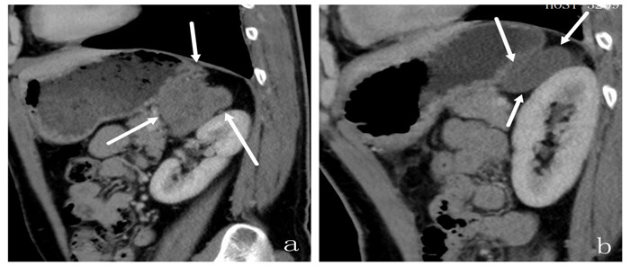
图 2 小肠GIST
A: 小肠肠壁肿块,病理为中度危险性GIST,最大径6.12 cm,内见坏死伴积气,为典型Torricelli-Bernoulli征,增强扫描静脉期不均匀明显强化; B: 手术切除肿块4月后复查,肿瘤复发,最大径3.54 cm,明显强化.
Figure 2. GIST of small bowel.

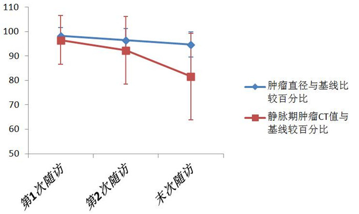
图 1 分子靶向治疗后MSCT随访的肿瘤直径、静脉期肿瘤CT值与基线比较百分比变化
Figure 1. The petcentage changes of tumor diameter and CT value of venous phase tumor compared with baseline after molecular targeted therapy by MSCT follow-up. *P < 0.05.

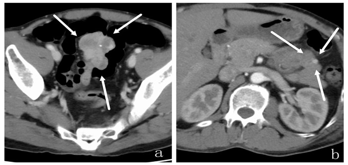
图 3 空肠远段GIST
A: 空肠远段肿块,病理为中度危险性GIST,最大径5.32 cm,分叶状,增强扫描静脉期明显强化; B: 手术切除4月后复查,左上腹空肠旁明显强化结节,考虑肿瘤复发.
Figure 3. GIST in the distal jejunum

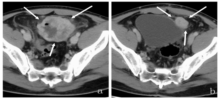
图 4 腹膜后GIST
A: 腹膜后肿块,病理为GIST,因粘连较多,仅取病理,未区分危险程度;治疗前静脉期肿瘤CT值为87 Hu,肿瘤最长径6.82 cm; B: 口服伊马替尼治疗12月后复查,肿瘤出现液化,静脉期肿瘤CT值为60 Hu(下降幅度31.03%),肿瘤最长径为6.35 cm(下降幅度6.89%),符合PR标准.
Figure 4. Retroperitoneal GIST
表 1 GIST患者经MSCT随访情况
Table 1. MSCT follow up GIST patients
治疗方式 MSCT随访频率 中位随访时间 随访次数(次) 总计随访次数(次) 手术患者 极低危险(n=6) 6月/次 9月 1~3 13 低危险(n=18) 6月/次 12月 1~4 41 中危险(n=7) 3~6月/次 12月 2~6 20 高危险(n=3) 3~6月/次 12月 2~7 9 分子靶向治疗 未区分(n=8) 每3月/次 12月 3~8 32 GIST:胃肠间质瘤;MSCT:多层螺旋CT.  下载: 导出CSV
表 2 GIST患者经MSCT随访的肿瘤最长径与静脉期肿瘤CT值比较
Table 2. Comparison of CT value of longest tumor diameter and venous tumor in GIST patients after MSCT follow-up(%, Mean±SD)
MSCT监测项目 基线 第1次随访 第2次随访 末次随访 肿瘤最长径(cm) 6.60±1.19 6.51±1.28 6.40±1.69 6.28±1.98 与基线比较百分比(%) - 98.35±3.26 96.51±4.68 94.72±5.19 静脉期肿瘤CT值(Hu) 73.38±6.26 70.50±5.26 67.38±8.81 59.88±11.13 与基线比较百分比(%) - 96.58±9.93 92.35±13.79 81.60±17.72
下载: 导出CSV
表 3 GIST患者经MSCT随访结果
Table 3. MSCT follow-up results of GIST patients (n, n=8)
随访次数 CR PR SD PD DCR[n(%)] 第1次 0 1 5 2 6(75.0) 第2次 0 2 4 2 6(75.0) 末次随访 0 4 1 3 5(62.5) CR:完全缓解;PR:部分缓解;PD:疾病进展;SD:病情稳定.
下载: 导出CSV
表 4 GIST转移灶情况
Table 4. GIST metastases
转移部位 转移病灶数量 病灶内是否出现液化坏死 单发 多发 是 否 肝脏转移(n=4) 0 4 3 2 腹腔转移(n=2) 1 1 1 1 多处转移(n=1) 0 2 1 0 盆腔转移(n=2) 0 1 1 0 总计(n=9) 1 8 6 3
下载: 导出CSV
-
[1] 于恒, 杜尚策, 徐恩, 等. 影响胃肠道间质瘤预后因素的研究进展[J]. 中华普外科手术学杂志: 电子版, 2020, 14(1): 97-100. doi: 10.3877/cma.j.issn.1674-3946.2020.01.028 [2] 崔仪, 韩振国. 胃肠道间质瘤治疗的研究进展[J]. 中国医药导报, 2020, 17(1): 42-5. https://www.cnki.com.cn/Article/CJFDTOTAL-YYCY202001011.htm [3] Bard V, Menasherov N, Morgenstern S, et al. Gastrointestinal stromal tumor of stomach: a gentle enemy of the surgeon. our experience in confronting the disease[J]. Surg Laparosc Endosc Percutaneous Tech, 2016, 26(5): 406-9. doi: 10.1097/SLE.0000000000000317 [4] 林振孟, 魏晟宏, 叶再生, 等. 评估不可切除或转移性胃肠间质瘤行伊马替尼治疗后CT征象变化临床意义研究[J]. 中国实用外科杂志, 2018, 38(5): 551-4. https://www.cnki.com.cn/Article/CJFDTOTAL-ZGWK201805015.htm [5] Shinagare AB, Jagannathan JP, Kurra V, et al. Comparison of performance of various tumour response criteria in assessment of regorafenib activity in advanced gastrointestinal stromal tumours after failure of imatinib and sunitinib[J]. Eur J Cancer, 2014, 50(5): 981-6. doi: 10.1016/j.ejca.2013.11.037 [6] 中国医师协会外科医师分会胃肠道间质瘤诊疗专业委员会, 中华医学会外科学分会胃肠外科学组. 胃肠间质瘤规范化外科治疗中国专家共识(2018版[) J]. 中国实用外科杂志, 2018, 38(9): 965-73. https://www.cnki.com.cn/Article/CJFDTOTAL-ZGWK201809001.htm [7] Boikos SA, Pappo AS, Killian JK, et al. Molecular subtypes of KIT/ PDGFRA wild-type gastrointestinal stromal tumors: a report from the national institutes of health gastrointestinal stromal tumor clinic [J]. JAMA Oncol, 2016, 2(7): 922-8. doi: 10.1001/jamaoncol.2016.0256 [8] Choi H, Charnsangavej C, Faria SC, et al. Correlation of computed tomography and positron emission tomography in patients with metastatic gastrointestinal stromal tumor treated at a single institution with imatinib mesylate: proposal of new computed tomography response criteria[J]. J Clin Oncol, 2007, 25(13): 1753- 9. doi: 10.1200/JCO.2006.07.3049 [9] 蒋颖. 胃肠道间质瘤术前及术后诊断的研究进展[J]. 南通大学学报: 医学版, 2020, 40(3): 272-5. https://www.cnki.com.cn/Article/CJFDTOTAL-NTYX202003021.htm [10] El-Menyar A, Mekkodathil A, Al-Thani H. Diagnosis and management of gastrointestinal stromal tumors: an up-to-date literature review [J]. J Cancer Res Ther, 2017, 13(6): 889-900. http://www.ncbi.nlm.nih.gov/pubmed/29237949 [11] Chen P, Song T, Wang X, et al. Surgery for duodenal gastrointestinal stromal tumors: a single-center experience[J]. Dig Dis Sci, 2017, 62 (11): 3167-76. doi: 10.1007/s10620-017-4742-4 [12] Herzberg M, Beer M, Anupindi S, et al. Imaging pediatric gastrointestinal stromal tumor (GIST)[J]. J Pediatr Surg, 2018, 53(9): 1862-70. doi: 10.1016/j.jpedsurg.2018.03.022 [13] 汪云, 刘洋. 胃肠道间质瘤的多层螺旋CT分析[J]. 医学影像学杂志, 2020, 30(3): 506-9. https://www.cnki.com.cn/Article/CJFDTOTAL-XYXZ202003046.htm [14] Khoshnood A. Gastrointestinal stromal tumor - A review of clinical studies[J]. J Oncol Pharm Pract, 2019, 25(6): 1473-85. doi: 10.1177/1078155219846955 [15] Nishida T, Blay JY, Hirota S, et al. The standard diagnosis, treatment, and follow-up of gastrointestinal stromal tumors based on guidelines [J]. Gastric Cancer, 2016, 19(1): 3-14. doi: 10.1007/s10120-015-0526-8 [16] 郁文英, 姚素, 孙婧, 等. 经腹与腔内超声联合检查与增强CT在胃肠间质瘤分子靶向药物疗效评估中的对比研究[J]. 重庆医科大学学报, 2020, 45(8): 1218-22. https://www.cnki.com.cn/Article/CJFDTOTAL-ZQYK202008026.htm [17] 黄婧颖, 张渺娜, 王新立, 等. 胃肠道间质瘤的MSCT表现与病理危险程度分级的关系[J]. 中国中西医结合影像学杂志, 2019, 17(1): 44-7, 51. doi: 10.3969/j.issn.1672-0512.2019.01.013 [18] 吕毛古, 秦叔逵, 吴建伟, 等. 多层螺旋CT评价伊马替尼治疗胃肠间质瘤疗效的价值[J]. 临床肿瘤学杂志, 2009, 14(6): 524-9. doi: 10.3969/j.issn.1009-0460.2009.06.010 [19] Cavnar MJ, Seier K, Curtin C, et al. Outcome of 1000 patients with gastrointestinal stromal tumor (GIST) treated by surgery in the preand post-imatinib eras[J]. Ann Surg, 2021, 273(1): 128-38. doi: 10.1097/SLA.0000000000003277 [20] Lin JX, Chen QF, Zheng CH, et al. Is 3-years duration of adjuvant imatinib mesylate treatment sufficient for patients with high-risk gastrointestinal stromal tumor? A study based on long-term followup[J]. J Cancer Res Clin Oncol, 2017, 143(4): 727-34. doi: 10.1007/s00432-016-2334-x [21] 徐锦锋, 邱士军, 宋裕娣, 等. 多层螺旋CT对胃肠道间质瘤的诊断价值及疗效评价[J]. 南方医科大学学报, 2010, 30(4): 875-7. https://www.cnki.com.cn/Article/CJFDTOTAL-DYJD201004059.htm [22] 刘芸, 方维东, 张姣, 等. 小肠间质瘤组织中Ki-67表达与MSCT征象、危险分级的相关性[J]. 分子影像学杂志, 2021, 44(4): 632-8. doi: 10.12122/j.issn.1674-4500.2021.04.11 -

 点击查看大图
计量
- 文章访问数: 261
- HTML全文浏览量: 151
- PDF下载量: 8
- 被引次数: 0