

 Find Duplicates
 Check Document
 Submission(new)
 Experts Office
 Editorial Office
2021 Vol. 44, No. 4
column
Display Method:
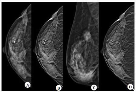
2021, 44(4): 567-573.
doi: 10.12122/j.issn.1674-4500.2021.04.01
Abstract:
ObjectiveTo investigate the relationship between molecular subtypes of breast cancer and the X-ray signs of digital breast tomosynthesis (DBT). MethodsThe DBT images and pathological data of 153 patients with breast cancer were retrospectively analyzed. According to the expression levels of estrogen receptor, progesterone receptor and human epidermal growth factor receptor 2 (HER2), they were divided into hormone receptor (HR) positive group (n=90), HER2 positive group (n=30) and triple negative group (n=33). DBT signs of different molecular types were compared according to the imaging criteria defined by ACR breast reporting and data system (BI-RADS). ResultsThe histological grade and Ki67 expression level in HER2+ and triple negative groups were higher than those in HR + group (P < 0.05). There were no significant differences among the three groups in simple mass, simple calcification, mass with calcification, asymmetries and architectural distortion (P > 0.05). The incidence of round/oval mass in HER2+ group and triple negative group was higher than that in HR+ group (P= 0.003). The incidence of lobulation in HR + group and HER2+ group was higher than that in triple negative group (P < 0.001). The incidence of spiculated sign in HR + group was higher than that in HER2 + and triple negative groups (P < 0.001). The incidence of fine line branched calcification and linear/segmental distribution in HER2+ group was higher than that in HR + and triple negative groups (P < 0.001). The incidences of architectural distortion of the surrounding glands and trabecular thickening in HER2 + group and triple negative group were higher than those in HR + group (P < 0.05). ConclusionThe molecular subtypes of breast cancer are related to the imaging signs of DBT. Understanding these signs is helpful to predict the molecular subtypes of breast cancer.

2021, 44(4): 574-582.
doi: 10.12122/j.issn.1674-4500.2021.04.02
Abstract:
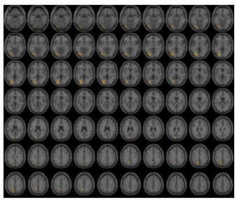
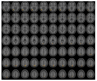
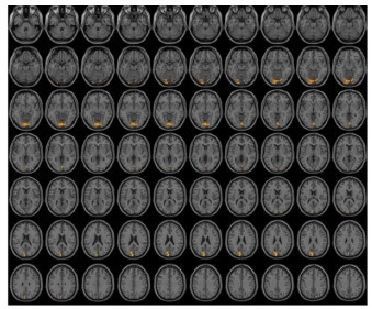
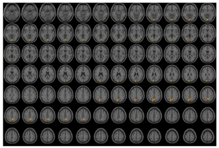
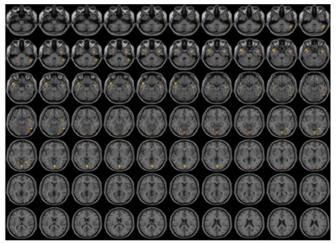
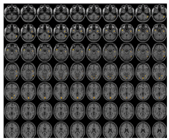
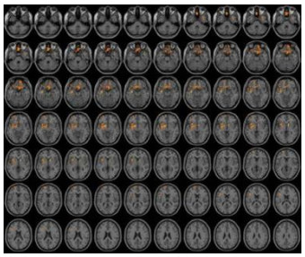
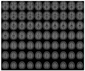
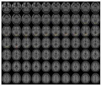
ObjectiveTo explore the functional network changes in patients with acute ischemic stroke in non-dominant cerebral hemisphere. MethodsIn this trial, 15 healthy subjects (HSs) were recruited as controls, 15 subjects with middle cerebral artery acute infarction in non-dominant cerebral hemisphere were recruited into stroke group. Rs-fMRI data of subjects were obtained by Siemens 3.0 T superconducting MRI. It was based on Matlab 2012a platform, using DPABI software for data analysis and mapping with functional connectivity as an outcome indicator. ResultsFinally, the changes of functional connectivity changes (P < 0.005) in ten subjects were as follows: Centered to the seed region of the dorsolateral superior frontal gyrus, FC increased at the left posterior cingulate cortex(T=6.5173). Centered to the seed region of the right orbital superior frontal gyrus, FC increased at the left BA18(T=6.0674). Centered to the seed region of the Rolandic_Oper_L(ALL), FC increased at the cortex around the left fissura calcarina(T=5.7831). Centered to the seed region of the right intraorbital superior frontal gyrus, FC increased at the cortex around the left lobule fusiform(T=5.7361). Centered to the seed region of the left insular lobe, FC increased at the cortex around the left lobule fusiform(T=4.5436). Centered to the seed region of the right insular lobe, FC increased at the cortex around the right posterior cerbellar lobe(T=4.9352). Centered to the seed region of the left cuneus, FC increased at the cortex around the left insular lobe(T=5.6924). Centered to the seed region of the right lobule fusiform, FC increased at the cortex around the left gyri orbitales(T=8.4505). Centered to the seed region of the left precuneus, FC increased at the cortex around the left lenticular putamen(T=5.1894). Centered to the seed region of the left paracentral lobule, FC increased at the cortex around the left lenticular putamen(T=7.9109). Centered to the seed region of the left gyri temporales transversi, FC increased at the cortex around the left gyrus lingualis(T=5.9146). ConclusionSpecific functional network changes present in patients with acute ischemic stroke in non-dominant cerebral hemisphere, and function connection enhancement among the brain regions related to visual, uditory, higher cognition and motor.
2021, 44(4): 583-588.
doi: 10.12122/j.issn.1674-4500.2021.04.03
Abstract:
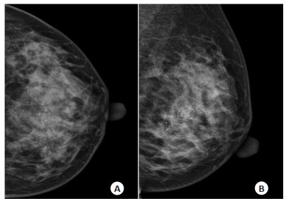
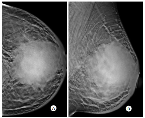
ObjectiveTo investigate the correlation between the ultrasonic features and immunohistochemical parameters in breast cancer. MethodsThe data of 710 cases of breast cancer proved by pathology were collected retrospectively. The sonographic features (the maximum diameter, position, aspect ratio, internal echo, posterior echo, boundary, margin, shape, calcification, Alder blood flow grading, lymph node metastasis) and immunohistochemical parameters (ER, PR, HER-2, Ki-67) were recorded. The correlation between ultrasonic features and immunohistochemical parameters was analyzed. ResultsThe positive rates of ER, PR, HER-2 and Ki-67 were 72.5%, 64.1%, 61.0% and 80.3% in 710 breast cancer masses, respectively. The positive rate of ER and PR was higher in the maximum diameter ≤2 cm, and that of Ki-67 was higher in the maximum diameter > 2 cm. When ER was positive, the ratio of breast cancer was more than 1(P < 0.001). When PR was positive, the ultrasonic features of breast cancer were irregular shape, unclear boundary, attenuation or disappearance of Posterior Echo. When HER-2 and Ki-67 were positive, the ratio of vertical to horizontal was≤1(P < 0.001), and the axillary lymph node metastasis was more likely. ConclusionThe ultrasonic features of breast cancer are correlated with the expressions of ER, PR, HER-2 and Ki-67, which can reflect the biological behavior of the tumor. It provides a powerful reference for the choice of clinical treatment plan and the evaluation of postoperative prognosis.
2021, 44(4): 589-593.
doi: 10.12122/j.issn.1674-4500.2021.04.04
Abstract:
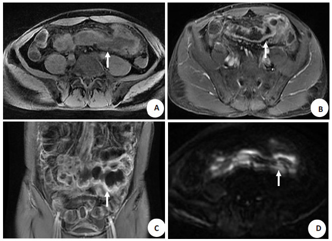
ObjectiveTo establish a rabbit sciatic nerve crush injury model suitable for MRI monitoring, and discuss the selection of b value of diffusion kurtosis imaging (DKI). MethodsTwenty-seven healthy New Zealand white rabbits were selected. The sciatic nerve injury model was established with self-made flat forceps with a width of about 8mm. The right posterior limb was selected as the injury side and the left was the sham surgical side. DKI was performed before and 1 d, 3 d, 1 week, 2 weeks, 4 weeks and 8 weeks after injury, with b values of 0, 750, 1500 s/mm2 and 0, 1000, 2000 s/mm2, respectively. Two rabbits were randomly selected at each time point for pathological examination. ResultsThe parameters of DKI1500 and DKI2000 showed similar trends. Both FA1500 and FA2000 decreased to the lowest level on the first day and then continued to increase at 3-8 weeks, with statistically significant differences among groups at each time point (P < 0.05). The MK1500 of both sides was significantly decreased on the first day, and then slowly increased. The differences in the MK1500 of both sides at the second week (P=0.022), the fourth week (P=0.018), the sixth week (P=0.016) and the eighth week (P=0.016) after injury were significant. The difference of MK2000 between groups was significant only at the fourth week (P=0.002). There was no significant difference between Rk and AK at most time points. ConclusionUsing flat forceps to clamp the middle segment of the rabbit sciatic nerve to make the injury model can facilitate the direct monitoring and quantitative measurement of the injured nerve by MRI. A maximum b value of 1500 s/mm2 for DKI imaging may be more suitable than 2000 s/mm2.
2021, 44(4): 594-601.
doi: 10.12122/j.issn.1674-4500.2021.04.05
Abstract:
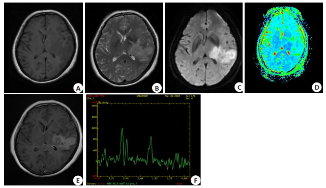
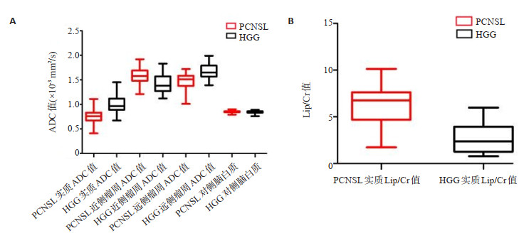
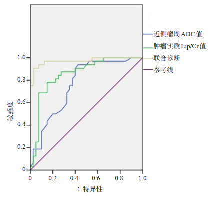
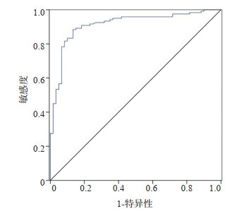
ObjectiveTo investigate the application value of the apparent diffusion coefficient (ADC) value of different tumor regions combined with the relative ratio of MRS metabolites in the diagnosis and differentiation of primary central nervous system lymphoma (PCNSL) under Logistic regression model. MethodsThe characteristics of conventional MRI, DWI and MRS in 32 cases of PCNSL and 40 cases of HGG confirmed by clinical and pathology were analyzed and detected. The ADC values of tumor parenchyma, proximal peritumor (edema area within 1cm around the tumor), distal peritumor (edema area beyond 1cm around the tumor) and contralateral normal white matter were measured. The average values were calculated. Two-dimensional multi-voxel 1H MRS imaging was used to measure and calculate the relative ratio of metabolites. The differences of ADC values and the relative ratio of MRS metabolites in different regions of the two groups were analyzed. The indicators with higher differential value of PCNSL and HGG were screened by binary logistic regression analysis, and the diagnostic efficacy of each indicator alone and combined in the diagnosis of tumors in the two groups was compared by ROC curve analysis. ResultsThe overall differences in ADC values of tumor parenchyma, proximal peritumor, distal peritumor and contralateral white matter between PCNSL group and HGG group were significant within the group (all P < 0.001), and the pair comparison was statistically rising significant (all P < 0.05).The variation trend of ADC values in different areas of PCNSL group and HGG group were as follows: first and then falling parabola type and breaking line rising type, respectively. The ADC value of tumor parenchyma had the highest diagnostic efficacy (AUC=0.880) for the differentiation of tumor parenchyma and tumor parenchyma. Choline complex (Cho)/creatine (Cr), Cho/ N-acetyl aspartic acid (NAA) and lipid (Lip)/Cr values in different regions of HGG group and PCNSL group were compared within the group (P < 0.001). Pair comparison within groups: the values of Cho/Cr, Cho/NAA and Lip/Cr in tumor parenchyma of PCNSL group and HGG group were all higher than those of proximal peritumor, distal peritumor and contralateral white matter. The LIP/CR in the tumor parenchyma between the two groups was significantly different (t=6.418, P < 0.001).Logistic regression equation model was as follows: Logistic(P) =-13.190 + 1.374 × ADC value of proximal peritumor +0.542×LIP/Cr value of tumor parenchyma. A high level of ADC value of proximal peritumor and LIP/Cr value of tumor parenchyma were risk factors for PCNSL. Under the Logistic regression equation model, the area under the joint diagnosis curve was the largest (AUC=0.973). The sensitivity and specificity of the two groups were 90.6% and 97.5% (P < 0.001), the combined diagnostic efficacy was the highest. ConclusionThe measurement technique of ADC value and relative ratio of different metabolites of MRS can provide pathological basis and molecular imaging basis for noninvasive diagnosis of tumor and peri-tumor micro-invasion state. Combined diagnosis under Logistic regression model can effectively improve the diagnostic efficiency, thus providing reliable basis for early diagnosis, differentiation and treatment of the two.

2021, 44(4): 602-607.
doi: 10.12122/j.issn.1674-4500.2021.04.06
Abstract:
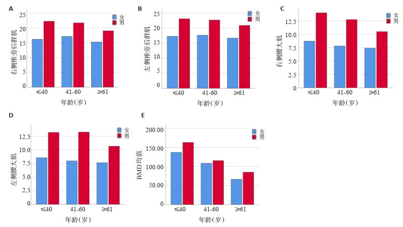
ObjectiveTo investigate the age-related changes of bone mineral density (BMD) and paravertebral muscle and their correlation by quantitative CT. MethodsThe patients who underwent QCT bone mineral density examination in our hospital from June to November 2020 were retrospectively analyzed. Quantitative CT was used to measure the cross-sectional area of the paravertebral posterior muscle groups (erector spinae and multifidus) at the central level of L3. The Philips Nebula Medical Image Postprocessing Platform V9.0 was used to measured the cross-sectional area of the psoas major (paraspinal anterior muscle group) in the L4-L5 intervertebral space. The change of paravertebral muscle and BMD with age and their correlation were analyzed. ResultsThe content of the paraspinal posterior muscle (cross-sectional area) grew considerably in women between the ages of 40 and 60, but declined with age in males, whereas the content of the right psoas major muscle decreased with age. The content of the left psoas major muscle increased significantly between the ages of 40 and 60 in men and under 40 in women. The paravertebral muscle content of males was greater than that of females with the increase of age, and the BMD also decreased with the increase of age. The BMD of males was greater than that of females(male r=-0.625, P= 0.000; female r=-0.600, P=0.000).The psoas major muscle and paravertebral posterior muscle group were positively correlated with BMD, the correlation was higher in male than in female, and the right was higher than the left. (Right paravertebral posterior muscle in male r=0.432, P=0.009;The left paravertebral posterior muscle r=0.401, P=0.017;Right psoas major r=0.510, P=0.002; left psoas major r=0.447, P=0.007;Right paravertebral posterior muscle in female r=0.319, P=0.000; The left paravertebral posterior muscle r=0.276, P=0.000; Right psoas major r=0.279, P=0.000; left psoas major r=0.220, P=0.004). ConclusionThe paraspinal muscle content in men is higher than in women. BMD is inversely proportional to age. BMD is favorably linked with the psoas major and posterior paraspinal muscles. The male-to-female correlation is greater than the female-to-female correlation. The right side of the building is higher than the left. Men's paraspinal group muscles have a lower correlation with BMD than women's psoas muscles, whereas women's paraspinal group muscles have a greater correlation with BMD than men's psoas muscles relevance.
2021, 44(4): 608-611.
doi: 10.12122/j.issn.1674-4500.2021.04.07
Abstract:
ObjectiveTo analyze MSCT and MRI findings of intracranial anaplastic meningioma, compare with the pathological results, and improve the diagnosis of the disease. MethodsWe collected cases of intracranial anaplastic meningioma confirmed by pathology in our hospital from January 2011 to December 2019. Seventeen patients were collected, including 15 males and 2 females. Aged from 1 to 67 years old, with a bipolar distribution, the patients included 7 patients under 10 years old and 7 patients over 50 years old. The imaging data of intracranial anaplastic meningioma cases were collected, including MSCT scan, T1WI, T2WI, FLAIR and enhanced examination of MRI. Their imaging characteristics were analyzed. ResultsThere were 16 tumors mainly located in the supra, 1 case was under the curtain, the main body was located in the fourth ventricle. The morphology of this group was divided into 2 types, 11 cases were cystic-solid and 6 cases were solid. The shape of cystic-solid tumors was mostly irregular. The ratio of cystic parts to solid parts was quite different. The tumor borders were unclear, and the peritumoral edema was light. Solid tumors were mainly round or lobulated, with clear borders, often accompanied by light or moderate peritumoral edema. The MSCT scan showed that the solid components of cystic-solid tumors and solid tumors were mainly of equal or slightly higher density. The cystic components of cystic-solid tumors showed cerebrospinal fluid-like density changes. MRI scans showed solid components of cystic-solid tumors and solid tumors with slightly lower or lower signals at T1WI, equal or slightly higher signals at T2WI and FLAIR, and enhanced MRI scans showed solid components of cystic-solid tumors and solid tumors were significantly unevenly enhanced, and the cyst wall of the cystic-solid tumors was obviously strengthened in a ring shape. There was no enhancement in the cyst. ConclusionMSCT and MRI manifestations of intracranial anaplastic meningioma had certain characteristics. Combining with the patient's gender, age and clinical history, it has higher clinical significance for its diagnosis and differential diagnosis.
2021, 44(4): 612-617.
doi: 10.12122/j.issn.1674-4500.2021.04.08
Abstract:
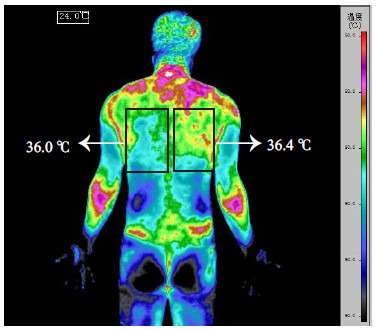
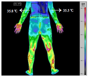
ObjectiveTo measure the temperature in the affected area using infrared thermal imaging.The warm area and the cold area were divided into two groups for silver needle heat conduction and nerve block treatment, then the efficacy was compared to choose the best treatment method for back myofascial pain. MethodsAfter patients who diagnosed with myofascial pain had been examed by infrared thermal imaging, we measured the temperature of the affected side and the relative healthy side(△T=the temperature of affected area - the temperature of healthy area). Fifty cases were selected in both warm group (group W, △T≥0.2 ℃) and the cold group (group C, △T≤-0.2 ℃). Then each group was randomly divided into 2 groups (WS, WN, CS, CN) with 25 patients. Group WS and CS received silver needle thermal conduction therapy (S) once in a month, and group WN and CN received nerve block treatment (N) once every 5 days, 2-3 times in total depends on how much the pain relieved. The change of patients' pain were inquired and compared with Visual Analogue Scale (VAS) at day 0 (before treatment), 1, 6, 15, 30 and 180 after the treatment. ResultsDuring the follow-up period, VAS of patients in group W at day 1, 30 and 180 showed that group WS was significantly higher than group WN. VAS of patients in group C at day 15, 30 and 180 showed that group CS was significantly lower than group CN(P < 0.05). ConclusionInfrared thermal imaging is used to judge the temperature of affected area, nerve block therapy used in warm and silver needle therapy is used in cold.
2021, 44(4): 618-623.
doi: 10.12122/j.issn.1674-4500.2021.04.09
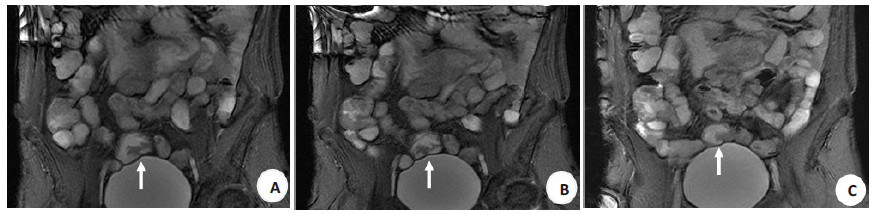
Abstract:
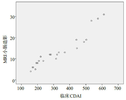
ObjectiveTo explore the correlation between magnetic resonance imaging enterography (MRE)on the classification of Crohn's disease activity and clinical Crohn's disease activity index (CDAI). MethodsTwenty-two patients with active Crohn's disease diagnosed clinically or pathologically and graded by clinical activity were examined by MRE. The scanned images were scored subjectively by 2 deputy director or above diagnostic physicians, and the classification of image activity was conducted according to their scores. Then the Results were compared with the clinical classification Results of the patient, and the consistency was observed. ResultsAll 22 patients with active Crohn's disease completed MRE, of which 7 were mild patients with a score of 6.5±1.7 and a median of 6.0. Nine were moderate patients with a score of 11.9±1.6. The median was 12.0 points; 6 severe patients had a score of 24.0±5.4 points and the median was 23.5 points. Spearman correlation analysis was used to compare Crohn's disease activity grading criteria with clinical CDAI calculation methods. The correlations were r=0.873、0.826、0.899, respectively(P < 0.05). ConclusionMRE has a significant correlation with CDAI. It can assess the activity grade of active Crohn's disease accurately and provide reference for clinical treatment.
2021, 44(4): 624-631.
doi: 10.12122/j.issn.1674-4500.2021.04.10
Abstract:
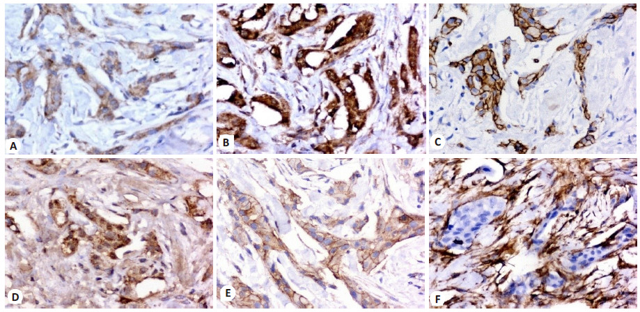
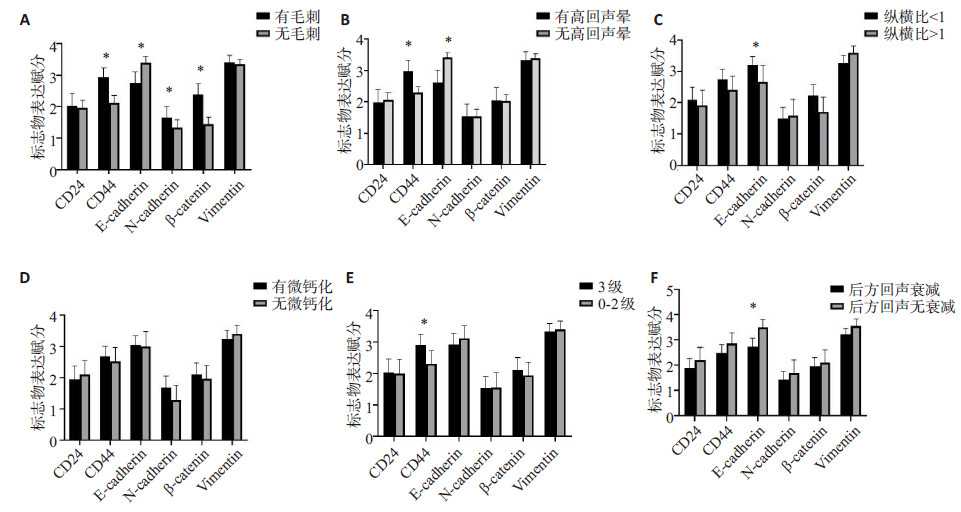
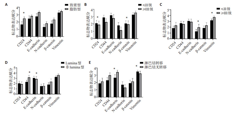
ObjectiveTo investigate the correlation and significance of ultrasonographic signs of breast cancer with the expression level of tumor stem cells and epithelial mesenchymal transformation markers. MethodsA total of 98 tissue specimens from patients undergoing breast cancer surgery in our hospital were selected. The corresponding breast ultrasound signs of each patient were analyzed, including whether there were burrs around the mass, whether there was hyperechoic halo at the edge, aspect ratio, posterior echo, microcalcification, and internal blood flow imaging grade. The expression of CD24, CD44, E-cadherin, N-cadherin, β-catenin, Vimentin were detected by immunohistochemistry. The correlation between these indexes and ultrasound findings were analyzed. Other factors that may affect the expression of tumor stem cells and epithelial mesenchymal transition markers were analyzed. Multivariate logistic regression analysis was used to analyze the correlation between the expression of tumor stem cells and epithelial mesenchymal transition markers. ResultsThe presence of burr around the tumor was significantly different with the expression levels of CD44, E-cadherin, N-cadherin and β-catenin (P < 0.05). The expression level of CD44 and E-cadherin were significantly different with the presence of hyperechoic halos at the edge of the mass (P < 0.05).The changes of aspect ratio and posterior echo of the tumor were significantly different with the expression level of E-cadherin (P < 0.05).There were significant differences in blood flow grade, gland type and CD44 expression level (P < 0.05). Gland background type was an independent factor associated with CD24 expression. Blood flow and grouping according to estrogen receptor expression were independent correlative factors of CD44 expression. The presence of hyperechoic halos, posterior echo characteristics of the mass, and axillary lymph node metastasis were independent related factors of E-cadherin expression. Clinical staging was an independent factor associated with N-cadherin expression. The presence of burr on the edge was an independent factor related to β-catenin expression. ConclusionUltrasound signs of breast cancer are associated with the expression of tumor stem cells and epithelial mesenchymal transformation markers. Ultrasound signs can be used as a way to noninvasively predict the expression level of tumor stem cells and epithelial mesenchymal transformation markers in breast cancer patients, and can provide more evidence for predicting the potential invasion ability of breast cancer.
2021, 44(4): 632-638.
doi: 10.12122/j.issn.1674-4500.2021.04.11
Abstract:
ObjectiveTo investigate the correlation of Ki-67 expression with MSCT findings and risk grade in small intestinal stromal tumor (SIST). MethodsThe clinicopathological, immunohistochemical and imaging data of 87 cases of SIST confirmed by surgery and pathology were retrospectively analyzed to explore the relationship between Ki-67 expression and CT findings and risk classification. ResultsIn one-way ANOVA, Ki-67 expression was significantly different in location, shape, size, invasion and metastasis, pathological group and mitotic image (P < 0.05); There was no significant difference in gender, age, clinical features, cystic degeneration or necrosis, ulcer, growth pattern, whether it was supplied by superior mesenteric artery, enhancement degree in arterial phase, enhancement mode in venous phase or delayed phase (P > 0.05). The Results of multivariate logistic regression analysis showed that the expression of Ki-67 was 5% to 10% in subgroups, the size was statistically significant (P < 0.05), and the control was less than 2 cm, of which the or value of 2-5 cm group was 2.179, or value of 5-10 cm group was 4.345, or value of greater than 10 cm group was 7.698. The or value of ileum was 17.622 in greater than 10% subgroup (P < 0.05), the or value of 2-5 cm subgroup was 2.179, 5-10 cm subgroup was 4.345, and greater than 10 cm subgroup was 7.698. ConclusionThe expression of Ki-67 in SIS is correlated with MSCT signs, and the expression of Ki-67 is higher in high-risk group and tissues with larger mitosis number, which has certain guiding significance for the selection of clinical treatment and prognosis of SIST.
2021, 44(4): 639-642.
doi: 10.12122/j.issn.1674-4500.2021.04.12
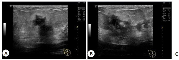
Abstract:
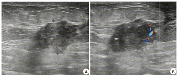
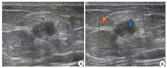
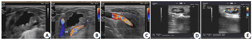
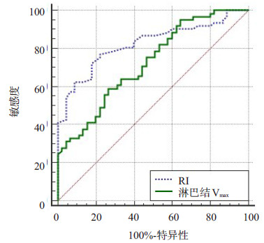
ObjectiveTo explore the application value of high-frequency ultrasound and its image features in the diagnosis of superficial lymph node lesions. MethodsThe clinical data of 106 patients with superficial lymph node lesions admitted to the hospital between June 2019 and February 2021 were reviewed. All patients underwent high-frequency ultrasound examination, and pathological examination Results were used as the gold standard. The patients were grouped based on the pathological diagnosis of benign and malignant results. The features of high-frequency ultrasound images [lymph node long diameter (L)/short diameter (S), vascularization score (VS), lymph node maximum blood flow velocity (Vmax), minimum flow velocity (Vmin), resistance index (RI)] were compared between benign group (n=45) and malignant group (n=61). The differences in Results of high-frequency ultrasound and pathological examination were compared. ROC curve was used to analyze the value of high-frequency ultrasound in evaluating superficial lymph node lesions. ResultsThe difference in lymph node L/S < 2, VS, lymph node Vmax, and RI between benign group and malignant group were significant(P < 0.05). The accuracy rate, sensitivity, specificity, positive predictive value and negative predictive value of high-frequency ultrasound in the diagnosis of superficial lymph node lesions were 85.85%, 90.16%, 80.00%, 85.94% and 85.71%. ROC curve Results showed that the diagnostic value of lymph node Vmax and RI were both high (P < 0.05). ConclusionHigh-frequency ultrasound is of higher value in evaluating superficial lymph node lesions, and can distinguish benign and malignant superficial lymph node lesions.
2021, 44(4): 643-647.
doi: 10.12122/j.issn.1674-4500.2021.04.13
Abstract:
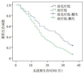
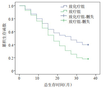
ObjectiveTo analyze the effect of intensity modulated radiation therapy combined with concurrent calcium folinate (CF) regimen on changes of levels of insulin-like growth factor 1 and vascular endothelial growth factor and prognosis of esophageal squamous cell carcinoma. MethodsA total of 110 patients with esophageal squamous cell carcinoma admitted to department of oncology of the hospital were selected between January 2015 and January 2018. They were randomly divided into radiotherapy group (treated with intensity modulated radiation therapy) and chemoradiotherapy group (given intensity modulated radiation therapy combined with concurrent CF chemotherapy regimen), with 55 cases in each group. The short-term efficacy, serum tumor markers, toxic and side effects and survival status were compared between the two groups after treatment. ResultsThe difference in the short-term efficacy between chemoradiotherapy group and radiotherapy group after treatment was not significant (P>0.05). The levels of insulin-like growth factor 1, vascular endothelial growth factor, squamous cell carcinoma and carcinoembryonic antigen after treatment were significantly decreased in the two groups (P < 0.05). The levels of chemoradiotherapy group were lower than those of radiotherapy group (P < 0.05). The differences in the incidence rates of toxic and side effects between the two groups was not significant(P>0.05). The 2-year and 3-year survival rates were significantly higher in chemoradiotherapy group compared with those in radiotherapy group (P < 0.05). The differences in the median progression-free survival time and overall survival time between the two groups were significant (P < 0.05). ConclusionIntensity modulated radiation therapy combined with concurrent CF chemotherapy regimen has a good short-term efficacy in the treatment of esophageal squamous cell carcinoma. It can reduce the levels of serum tumor markers, do not increase the occurrence of toxic and side effects, improve the survival rate and prolong the survival time of patients.
2021, 44(4): 648-652.
doi: 10.12122/j.issn.1674-4500.2021.04.14
Abstract:
ObjectiveTo investigate the value of dual source CT (DSCT) in the evaluation of in stent restenosis in elderly patients with acute ST segment elevation myocardial infarction (STEMI) after drug-eluting stent treatment and its influencing factors. MethodsA total of 210 elderly patients with STEMI drug-eluting stents who were treated in our hospital from May 2017 to May 2020 were selected. The value of DSCT in the diagnosis of in-stent restenosis were analyzed, the clinical data of patients with in stent restenosis and patency were compared. The Logistic regression was used to analyze the influencing factors. ResultsThirty-two patients were diagnosed with in-stent restenosis by CAG examination, and the incidence of in-stent restenosis was 15.24%. The sensitivity, specificity, accuracy, positive predictive value and negative predictive value of DSCT in the diagnosis of stent stenosis were 91.07%, 95.00%, 94.20%, 82.26% and 97.66%, respectively. The accuracy rate of DSCT in determining the conditions in stents with diameter ≥3.0 mm was significantly higher than that with diameter < 3.0 mm (P < 0.05). The age, proportion of diabetes mellitus, proportion of smoking history, LDL-C, number of stents and Gensini score of patients with stent restenosis were ignificantly higher than those with stent patency (P < 0.05). Logistic regression analysis showed that age, diabetes mellitus, smoking history and Gensini score were risk factors for in-stent restenosis after STEMI drug-eluting stents in the elderly (OR=1.409, 1.754, 1.842 and 2.512, P < 0.05). ConclusionDSCT is of high application value in the assessment of in-stent restenosis after STEMI drug-eluting stent treatment in the elderly, which is influenced by the patient's age, diabetes mellitus, smoking history, and Gensini score.
2021, 44(4): 653-658.
doi: 10.12122/j.issn.1674-4500.2021.04.15
Abstract:
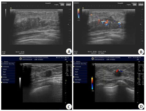
ObjectiveTo explore the value of color Doppler ultrasonography (CDFI) in evaluating the effect of adjuvant chemotherapy before breast cancer surgery. MethodsFrom January 2015 to January 2021, 88 patients with breast cancer who were treated with adjuvant chemotherapy before surgery in our Hospital were selected. According to the effect of chemotherapy, the patients were divided into effective group of 60 cases and ineffective group of 28 cases. The two groups of chemotherapy were compared. The size of the primary lesion, the grading characteristics of blood flow in the lesion, and the characteristics of ultrasound imaging before and after were compared. ResultsBefore chemotherapy, the differences of the length, width, thickness, lesion area, and volume of the lesion between two groups were not significant (P>0.05). After chemotherapy, the length and width of the lesion, thickness, lesion area, and lesion volume in effective group were lower than those of ineffective group (P < 0.05). The measured values of lesion length, width, thickness, lesion area, and lesion volume of the two groups were all lower than those before chemotherapy (P < 0.05). Before chemotherapy, the differences of the morphology, border, hyperechoic band, and posterior echo composition between two groups were not significant (P>0.05). After chemotherapy, the tumor lesions in the effective group had regular morphology and borders. The proportions of clear and no abnormal back echoes were higher than those in the chemotherapy-ineffective group(P < 0.05). Before chemotherapy, there was no difference of the blood flow classification of tumor lesions between two groups (P>0.05). After chemotherapy, the proportion of patients in the effective group with tumor lesion blood flow classification (grade 0+Ⅰ) was higher than that in the ineffective group(P < 0.05). ConclusionColor Doppler ultrasonography can evaluate the effect of adjuvant chemotherapy before surgery for breast cancer from three aspects: lesion size, blood flow grade, and changes in acoustic and image characteristics. It is of great value for guiding clinical treatment and surgery.
2021, 44(4): 659-663.
doi: 10.12122/j.issn.1674-4500.2021.04.16
Abstract:
ObjectiveTo explore the application effect of high-resolution magnetic resonance imaging (HR-MRI) in the rupture risk assessment of unruptured intracranial aneurysms. MethodsThe clinical data of 88 patients (101 aneurysms) diagnosed as intracranial aneurysms by three-dimensional digital subtraction angiography in the hospital were retrospectively analyzed between January 2017 and December 2020. According to whether the artery was ruptured, the patients were divided into ruptured group (n=30, 35 aneurysms) and unruptured group (n=58, 66 aneurysms), and the patients in the unruptured group were classified as enhanced subgroup (n=22, 26 aneurysms) and unenhanced subgroup (n=36, 40 aneurysms). All patients underwent HR-MRI imaging to observe the characteristics of aneurysms in each group. ResultsCompared with the diagnostic result of digital subtraction angiography, the accuracy rate of HR-MRI imaging examination was 79.55%. The enhancement rate of ruptured group was significantly higher than that of unruptured group (P < 0.05). There was no statistical significance in aneurysm location, aneurysm size, presence or absence of wide-necked aneurysm or size ratio between ruptured group and unruptured group (P>0.05). The ratios of ascus and aspect ratio≥2 in ruptured group were significantly higher than those in unruptured group (P < 0.05). The differences in the location of aneurysm, the size of aneurysm, and presence or absence wide-necked aneurysm were not significant between enhanced group and unenhanced group (P>0.05). The ratios of ascuss, aspect ratio≥2, and size ratio≥2 were significantly higher in enhanced group than those in unenhanced group (P < 0.05). ConclusionHR-MRI imaging has a good application effect in the rupture risk assessment of unruptured intracranial aneurysms. The risk of tumor rupture has a certain relationship with the enhancement of the tumor wall. And the factors affecting the enhancement of the tumor wall are ascus status and morphological characteristics of aneurysm such as aspect ratio and size ratio.

2021, 44(4): 664-667.
doi: 10.12122/j.issn.1674-4500.2021.04.17
Abstract:
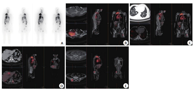
ObjectiveTo investigate the gain value of single photon emission computed tomography and computed tomography (SPECT/CT) in the diagnosis and treatment of differentiated thyroid cancer and the therapeutic effect of high-dose 131Ⅰ. MethodsA total of 125 patients with differentiated thyroid cancer who were treated in our hospital from January 2018 to January 2020 were selected. The patients were performed with thyroidectomy and debridement in our hospital, and SPECT/CT and 131Ⅰ-whole body scan (WBS) were performed. ResultsA total of 682 metastatic foci were finally diagnosed in 125 patients, including 589 metastatic foci taking iodine and 93 metastatic foci not taking iodine. The sensitivity, accuracy and negative predictive values of SPECT/CT in diagnosis of metastases were 89.74%, 88.77% and 46.15%, respectively, which were higher than those of 131Ⅰ-WBS (P < 0.05). There was no significant difference in the specificity and positive predictive value between SPECT/CT and 131Ⅰ-WBS in the diagnosis of metastasis (P>0.05). SPECT/CT changed the treatment plan of 32 patients (25.60%). The total effective rate of high dose 131Ⅰ after operation was 71.20%.There was no significant difference in total treatment rate among patients with different genders, operation methods and body mass index (P>0.05). The treatment total effective rate in patients with age < 50 years old, clinical stage Ⅰ-Ⅱ were significantly higher than the age ≥50, Ⅲ-Ⅳ patients (P < 0.05). ConclusionSPECT/CT is helpful for the diagnosis and treatment of differentiated thyroid cancer, the therapeutic effect of high-dose 131Ⅰ is related to the age and clinical stage of patients.
2021, 44(4): 668-672.
doi: 10.12122/j.issn.1674-4500.2021.04.18
Abstract:
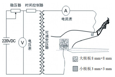
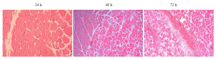
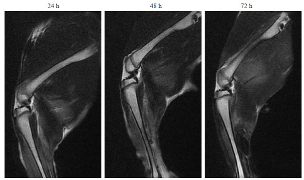
ObjectiveTo explore the evaluation effect of conventional magnetic resonance imaging sequence and multi-B-value diffusion weighted imaging (DWI) sequence on electric shock injury model. MethodsAccording to the electric shock time, 30 rabbits were randomly divided into three groups: Group A (0.2 s), Group B (0.5 s) and Group C (1 s), with 10 rabbits in each group. After electric injury, the limb injury of rabbits was graded according to the standard. H&E staining was used to observe the histopathological changes of lower limb muscles in rabbits at 24 h, 48 h and 72 h after electric injury. The lower limb injuries of rabbits after electric injury at 24 h, 48 h and 72 h were observed by using MRI T2WI plain scan and T1WI-FS enhanced scan. Using DWI scanning, the ADC values of rabbits with different degrees of electrical injury under different B values were observed. ResultsUnder three different electric shock time conditions, 7 rabbits suffered from mild injury, 7 rabbits suffered from moderate injury, 6 rabbits suffered from severe injury and 10 rabbits suffered from severe injury (χ2= 21.486, P=0.002). Histological results showed that there were bleeding, coagulation necrosis, myolysis and inflammatory infiltration in muscle interstitium of lower limbs of rabbits 24 h after electric shock. After 48 h, swelling of muscle interstitial blood vessels and thrombosis were observed. After 72 h, the necrotic tissue of muscle was further enlarged and aggravated. The signals of T2WI plain scan and T1WI-FS enhanced scan showed progressive necrosis of rabbits after electric shock, which was consistent with the histological results. DWI scanning results with different B values showed that ADC values of "light", "medium", "heavy" and "extra-heavy" injuries were significantly different when B values were equal to 600 s/mm2 and 800 s/mm2 (P < 0.01). ConclusionT2WI plain scan and T1WI-FS enhanced MRI combined with multi-B-value DWI sequence scan have important reference significance for early diagnosis of clinical electric shock injury.
2021, 44(4): 673-677.
doi: 10.12122/j.issn.1674-4500.2021.04.19
Abstract:
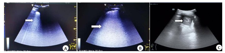
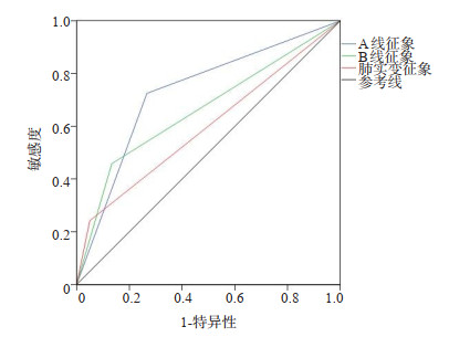
ObjectiveTo explore the clinical value of bedside ultrasound in diagnosing elderly patients with community-acquired pneumonia. MethodsA total of 120 elderly patients with community-acquired pneumonia admitted to Chongqing Dazu District People's Hospital from August 2019 to June 2020 were selected as the infection group and 60 healthy volunteers as the control group. The patients received bedside ultrasound examinations. The lung ultrasound signs (A-line signs, B-line signs, lung consolidation signs) and lung ultrasound scores, and stratified comparison according to the patient's condition between two groups were compared. ROC curve was used to analyze the value of pulmonary ultrasound score in the diagnosis of pulmonary infection. ResultsThe detection rate of A-line signs, B-line signs, lung consolidation signs and lung ultrasound scores of the infection group were significantly higher than those of the control group(P < 0.05). The detection rate of lung consolidation signs and lung ultrasound scores of patients in the infection group with a CPIS score >6 points were significantly higher than those of patients with a CPIS score ≤6 points(P < 0.05). The A-line signs and B-line signs of patients in the infection group with a CPIS score >6 were compared with those with a CPIS score of ≤6, (P>0.05). The detection rate of B-line signs, lung consolidation signs, and lung ultrasound scores of patients in the death group were significantly higher than those of the surviving patients (P < 0.05). The sensitivity of lung ultrasound scoring to diagnose lung infection was 89.87%, specificity was 83.36%, missed diagnosis rate was 10.13%, misdiagnosis rate was 16.64%, and the AUC value of the area under the ROC curve was 0.913. ConclusionBedside ultrasound examination has high clinical value for diagnosing elderly patients with community-acquired pneumonia and judging their condition.
2021, 44(4): 678-681.
doi: 10.12122/j.issn.1674-4500.2021.04.20
Abstract:
ObjectiveTo investigate the value of 64 slice spiral CT (MSCT) virtual endoscopy combined with CT target scan in the diagnosis of central small cell lung cancer (SCLC). MethodsA total of 72 children with central SCLC in our hospital from January 2018 to February 2021 were selected as the observation group. Seventy patients with central benign lesions were selected as the control group. The MSCT virtual endoscopy and CT target scanning images were analyzed. ResultsThe age of observation group was significantly higher than that of control group (P < 0.05). The proportions of burr sign, pleural depression sign and vacuolar sign in the CT target scanning features of the observation group were significantly higher than those in the control group (P < 0.05). There was no significant difference between the observation group and the control group in the proportion of irregular morphology and air bronchial sign of the CT target scanning features (P>0.05). The consistent Kappa value of MSCT virtual endoscopy and fiberoptic bronchoscope was 0.782(P < 0.05). The sensitivity, accuracy and negative predictive values of MSCT virtual endoscopy combined with CT target scan in the diagnosis of central SCLC were significantly higher than those of CT target scan (P < 0.05). There was no significant difference in specificity and positive predictive value between MSCT virtual endoscopy combined with CT target scan and CT target scan in the diagnosis of central SCLC (P>0.05). ConclusionMSCT virtual endoscopy combined with CT target scan has a good application value in the diagnosis of central SCLC.
2021, 44(4): 682-685.
doi: 10.12122/j.issn.1674-4500.2021.04.21
Abstract:
ObjectiveTo explore the risk factors of death in elderly patients with hip fracture within 1 year after surgery. MethodsA total of 204 elderly hip fractures patients who underwent surgery treatment from 2016 to 2019 were selected. The clinical data were retrospectively collected, including age, gender, preoperative comorbidities, fracture type, anesthesia method, American Society of Anesthesiologists (ASA) score, preoperative hemoglobin, preoperative albumin, preoperative serum calcium, length of hospital stay were recorded, and death within 1 year after operation as the clinical outcome. Multivariate logistic regression model was used to analyze the independent risk factors of death within 1 year after surgery. ResultsA total of 82 patients died within 1 year after surgery, with a mortality rate of 40.2%. Gender, BMI, fracture type, anesthesia methods, preoperative hemoglobin, preoperative serum calcium and length of hospital stay did not affect the postoperative mortality of the hip in 80- year- old patients, and the difference was not significant (P>0.05). Age, number of preoperative complications, ASA score, and preoperative albumin were the independent risk factors for death within 1 year after surgery in elderly patients with hip fracture. ConclusionThe mortality rate of the elderly patients with hip fracture within 1 year after operation is relatively high. Age, preoperative comorbidities, ASA score, and preoperative albumin are the independent risk factors of 1-year mortality in elderly patients with hip fracture.
2021, 44(4): 686-690.
doi: 10.12122/j.issn.1674-4500.2021.04.22
Abstract:
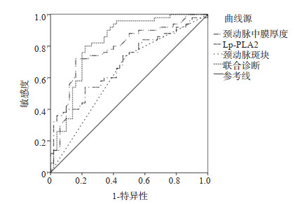
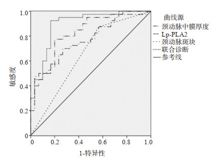
ObjectiveTo investigate the application value of carotid artery color Doppler ultrasound combined with serum lipoprotein associated phospholipase A2 (Lp-PLA2) in high-risk population of coronary heart disease. MethodsA total of 300 patients with high risk of coronary heart disease in our hospital from January 2018 to January 2021 were selected. The differences of carotid artery media thickness, plaque and serum Lp-PLA2 between coronary heart disease and non coronary heart disease patients were analyzed. The receiver operating characteristic curve (ROC) was used to analyze the value of carotid artery intima-media thickness, plaque and serum Lp-PLA2 in predicting coronary heart disease and multivessel disease. ResultsThere were 73 patients with coronary heart disease diagnosed by coronary angiography, and the incidence of coronary heart disease was 24.33%. The carotid media thickness, carotid plaque ratio and serum Lp-PLA2 in coronary heart disease patients were significantly higher than that in patients without coronary heart disease (P < 0.05). In patients with multi vessel coronary artery disease, carotid artery intima-media thickness, carotid artery plaque ratio and serum Lp-PLA2 were significantly higher than that in patients with single vessel disease (P < 0.05). The area under the ROC curve of carotid media thickness, plaque and Lp-PLA2 joint in predicting coronary heart disease was 0.821 (P < 0.05), the sensitivity and specificity were 80.00% and 78.50% respectively. The area under the ROC curve of carotid intima-media thickness, plaque and Lp-PLA2 joint in predicting multi vessel lesions of coronary heart disease was 0.894 (P < 0.05), the diagnostic sensitivity and specificity were 92.00% and 82.50% respectively. ConclusionCarotid artery color Doppler ultrasound combined with serum Lp-PLA2 has good application value in the screening of high-risk population of coronary heart disease.It has high value in the diagnosis of coronary heart disease and multi vessel disease.
2021, 44(4): 691-694.
doi: 10.12122/j.issn.1674-4500.2021.04.23
Abstract:
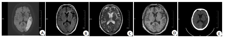
ObjectiveTo explore the comparative analysis of magnetic resonance imaging (MRI), multi-slice spiral CT (MSCT) and digital subtraction angiography (DSA) in the clinical diagnosis of acute cerebral infarction (ACI). MethodsThe clinical data of 100 patients with suspected ACI in the hospital from March 2019 to December 2020 were retrospectively analyzed. All patients underwent MRI, MSCT and DSA. Taking DSA results as the golden standard, consistency of diagnosis results between MRI, MSCT and DSA were analyzed. The accuracy, specificity and sensitivity of MRI and MSCT in the diagnosis of ACI were calculated. ResultsThe diagnosis results of DSA showed that among the 100 patients with suspected ACI, 79 cases with ACI and 21 cases without. The diagnosis results of MRI showed that 77 cases with ACI and 23 cases without. Consistency analysis between DSA and MRI showed that Kappa value was 0.637, indicating good consistency. The diagnosis results of MSCT showed that 71 cases with ACI and 29 cases without. Consistency analysis between DSA and MSCT showed that Kappa value was 0.524, indicating good consistency. The accuracy and sensitivity of MRI in the diagnosis of ACI were higher than those in control group (P < 0.05). The detection rates of onset within 24 h and 72 h by MRI were higher than those by MSCT(P < 0.05). ConclusionThe consistency of diagnosis results between MRI, MSCT and DSA is good for ACI. However, diagnosis advantages of MRI are more significant, which can provide reference for developing clinical treatment regimens as early as possible.
2021, 44(4): 695-700.
doi: 10.12122/j.issn.1674-4500.2021.04.24
Abstract:
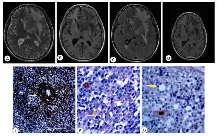
ObjectiveTo analyze the correlation between MRI findings of intracranial diffuse large B-cell lymphoma (DLBCL) and vasculogenic mimicry(VM), reticular fiber. MethodsNinety-three patients with intracranial DLBCL who were diagnosed and underwent MRI examination in our hospital from March 2017 to April 2020 were collected. The MRI signs of all patients were recorded, and the tumor specimens were stained and analyzed. We analyzed the enhancement index of VM and reticular fiber with different properties, the relationship between VM, reticular fiber and enhancement index, and the relationship between MRI findings and vasculogenic mimicry, reticular fiber. ResultsThe density of microvessels in the tumor was 42.42± 18.78 mm2, and the number of mature vessels in the lesion was 9.65±4.02 mm2. Among all intracranial patients, there were 62 single cases and 31 multiple cases, with a total of 149 lesions. Among the 93 MRI results, 53 T1WI showed isointensity and 40 slightly hypointensity. 44 T2WI showed equal signal and 49 showed slightly higher signal. All lesions showed high DWI signal. There were 62 lobulation sign lesions, 47 umbilical concave sign lesions, 31 sharp Angle sign lesions and 26 clenched fist sign lesions.18 meningeal infiltrating lesions. PAS staining was positive in all lesions, including 53 strong positive lesions and 40 weak positive lesions. Reticular fiber staining: 49 lesions (strongly positive) had more reticular fibers. Reticular fibers were scattered in 44 lesions (weakly positive). All patients had enhancement, enhancement index 0.996 ± 0.368. The microvessel density of patients was positively correlated with the enhancement index(P < 0.05), and the enhancement index of patients was positively correlated with the number of mature vessels (P < 0.05). The enhancement index of weak positive VM group was lower than that of strong positive VM group (P < 0.05). The strengthening index of weak positive reticular fiber group was lower than that of strong positive group (P < 0.05). VM, reticular fiber and enhancement index were positively correlated(P < 0.05). MRI findings of necrosis were negatively correlated with VM (P < 0.05). ConclusionThe MRI manifestation necrosis of DLBCL is negatively correlated with VM. DLBCL has certain characteristics in MRI manifestation and can be diagnosed according to its manifestation.
2021, 44(4): 701-705.
doi: 10.12122/j.issn.1674-4500.2021.04.25
Abstract:
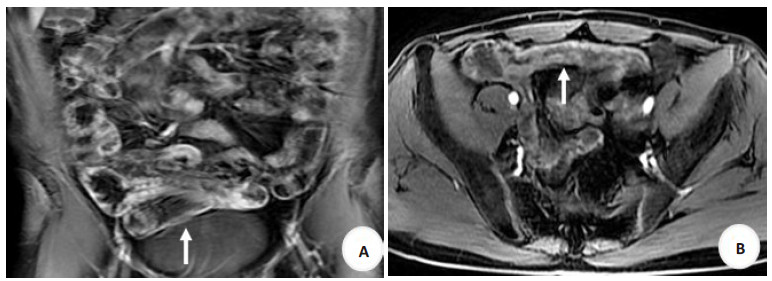
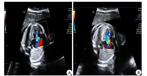
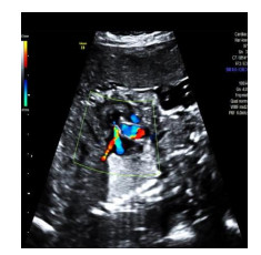
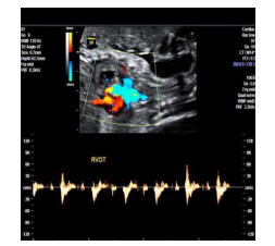
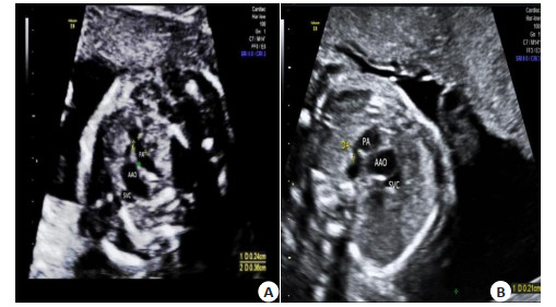
ObjectiveTo investigate the diagnostic value of fetal echocardiography for obstructive heart malformations of the right ventricular system with low lung blood. MethodsFrom March 2017 to March 2020, 1036 pregnant women who had a high-risk tendency or voluntarily requested fetal echocardiography during their pregnancy period of 20 to 28 weeks in our hospital were reviewed. The position of the fetal heart, the images of each segment and the combined malformations were obtained and analyzed, and the accuracy of echocardiography with relevant statistical data were analyzed. ResultsThere were 86 cases of dextrocardia fetus, 39 cases of dextrocardia mirror images (45.35%), 37 cases of dextrorotation heart (43.02%), 4 cases of right atrial heterogeneity (4.65%), and 6 cases of left atrial heterogeneity (6.98%) detected in 1036 pregnant women. Echocardiography had diagnostic significance for the connection of various segments of the heart, ventricle and aorta (P < 0.05), and it was consistent with the diagnosis of cardiovascular angiography (Kappa > 0.5). Compared with those with pulmonary artery stenosis or atresia in dextrorotation heart and right-sided heart, the difference was significant (P < 0.05). The sensitivity of echocardiography to the diagnosis of cardiac malformations was 95.00% (76/80), specificity was 83.33% (5/6), accuracy was 94.19% (81/86), and the consistency between echocardiography and cardiovascular angiography was strong (Kappa=0.503). ConclusionEach segment of right heart system obstructive cardiac malformation can be abnormal, and most of them are complicated with abnormal pulmonary blood deficiency. Echocardiography is more intuitive and comprehensive to observe the changes of cardiac anatomy and hemodynamics, which has high diagnostic value in judging the complicated malformation with pulmonary blood deficiency.
2021, 44(4): 706-709.
doi: 10.12122/j.issn.1674-4500.2021.04.26
Abstract:
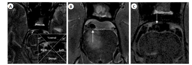
ObjectiveTo analyze the distribution, composition and related characteristics of plaque in patients with symptomatic and asymptomatic basilar artery stenosis using high-resolution MRI imaging. MethodsA retrospective analysis of 106 patients with basilar artery stenosis who underwent high-resolution MRI examinations in our hospital was conducted from March 2018 to March 2021. They were divided into symptomatic group (n=45) and asymptomatic group (n=61) according to the presence or absence of posterior circulation stroke with the imaging data collected. The distribution, composition, reconstruction and other related characteristics of the two groups of plaques were analyzed. ResultsThe plaques of the symptomatic group mainly localized on the dorsal wall, the plaques of the asymptomatic group mainly localized on the ventral wall, the distribution and composition of the plaques in the two groups were significantly different (P < 0.05). The probability of bleeding and fiber cap rupture in symptomatic group was higher than that in asymptomatic group (P < 0.05). The vascular area, wall area, lumen area, maximum wall thickness, stenosis rate, and vascular remodeling index of the most stenosis layer in the symptomatic group were higher than those in the asymptomatic group (P < 0.05). There was no significant difference in plaque load between the two groups (P > 0.05). There was a significant difference in the distribution of plaque enhancement grades between the two groups, and the level 2 plaque enhancement rate of the symptomatic group was higher than that of the asymptomatic group (P < 0.05). ConclusionThere are massive differences between symptomatic and asymptomatic patients with basilar artery stenosis in plaque distribution, composition, and remodeling. High-resolution MRI imaging can give a reliable imaging reference for identifying plaque characteristics and early intervention.
2021, 44(4): 710-713.
doi: 10.12122/j.issn.1674-4500.2021.04.27
Abstract:
With the progress of nanobiotechnology and the rise of optical imaging technology recently, molecular imaging has shown a development trend of close integration with many disciplines such as materials science, chemistry, medical physics, biomedical engineering and genomics, etc. New molecular imaging agents based on nanotechnology are developing rapidly, and nanoparticles modified with small molecules, peptides, antibodies and aptamers have been widely used in preclinical research and clinical translation. Multimodal molecular imaging technology has emerged as a key component of precision medicine, and a new wave of imaging technology upgrades can obtain more information at the tissue and molecular levels, further promoting cross-fertilization between disciplines. In this paper, we review the cutting-edge technologies and clinical applications of optical and photoacoustic molecular imaging, magnetic resonance molecular imaging, and positron emission tomography molecular imaging.
With the progress of nanobiotechnology and the rise of optical imaging technology recently, molecular imaging has shown a development trend of close integration with many disciplines such as materials science, chemistry, medical physics, biomedical engineering and genomics, etc. New molecular imaging agents based on nanotechnology are developing rapidly, and nanoparticles modified with small molecules, peptides, antibodies and aptamers have been widely used in preclinical research and clinical translation. Multimodal molecular imaging technology has emerged as a key component of precision medicine, and a new wave of imaging technology upgrades can obtain more information at the tissue and molecular levels, further promoting cross-fertilization between disciplines. In this paper, we review the cutting-edge technologies and clinical applications of optical and photoacoustic molecular imaging, magnetic resonance molecular imaging, and positron emission tomography molecular imaging.
2021, 44(4): 714-717.
doi: 10.12122/j.issn.1674-4500.2021.04.28
Abstract:
Stroke is a disease which affects brain networks. Functional magnetic resonance imaging is widely used to explore the functional changes and brain reorganization after stroke injury. Dynamic functional network connectivity is a new method to describe the dynamic characteristics of brain functional connectivity at rest stage. In recent 5 years, the application of dynamic functional network connectivity analysis in the field of stroke was mainly used to explore the functional connectivity properties of different brain networks, sensorimotor networks and language networks. The results showed that the functional connections of the brain network presented a series of repeated states of dense or sparse connections which have different temporal variation characteristics after stroke. It provides a new perspective and shows its potential advantages in study of stroke. This paper reviewed the current status of dynamic functional network connectivity which mainly focus on the functional connectivity properties of different brain networks, sensorimotor networks and language networks after stroke.
Stroke is a disease which affects brain networks. Functional magnetic resonance imaging is widely used to explore the functional changes and brain reorganization after stroke injury. Dynamic functional network connectivity is a new method to describe the dynamic characteristics of brain functional connectivity at rest stage. In recent 5 years, the application of dynamic functional network connectivity analysis in the field of stroke was mainly used to explore the functional connectivity properties of different brain networks, sensorimotor networks and language networks. The results showed that the functional connections of the brain network presented a series of repeated states of dense or sparse connections which have different temporal variation characteristics after stroke. It provides a new perspective and shows its potential advantages in study of stroke. This paper reviewed the current status of dynamic functional network connectivity which mainly focus on the functional connectivity properties of different brain networks, sensorimotor networks and language networks after stroke.
Progresses in molecular imaging diagnosis, prevention and treatment for contrast-induced nephropathy
2021, 44(4): 718-724.
doi: 10.12122/j.issn.1674-4500.2021.04.29
Abstract:
With the widespread application of contrast agents in modern medicine, contrast induced nephropathy (CIN) has received more attention. As the third most common cause of iatrogenic acute kidney injury after renal hypoperfusion and nephrotoxic medications, CIN refers to acute renal failure due to the administration of the iodinated contrast agents in angiography or other medical procedures. Patients with CIN show prolonged hospitalization, chronic renal insufficiency, other vascular events and higher mortality. At present, there is no effective treatment for CIN and therefor early evaluation and prevention have been the leading focus of CIN treatment. This article reviews the progress in prevention and treatment of CIN since 2014, including pathophysiological mechanisms, molecular markers for early diagnosis, molecular imaging diagnosis, risk factors and risk prediction, contrast agent selection, contrast agent metabolism monitoring, drug combination hydration therapy, gene therapy, traditional Chinese medicine and integrated traditional Chinese medicine and Western medicine.
With the widespread application of contrast agents in modern medicine, contrast induced nephropathy (CIN) has received more attention. As the third most common cause of iatrogenic acute kidney injury after renal hypoperfusion and nephrotoxic medications, CIN refers to acute renal failure due to the administration of the iodinated contrast agents in angiography or other medical procedures. Patients with CIN show prolonged hospitalization, chronic renal insufficiency, other vascular events and higher mortality. At present, there is no effective treatment for CIN and therefor early evaluation and prevention have been the leading focus of CIN treatment. This article reviews the progress in prevention and treatment of CIN since 2014, including pathophysiological mechanisms, molecular markers for early diagnosis, molecular imaging diagnosis, risk factors and risk prediction, contrast agent selection, contrast agent metabolism monitoring, drug combination hydration therapy, gene therapy, traditional Chinese medicine and integrated traditional Chinese medicine and Western medicine.
2021, 44(4): 725-728.
doi: 10.12122/j.issn.1674-4500.2021.04.30
Abstract:
Intravoxel incoherent motion (IVIM) is a translational motion that presents a directional or amplitude velocity distribution within a given voxel and at a given measurement time. IVIM is a new magnetic resonance technique for measuring microcirculatory perfusion using multi-B value DWI. The related parameters of the apparent diffusion coefficient, pure diffusion coefficient, the pseudo diffusion coefficient, filling fraction quantitative indexes such as accurate and timely reflect the features of the different parameters of various diseases, and based on IVIM perfusion magnetic resonance imaging, do not need to use contrast agents, make it's in recent years in the study of the uterus. Its related diseases reflect its unique advantages, especially in the survey of preoperative staging of malignant uterine tumors and the detection of therapeutic effects. This new technology will provide a basis for us to provide better clinical diagnosis and select the best treatment plan. This article reviews the unique features of IVIM in the diagnosis of uterine diseases.
Intravoxel incoherent motion (IVIM) is a translational motion that presents a directional or amplitude velocity distribution within a given voxel and at a given measurement time. IVIM is a new magnetic resonance technique for measuring microcirculatory perfusion using multi-B value DWI. The related parameters of the apparent diffusion coefficient, pure diffusion coefficient, the pseudo diffusion coefficient, filling fraction quantitative indexes such as accurate and timely reflect the features of the different parameters of various diseases, and based on IVIM perfusion magnetic resonance imaging, do not need to use contrast agents, make it's in recent years in the study of the uterus. Its related diseases reflect its unique advantages, especially in the survey of preoperative staging of malignant uterine tumors and the detection of therapeutic effects. This new technology will provide a basis for us to provide better clinical diagnosis and select the best treatment plan. This article reviews the unique features of IVIM in the diagnosis of uterine diseases.