


Ultrasonographic features and immunohistochemical parameters of mucinous Carcinoma of breast
-
摘要:
目的 分析乳腺粘液癌的超声图像特征及免疫组化指标,提高诊断粘液癌的准确性。 方法 回顾性分析56例乳腺癌肿块的超声特征(肿块最大径、纵横比、高回声晕、转移情况、弹性评分等)、剪切波速度(SWVmax、SWVratio、SWVmean)及免疫组化检测结果(雌激素受体、孕激素受体、人表皮生长因子受体2、Ki-67),将粘液癌患者作为实验组(n=24),将浸润性导管癌患者作为对照组(n=32),对比两组超声特征及免疫组化指标表达的差异性,并对超声特征及免疫组化指标进行相关性分析。 结果 与浸润性导管癌相比,粘液癌肿块多表现为边界清晰、有高回声晕、后方回声增强、弹性评分 < 4分且BI-RADS分类 < 4b类(χ2=4.419、5.171、6.162、4.743、14.000,P < 0.05),SWVmax及雌激素受体、人表皮生长因子受体2的阳性率在两组中的差异具有统计学意义(t=- 2.069,χ2=4.371、4.051,P < 0.05)。 结论 乳腺粘液癌超声特征及免疫组化指标的表达与浸润性导管癌相比具有一定的差异,可为超声诊断乳腺癌类型及治疗方案的选择提供参考。 Abstract:Objective To analyze the ultrasonographic features and immunohistochemical indexes of mucinous carcinoma of the breast, and improve the accuracy in the diagnosis of mucinous carcinoma. Methods The ultrasonographic features (maximum diameter, aspect ratio, high echo halo, metastasis, elastic score), shear wave velocity (SWVmax, SWVratio, SWVmean) and immunohistochemical detection (ER, PR, HER-2, Ki-67) of 56 cases were analyzed retrospectively. 24 cases with mucinous carcinoma were used as experimental group and 32 cases with invasive ductal carcinoma were used as control group.The differences of ultrasonic features and immunohistochemical indexes between the two groups were compared. The correlation between ultrasonic features and immunohistochemical indexes was analyzed. Results Compared with invasive ductal carcinoma, mucinous carcinoma showed clear boundary, high echo halo, posterior echo enhancement, less than 4 score and BI-RADS classification < 4b (χ2=4.419, 5.171, 6.162, 4.743, 14.000, P < 0.05). The positive rates of SWVmax, ER and HER-2 were significantly different between the two groups (t=-2.069, χ2=4.371, 4.051, P < 0.05). Conclusion Compared with invasive ductal carcinoma, the expression of ultrasound features and immunohistochemical markers in mucinous carcinoma of breast are different, which can provide reference for the diagnosis and treatment of mucinous Carcinoma of breast. -

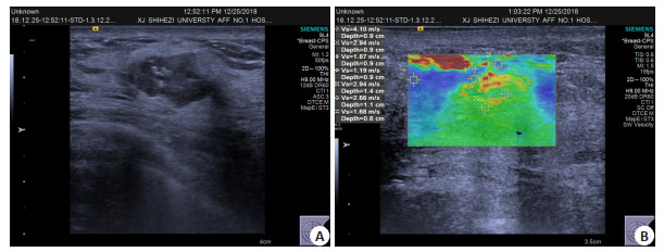
图 1 患者女,50岁,乳腺粘液癌
A: 常规超声图像:肿块最大径1.96 cm;B: VTIQ技术速度模式图像,SWVmax为4.10 m/s;ER(强阳性,95%)、PR(强阳性,90%)、Her-2(1+)、Ki-67(40%+)
Figure 1. A 50-year-old female patient with mucosal carcinoma of breast.

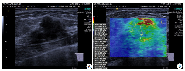
图 2 患者女,50岁,乳腺浸润性导管癌
A: 常规超声图像:肿块最大径2.08 cm;B: VTIQ技术速度模式图像:SWVmax为9.47 m/s;ER(弱阳性,70%+)、PR(强阳性,95%+)、Her-2(3+)、Ki-67(50%+)
Figure 2. A 50-year-old female patient with invasive ductal carcinoma of the breast.
表 1 粘液癌及浸润性导管癌常规超声特征的比较
Table 1. Comparison of routine ultrasonographic features of mucinous carcinoma and invasive ductal carcinoma[n(%)]
特征 粘液癌 浸润性导管癌 χ2 P 最大径(cm) 0.875 0.350 ≤2 12(37.5) 20(62.5) >2 12(50.0) 12(50.0) 位置 6.017 0.111 内上 6(54.5) 5(45.5) 内下 0(0.0) 0(0.0) 外上 9(29.0) 22(71.0) 外下 6(60.0) 4(40.0) 乳晕区 3(75.0) 1(25.0) 纵横比 1.864 0.172 >1 2(22.2) 7(77.8) ≤1 22(46.8) 25(53.2) 内部回声 1.427 0.232 均匀 0(0.0) 4(100.0) 不均匀 22(44.0) 28(56.0) 后方回声 6.162 0.046 衰减 8(30.8) 18(69.2) 混合 4(33.3) 8(66.7) 增强 12(66.7) 6(33.3) 边界 5.419 0.020 不清晰 9(29.0) 22(71.0) 清晰 15(60.0) 10(40.0) 边缘 0.021 0.884 不规整 20(44.4) 25(55.6) 规整 4(36.4) 7(63.6) 高回声晕 5.171 0.023 无 10(30.3) 23(69.7) 有 14(60.9) 9(39.1) 钙化 1.556 0.212 无 20(47.6) 22(52.4) 有 4(28.6) 10(71.4) 血流 1.641 0.200 0~1级 9(56.3) 7(43.7) 2~3级 15(37.5) 25(62.5) 弹性评分(分) 4.743 0.029 <4 10(66.7) 5(33.3) ≥4 14(34.1) 27(65.9) BI-RADS分类 14.000 < 0.001 <4b 12(85.7) 2(14.3) ≥4b 12(28.6) 30(71.4) 转移情况 0.570 0.450 未转移 18(46.2) 21(53.8) 转移 6(35.3) 11(64.7)  下载: 导出CSV
表 2 粘液癌及浸润性导管癌剪切波速度值的比较
Table 2. Comparison of shear wave velocity values between mucinous carcinoma and infiltrating ductal carcinoma (Mean±SD)
乳腺癌类型 VTIQ剪切波速度 SWVmax SWVratio SWVmean 粘液癌(n=8) 5.59±2.25 3.58±2.49 4.21±1.52 浸润性导管癌(n=21) 7.23±1.78 3.73±1.20 4.48±1.31 t -2.069 -0.224 -0.520 P 0.048 0.824 0.607 SWVmax:剪切波速度最大值;SWVratio:病灶与同高度正常腺体的剪切波速度比值;SWVmean:剪切波速度平均值.
下载: 导出CSV
表 3 粘液癌及浸润性导管癌免疫组化指标表达的比较
Table 3. Comparison of the expression of immunohistochemical indexes between mucinous carcinoma and invasive ductal carcinoma[n(%)]
病理类型 ER PR HER-2 Ki-67 (+) (-) (+) (-) (+) (-) (+) (-) 粘液癌(n=24) 21(87.5) 3(12.5) 22(91.7) 2(8.3) 5(20.8) 19(79.2) 17(70.8) 7(29.2) 浸润性导管癌(n=32) 20(62.5) 12(37.5) 25(78.1) 7(21.9) 15(46.9) 17(53.1) 25(78.1) 7(21.9) χ2 4.371 1.864 4.051 0.389 P 0.037 0.172 0.044 0.533 ER:雌激素受体;PR:孕激素受体;HER-2:人表皮生长因子受体2.
下载: 导出CSV
-
[1] Siegel RL, Miller KD, Jemal A. Cancer statistics, 2020[J]. CA A Cancer J Clin, 2020, 70(1): 7-30. doi: 10.3322/caac.21590 [2] 焦丹, 刘越. p53和Bcl-2阳性表达与乳腺癌患者临床特征的关系[J]. 癌症进展, 2019, 17(12): 1445-8. [3] 代妮娜, 张文君, 张忠磊, 等. 不典型乳腺癌的超声诊断价值[J]. 实用肿瘤杂志, 2018, 33(4): 367-70. [4] Larribe M, Thomassin-Piana J, Jalaguier-Coudray A. Breast cancers with round lumps: correlations between imaging and anatomopathology[J]. Diagn Interv Imaging, 2014, 95(1): 37-46. doi: 10.1016/j.diii.2013.04.003 [5] 罗葆明, 欧冰, 智慧, 等. 改良超声弹性成像评分标准在乳腺肿块鉴别诊断中的价值[J]. 现代临床医学生物工程学杂志, 2006(5): 396-8. doi: 10.3760/cma.j.issn.1674-1927.2006.05.004 [6] 杨文涛, 步宏. 乳腺癌雌、孕激素受体免疫组织化学检测指南[J]. 中华病理学杂志, 2015, 44(4): 237-9. doi: 10.3760/cma.j.issn.0529-5807.2015.04.005 [7] 《乳腺癌HER检测指南(版)》编写组. 乳腺癌HER2检测指南(2014版) [J]. 中华病理学杂志, 2014, 43(4): 262-7. [8] 水若鸿, 杨文涛. 乳腺癌Ki-67阳性指数的检测和评估[J]. 中华病理学杂志, 2013, 42(6): 420-3. doi: 10.3760/cma.j.issn.0529-5807.2013.06.019 [9] 牛乐军, 王巧玲, 梁凯迪. 乳腺黏液腺癌超声表现及其临床病理特征分析[J]. 临床军医杂志, 2020, 48(4): 413-4. [10] 刘锋. 73例乳腺黏液腺癌的临床特征及预后分析[D]. 重庆: 重庆医科大学, 2017. [11] Tang F, Kang YF, Lu GL, et al. A comparative study of three ultrasonic elastographic methods for the differentiation and diagnosis of benign and malignant breast masses[J]. Clin Exp Obstet Gyn, 2019, 46(4): 587-92. [12] Padmanaban V, Krol I, Suhail Y, et al. E-cadherin is required for metastasis in multiple models of breast cancer[J]. Nature, 2019, 573 (7774): 439-44. doi: 10.1038/s41586-019-1526-3 [13] 王雅婷. 超声造影评价乳腺肿块及其与免疫组化因子相关性[D]. 乌鲁木齐: 新疆医科大学, 2018. [14] 杨律, 郭丽苹. 22例乳腺黏液腺癌超声图像特征及误诊原因分析[J]. 中国妇幼保健, 2018, 33(6): 1421-3. [15] 裴蓓. 剪切波弹性模量参数与乳腺癌临床病理、免疫组化特征关系的研究[D]. 合肥: 安徽医科大学, 2020. [16] Liu YB, Huang YN, Han J, et al. Association between shear wave elastography of virtual touch tissue imaging quantification parameters and the ki-67 proliferation status in luminal-type breast cancer[J]. J Ultrasound Med, 2019, 38(1): 73-80. doi: 10.1002/jum.14663 [17] 曹春莉. 超声弹性技术对乳腺癌的诊断价值及与生物学特征的相关性[D]. 石河子: 石河子大学, 2020. [18] Spak DA, Plaxco JS, Santiago L, et al. BI-RADS® fifth edition: a summary of changes[J]. Diagn Interv Imaging, 2017, 98(3): 179-90. doi: 10.1016/j.diii.2017.01.001 [19] Kaoku S, Konishi E, Fujimoto Y, et al. Sonographic and pathologic image analysis of pure mucinous carcinoma of the breast[J]. Ultrasound Med Biol, 2013, 39(7): 1158-67. doi: 10.1016/j.ultrasmedbio.2013.02.014 [20] 徐乐. 乳腺癌超声图像特征与ER、PR、CerbB-2的相关性研究[D]. 重庆: 重庆医科大学, 2016. [21] Zhou J, Tan H, Bai Y, et al. Evaluating the HER-2 status of breast cancer using mammography radiomics features[J]. Eur J Radiol, 2019, 121: 108718. doi: 10.1016/j.ejrad.2019.108718 [22] 赵枫, 肖际东, 文欢, 等. 计算机辅助诊断技术可提高肿块最大径≤ 10 mm早期乳腺癌的超声诊断[J]. 分子影像学杂志, 2021, 44(2): 226-31. doi: 10.12122/j.issn.1674-4500.2021.02.03 [23] 杨红玲, 杨清. 高频超声联合磁共振可提高对乳腺癌早期诊断的价值[J]. 分子影像学杂志, 2020, 43(3): 520-4. doi: 10.12122/j.issn.1674-4500.2020.03.32 -

 点击查看大图
计量
- 文章访问数: 793
- HTML全文浏览量: 199
- PDF下载量: 12
- 被引次数: 0