

 Find Duplicates
 Check Document
 Submission(new)
 Experts Office
 Editorial Office
2021 Vol. 44, No. 2
column
Display Method:
2021, 44(2): 213-218.
doi: 10.12122/j.issn.1674-4500.2021.02.01
Abstract:
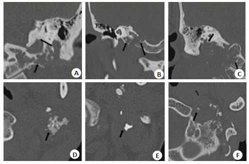
ObjectiveTo explore the diagnostic value of high resolution CT (HRCT), routine and functional MRI in low grade chondrosarcoma of skull base. MethodsThe HRCT and MR images of 10 patients with pathologically proved low grade chondrosarcoma of the skull base (grade Ⅰ-Ⅱ) were retrospectively analyzed. The sequences of MRI scan included conventional T1WI, T2WI, DWI, DCE-MRI and MR spectroscopy (MRS). Bone destruction and calcification were mainly observed on HRCT. The shape, size, location, involvement range and signal characteristics of the lesions were analyzed on routine MRI. Apparent diffusion coefficient (ADC) and DCE-MRI parameters including time intensity curve (TIC), maximum ratio of enhancement (ERmax) and time to peak (TTP) were measured on functional MRI. The Cho/Cr peak ratio and the presence or absence of NAA peak were observed on MRS. Results Bone destruction characterized by mouse nibbled was found in all tumors (10/10) on HRCT. 6 cases showed calcification which was punctuate and irregular patchy. All tumors (10/10) were irregular with an average size of 3.5 cm ×3.0 cm ×4.2 cm. Eight of the 10 cases were located in jugular foramen and two in temporal mastoid region. The surrounding structures were mainly involved in jugular foramen (n=8), tympanic and mastoid segments of facial nerve canal (n=8), mastoid portion of temporal bone (n=7), middle ear (n=5), hypoglossal canal (n=4), parapharyngeal space and deep parotid lobe (n=4), carotid artery space (n=3), and cerebellopontine angle area (n=1). Ten lesions showed homogeneous isointensity on T1WI and inhomogeneous hyperintensity on T2WI. And after enhancement, the lesions showed inhomogeneous mild to moderate enhancement. The average ADC value of 10 lesions was (1.96±0.10)×10-3 mm2/s. The TIC of DCE-MRI in 10 cases showed a continuous ascending pattern (type Ⅰ), with ERmax=1.20±0.44 and TTP (≥210 s). The ratio of Cho peak to Cr peak on MRS of 10 lesions was less than 1, and no obvious NAA peak was found. ConclusionSkull base bone destruction characterized by mouse nibbled with irregular soft tissue mass, which showed calcification on HRCT and obviously inhomogeneous high signal on T2WI, inhomogeneous mild to moderate enhancement after enhancement, unlimited dispersion on DWI, type Ⅰ TIC and the value of Cho/Cr on MRS was less than 1, no obvious NAA peak suggested the possibility of diagnosis of chondrosarcoma.
2021, 44(2): 219-225.
doi: 10.12122/j.issn.1674-4500.2021.02.02
Abstract:
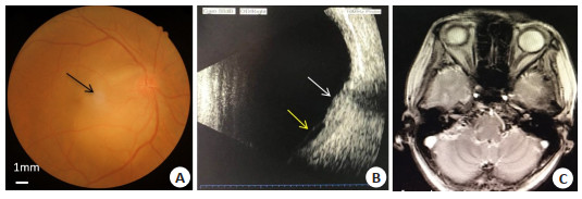
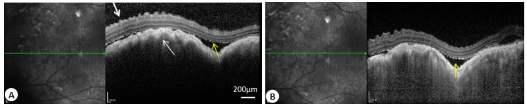
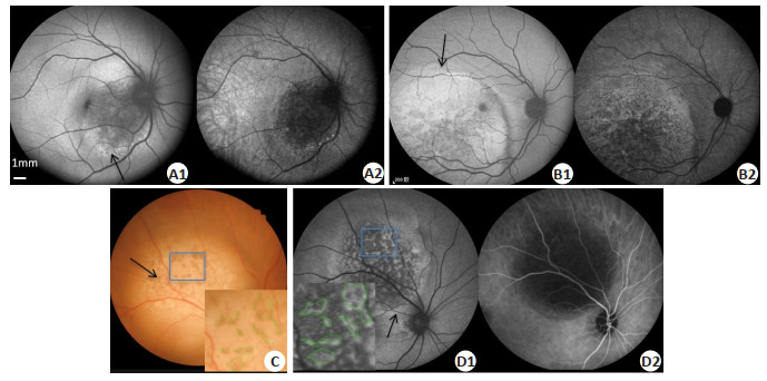
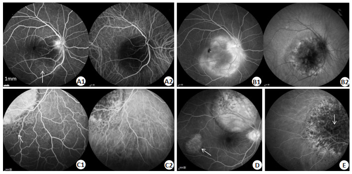
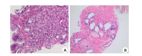
ObjectiveTo discuss the clinic diagnosis of choroidal metastasis (CM) through the investigation of its clinical and imaging characteristics. MethodsThe clinical data was collected from 10 patients with CM, in our hospital from 2012 to 2019. There were 4 males and 6 females, with the mean age of 61±5.8 years. The data included detailed medical history, BCVA, slit lamp biomicroscopy, fundus photography, SD-OCT, fundus fluorescein angiography (FFA), fundus autofluorescence (FAF) and type-B ultrasound, MRI and tumor biomarkers. ResultsTotally 12 eyes of 10 patients with CM were examined, with primary cancer site in the lung in 8 cases and in breast in 2 cases. Yellow isolated bulge lesion was found in 11 eyes(91.67%), while pigmentation being visible on the surface of tumor in 6 eyes (50%). FAF showed high autofluorescence in dots or circles around the tumor, or high autofluorescence in the whole tumor. The inside part demonstrated non-uniform autofluorescence performance. The area of FAF showed high autofluorescence was consistent with the pigment on the tumor surface (100%, P < 0.05). SD-OCT was manifested by wavy ridges in the choroid-pigment epithelium layer and the neuroepithelial layer. A large number of fine granular strong reflective materials were observed between the retinal neuroepithelial layer and the RPE layer, combined with exudative retinal detachment. The tumors showed low fluorescence with unclear borders in the early stage, needle-like spotted high fluorescence in the middle stage and diffusive strong fluorescence in the late stage 5 eyes(41.67%), relative low fluorescence in the center of tumors in 5 eyes. ICGA revealed that large areas of tumor show low fluorescence in the early stage, followed by spot-like high fluorescence in the surrounding area and slowly expanding and blurring over time. Most of the tumor body was still low fluorescence, and the diameter was reduced in comparison with the earlier phase. B-type ultrasound scan showed uniform and substantial uplift. ConclusionColor photography of the fundus shows the location, size, pigmentation and retinopathy of tumors. The situation of retinal pigment epithelium in autofluorescence is significantly related to the pigment on the tumor surface. SD-OCT reflects the height of tumors and retinal detachment. ICGA and FFA complement each other to help the differential diagnosis of choroidal tumors.
2021, 44(2): 226-231.
doi: 10.12122/j.issn.1674-4500.2021.02.03
Abstract:
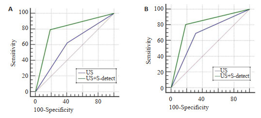
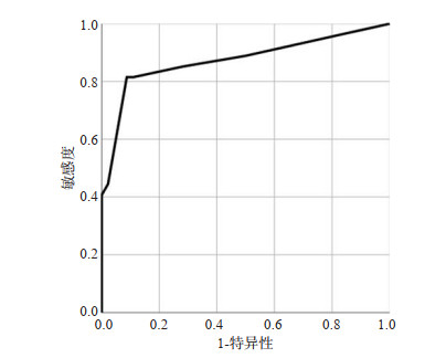
ObjectiveTo investigate the application of computer-aided ultrasonography assisting radiologists in differential diagnosis for early breast cancer. MethodsThe representative images of 120 breast masses (maximum diameter ≤20 mm, 50 breast cancer nodules, 70 benign nodules) pathologically proved were reviewed retrospectively. According to the size of the mass, patients were divided into maximum diameter ≤10 mm group (56 nodules) and maximum diameter 11-20 mm group (64 nodules). Two radiologists made their diagnosis with reference to the breast imaging report and data system ultrasonography (BI-RADS-US) classification. Combined with computer-aided ultrasonography results, all the masses were re-diagnosed. Pathological results were used as gold standard. Diagnostic performance for US and the combination of US and computer-aided diagnosis was compared. ResultsFor the maximum diameter ≤10 mm group, conventional US diagnostic sensitivity, specificity and accuracy of radiologists were 62.5%, 59.4% and 60.7%, respectively, and the area under the receiver operating characteristic (ROC) curve (AUC) were 0.61. And the sensitivity, specificity and accuracy of the combination of US and computer-aided technology were 79.2%, 81.3% and 80.4%, AUC was 0.80. For the maximum diameter 11-20 mm group, conventional US diagnostic sensitivity, specificity and accuracy of the radiologists were 69.2%, 68.4% and 68.8%, respectively, and the AUC were 0.69. And the sensitivity, specificity and accuracy of the combination of US and CAD were 80.8%, 81.6% and 81.3%, AUC was 0.81. After computer-aided diagnosis, the sensitivity, specificity, accuracy and AUC of two groups with different sizes of breast mass were increased, especially significant promotion of the accuracy and AUC in maximum diameter ≤10 mm group (P < 0.05). ConclusionIt is demonstrated that computer-aided diagnosis can help radiologists improving their diagnostic efficacy, especially for early breast cancer with maximum diameter ≤10 mm.
2021, 44(2): 232-238.
doi: 10.12122/j.issn.1674-4500.2021.02.04
Abstract:
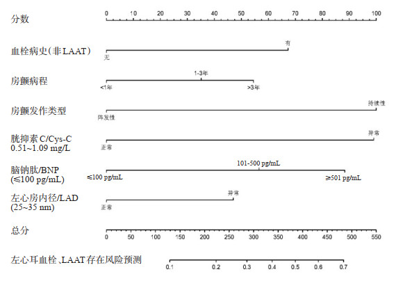
ObjectiveTo construct a model for predicting the risk of left atrial appendage thrombus (LAAT) in patients with nonvalvular atrial fibrillation (NVAF) to provide a reference for individualized clinical judgment. MethodsThe data of all patients hospitalized in the Department of Cardiology of the First Affiliated Hospital of Xi'an Jiaotong University between March 1, 2018 and November 30, 2020 were collected. Then a total of 530 patients with NVAF were obtained according to the screening criteria. It is divided into the non-LAAT group (n=428) and the LAAT group (n=102) based on the presence or absence of LAAT status. Relevant advances in global research and the characteristics of our institution's cases were reviewed, and risk factors that may be associated with LAAT formation in NVAF patients were collected extensively. Independent risk factors associated with LAAT formation were identified by binary logistic regression analysis. A model to predict the risk of LAAT occurrence in NVAF patients was constructed based on nomogram, and the accuracy of the model was evaluated and validated internally. ResultsSix factors, including history of thrombosis, duration of AF, type of AF, BNP, Cys-C, and LAD, were associated with the presence or absence of LAAT status in patients with NAVF (all P < 0.05) and could be used as independent risk factors to predict LAAT formation. Accordingly, a model was constructed to predict the risk of LAAT occurrence in NVAF patients based on nomogram, with a C-index of 0.74 (95%CI: 0.69-0.79). In addition, the prediction accuracy of the model is also shown in the calibration curve. ConclusionThe model for predicting the risk of LAAT in patients with NVAF based on Nomogram has good accuracy and practicability, and can be used to predict the risk of LAAT in patients with NVAF accurately and intuitively.
2021, 44(2): 239-243.
doi: 10.12122/j.issn.1674-4500.2021.02.05
Abstract:
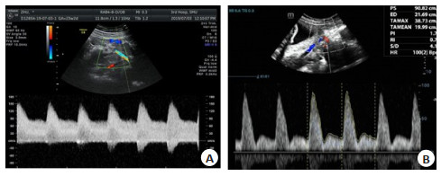
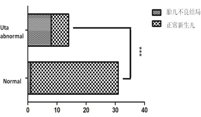
ObjectiveTo explore the predictive value of the uterine artery Doppler in systemic lupus erythematosus (SLE) pregnancies. MethodsData of 45 pregnancies in SLE patients from January 2010 to December 2020 were analysed retrospectively. Pregnancy and neonatal outcomes were collected. Adverse pregnancy outcome was defined as one of the following: pre-eclampsia, lupus nephritis, APS, APA(+), pre-term birth, small for gestational age, fetal growth restriction and placental abruption. Pulsatility index (PI) of the uterine artery were monitored by Doppler ultrasound. An increased pulsatility index and an early diastolic notch of uterine artery is consider with abnormal. ResultsAmong the 45 pregnant women, 15 (33%) developed preeclampsia, lupus nephritis, and antiphospholipid syndrome (APS), and 9(20%) fetuses had adverse outcomes. Complications include preeclampsia, lupus nephritis, antiphospholipid antibody (+) and antiphospholipid syndrome. In the 31 (69%) normal cases uterine artery spectrum group, 5 cases (11%) of pregnant women developed preeclampsia, and 1 case (2%) of fetus developed small for gestational age.14 women (31%) in the spectrum abnormality group and complications occurred in 10 women (22%). 8 cases (18%) had adverse fetal outcomes, including small for gestational age, fetal growth restriction and fetal loss. 3 patients (7%) with new SLE during pregnancy showed abnormal uterine artery waveforms (waveforms showed incisions and increased PI), and all three fetals died in perinatal period. ConclusionAn increased pulsatility index and an early diastolic notch in the waveform has been associated with an increased risk for adverse pregnancy outcomes.
2021, 44(2): 244-248.
doi: 10.12122/j.issn.1674-4500.2021.02.06
Abstract:
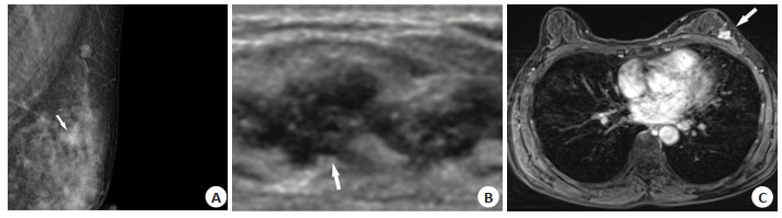
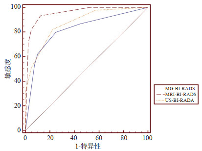
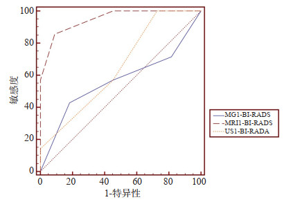
ObjectiveTo compare the efficacy of conventional ultrasound (US), conventional mammography (MG) and dynamic contrast-enhanced magnetic resonance imaging (DCE-MRI) in the diagnosis of breast lesions with calcification categorized 3-5. MethodsBetween January 2010 and February 2019, a total of 85 untreated breast lesions with calcification from 82 patients who underwent US, MG and MRI at the same time were retrospectively analyzed. All the lesions were confirmed by histopathological assessment. The sensitivity, specificity, accuracy, positive predictive value and negative predictive value were evaluated, and the diagnostic value of the three modalities were compared. Lesions were divided into consistent or inconsistent group on the basis of BI-RADS MG and US assessment. The diagnostic efficacy of three methods in the overall and inconsistent group were assessed by receiver operating characteristic (ROC) curve analysis. ResultsThe sensitivity, specificity, accuracy, positive predictive value and negative predictive value of MG, US and MRI in the diagnosis of breast lesions with calcification were 80.0% vs 82.2% vs 93.3%、75.0% vs 77.5% vs 87.5%、77.6% vs 80.0% vs 90.6%、78.3% vs 80.4% vs 89.4%、77.0% vs 79.5% vs 92.1%, respectively, with significant difference (P=0.022、0.019). The AUCs of MG and US were 0.821, 0.872 for overall group and 0.565, 0.649 for inconsistent group. The corresponding values of MRI were 0.956 and 0.948, which were significantly prior to those of MG and US (P < 0.05). ConclusionThe DCE-MRI has a better diagnostic efficiency than MG or US alone in the differentiation of the breast lesions with calcification especially when disagreement occurred.
2021, 44(2): 249-254.
doi: 10.12122/j.issn.1674-4500.2021.02.07
Abstract:
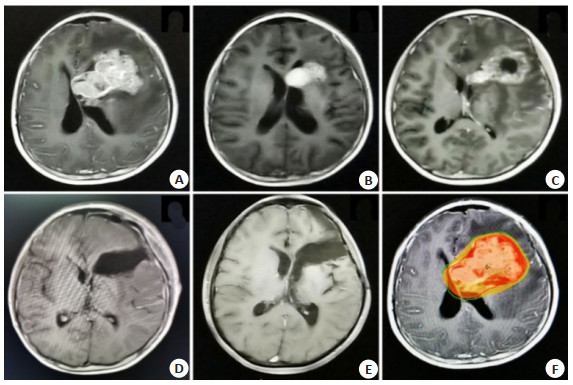
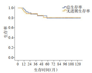
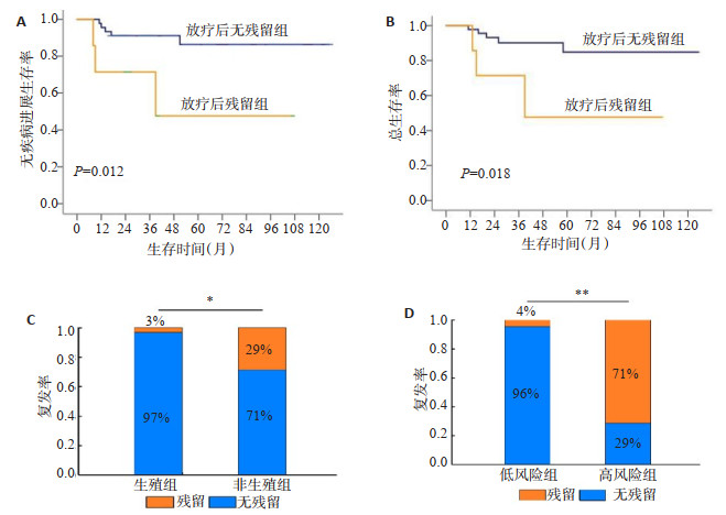
ObjectiveTo explore the efficacy of combined radiotherapy and chemotherapy on germ cell tumors in the basal ganglia and analysis of influencing factors. MethodsFrom January 2009 to January 2019, 52 cases of germ cell tumors in the basal ganglia were first diagnosed in the Cancer Center of Guangdong Sanjiu Brain Hospital. Survival analysis was performed on the whole group of patients; the residual and non-residual after radiotherapy and chemotherapy were divided to 2 groups. The clinical factors which would affect the efficacy were analyzed between 2 groups. ResultsOn June 15, 2020, the median follow-up time was 38 months (range 13-125 months), 49 cases were followed up, and 3 cases were lost. Of these, 40 survived and 9 died. The 3-year overall survival rate was 83%, and the 3-year progression-free survival rate was 84%. Six patients had significant residues (tumor stability or progression) after receiving chemotherapy and radiotherapy, and 46 patients had no significant residues (partial remission or complete remission). There were 30 patients (57.7%) diagnosed with germ cell tumors, and 22 patients (42.3%) with non-germinomaus germ cell tumors. Non-germinomaus germ cell tumors were more likely to remain after treatment than germ cell tumors. There was a significant statistical difference between the two groups (P=0.013), accompanied by abnormally increased tumor markers (AFP greater than 500 ng/mL or HCG greater than 1000 mU/mL). High risk patients were more likely to have residual after treatment than the low-risk group (P=0.000). Both univariate and multivariate analysis found that the presence or absence of significant tumor residue at the end of treatment was an independent prognostic factor affecting survival, P=0.024 (HR: 0.306, 95%CI: 0.024-1.488). ConclusionGerm cell tumors in the basal ganglia have different response to treatment with different tumor types and tumor markers. Obvious residual after treatment is an independent prognostic factor affecting survival. For the obvious residual non-germinomaus germ cell tumors, surgical treatment is recommended again to facilitate local control.
2021, 44(2): 255-258.
doi: 10.12122/j.issn.1674-4500.2021.02.08
Abstract:
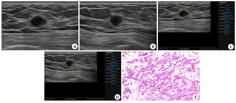
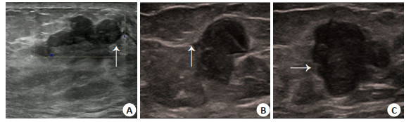
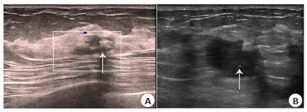
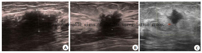
ObjectiveTo explore the reasons of up-regulation of BI- RADS 4 breast nodules by two-dimensional gray-scale ultrasound and improve the ability of ultrasound to identify benign breast nodules so as to reduce over-diagnosis and treatment. MethodsA total of 380 cases of breast nodules hospitalized in our hospital from December 2019 to November 2020 were analyzed retrospectively. According to the pathological results, the ultrasound BI-RADS 4 breast nodules were divided into two groups: the nodule up-regulation group and the nodule consistent group. Breast nodules are benign, but ultrasound BI-RADS 4 nodules were considered as up-regulation group, including 68 females and 2 males, aged from 26 to 76 years, with an average age of 48.43±11.52 years. The breast nodules were considered as malignant and the ultrasound BI-RADS 4 types were consistent, including 310 females and 0 males, aged from 26 to 87 years, with an average of 52.84 ± 11.28 years. The ultrasonographic features of the two groups were analyzed, and the reasons for the up-regulation of ultrasound evaluation of BI-RADS 4 breast nodules were summarized for differential diagnosis. ResultsFrom December 2019 to November 2020, there were 380 patients (100.00%) with Bi-RADS type 4 nodules, including 70 patients (18.42%) with up-regulated nodules and 310 patients (81.58%) with consistent nodules. There were 13 breast fibroadenomas (18.57%), 12 intraductal papillomas (17.14%), 7 breast adenopathy (10.00%), 5 non-lactation mastitis (7.14%), 3 benign phyllodes tumors (4.29%), 2 male breast development (2.86%), and 1 breast lobular atrophy(1.43%), 27 cases with more than two kinds of benign nodules. A total of 310 cases (100%) of breast cancer were in the same nodule group. Ultrasonographic signs: in the up-regulated nodule group, the boundary was clear, the aspect ratio was less than 1, the edge was smooth or lobulated, and coarse calcification was more common. In the consistent nodule group, the boundary was unclear, the aspect ratio was more than 1, the edge was angular or lobulated, and microcalcification was more common. There was significant difference between the two groups (P < 0.05). ConclusionCorrect identification of benign and malignant signs of ultrasound breast nodules and reduction of subjective diagnosis can effectively improve the accuracy of ultrasound benign nodules and improve diagnostic confidence.
2021, 44(2): 259-263.
doi: 10.12122/j.issn.1674-4500.2021.02.09
Abstract:
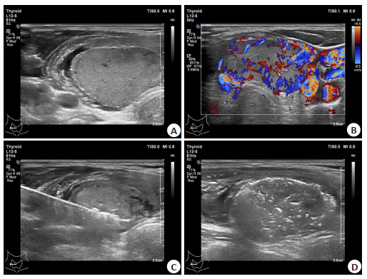
ObjectiveTo explore the effect and safety of ultrasound- guided microwave ablation in the treatment of benign thyroid lesions. MethodsA total of 230 patients with benign thyroid lesions undergoing ultrasound-guided microwave ablation from November 2019 to April 2020 in department of ultrasound medicine, West China Hospital, Sichuan University were enrolled. Ultrasound re-examination was conducted at 1 month, 3 month and 6 months after treatment to observe the changes in volume of thyroid nodules. The thyroid function indexes [free triiodothyronine (FT3), free thyroxine (FT4), thyroidstimulating hormone (TSH)] and thyroid immune antibody indexes [anti-thyroglobulin antibody (TGAb), anti-thyroid peroxidase antibody (TPOAb), anti-thyrotropin receptor antibody (TRAb)] were detected. And postoperative complications were observed. ResultsCompared with those before treatment, the volume of thyroid nodules gradually decreased at 1 month, 3 months and 6 months after treatment (P < 0.05). Compared with those before treatment, there was no significant difference in levels of serum FT3, FT4, TSH, TGAb, TPOAb and TRAb at 1 month, 3 months and 6 months after treatment (P>0.05). After microwave ablation, there were no severe complications (recurrent laryngeal nerve injury, parathyroid injury, intrathyroid hemorrhage, infection). There were 20 cases (8.70%) with mild to moderate neck pain, and 26 cases (11.30%) with neck swelling. ConclusionThe effect and safety of ultrasound-guided microwave ablation are good in the treatment of benign thyroid lesions, with few complications and basically without damage to thyroid function.
2021, 44(2): 264-269.
doi: 10.12122/j.issn.1674-4500.2021.02.10
Abstract:
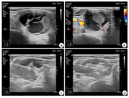
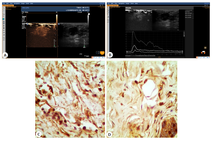
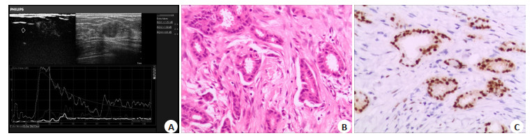
ObjectiveTo analyze the correlation between clinic-pathological and CEUS indicators in breast cancer anf explored the correlation between factors with good correlation with CEUS qualitative indicators and the characteristics of TAF in breast cancer. MethodsA total of 119 breast cancer patients were included in the study. CEUS was used to record the characteristics of CEUS. Marked the active marginal band of breast cancer mass and the normal band beside the lesion and marked the study section of mass. Immunohistochemical examination was performed. Target cells were labeled and combined with morphological characteristics to differentiate TAF cells. SPSS22.0 was adopted for statistical analysis. ResultsWhen the mass of breast cancer was more than 2 cm, the histological grade and clinical stage were higher, and the mass range, high enhancement, the probability of perfusion defect and perforator blood flow in contrast-enhanced were higher (P < 0.05). If there was filling defect, the distribution of TAF in the normal adjacent zone increased (P < 0.05). If there was high enhancement after mammography, the distribution of TAF in the active marginal zone of breast cancer increased (P < 0.05). When the lesions were large, TAF in the active marginal zone was more distributed (P < 0.05). When the clinical stage and histological grade were higher, the TAF of the two regions were higher (P < 0.05). But there was no correlation between the enhancement range and the distribution of TAF in the active marginal zone and the normal adjacent zone (P>0.05). There was no correlation between the occurrence of filling defect and the distribution of TAF in the active marginal zone (P>0.05). So the degree of contrast had nothing to do with the distribution of TAF in the normal zone (P>0.05). ConclusionTAF is a factor for poor prognosis of breast cancer. The number of TAF reflected the differentiation and metastasis of breast cancer to a certain extent.
2021, 44(2): 270-275.
doi: 10.12122/j.issn.1674-4500.2021.02.11
Abstract:
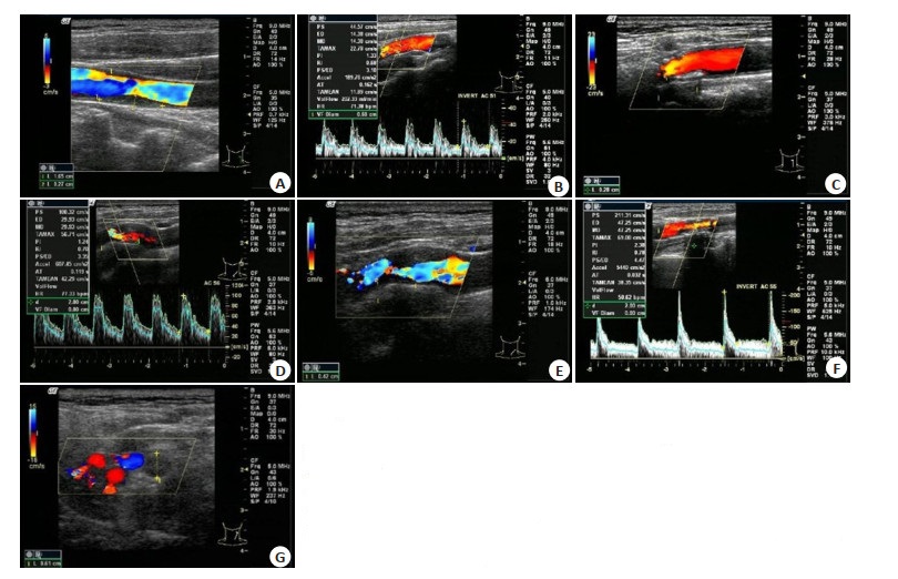
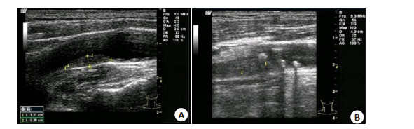
ObjectiveTo investigate the value of carotid duplex ultrasound and transcranial Doppler (TCD) in the diagnosis of carotid artery stenosis, vertebro-basilar artery stenosis and atherosclerotic plaque, and analyze their consistencies with cerebral angiography. Methods106 patients with atherosclerotic cerebral infarction who were treated between January 2019 and June 2020 were selected as the observation group. Meanwhile, 53 patients with atherosclerotic hypertension and/or hyperlipidemia and without cerebral infarction were selected as the control group. The detection rates of severer than moderate to severe stenosis of carotid artery and vertebro-basilar artery and unstable plaques by carotid duplex ultrasound and TCD were compared between the 2 groups. Cerebral angiography was taken as the golden standard to analyze the diagnostic consistency of carotid duplex ultrasound and TCD. ResultsThe detection rates of moderate stenosis, severe stenosis and complete occlusion of carotid artery and vertebro-basilar artery, and the detection rate of carotid atherosclerotic unstable plaques in the observation group were higher than those in the control group (P < 0.05). The sensitivity, specificity and accuracy of carotid duplex ultrasound combined with TCD in detection of severer than moderate to severe stenosis were 93.75%, 88.46% and 92.45%, with high consistency (Kappa=0.801). For detection of unstable plaques, the sensitivity, specificity and accuracy were 87.67%, 87.88% and 87.74%, with high consistency (Kappa=0.725). ConclusionCarotid duplex ultrasound and TCD are accurate, non-invasive and convenient in the diagnosis of carotid artery stenosis, vertebro-basilar artery stenosis and atherosclerotic plaque, which is of great significance for prevention, diagnosis and cause analysis of cerebral infarction.
2021, 44(2): 276-280.
doi: 10.12122/j.issn.1674-4500.2021.02.12
Abstract:
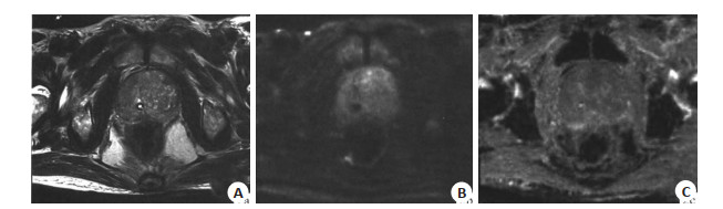
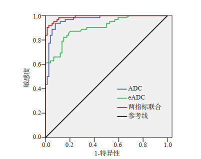
ObjectiveTo investigate the application value of 1.5 T magnetic resonance diffusion weighted imaging (MR DWI) in evaluating benign and malignant prostate lesions. MethodsThe relevant data of 136 patients with prostate lesions admitted to the hospital from October 2017 to August 2020 were retrospectively analyzed. All patients were subjected to MR DWI examination, and the diagnostic value of DWI in benign and malignant prostate lesions was analyzed. The ADC value, eADC value, and DWI semi-quantitative classification of patients with benign and malignant prostate lesions were compared. ResultsTaking pathological examination as the golden standard, the sensitivity, specificity and accuracy of DWI for diagnosing malignant prostate lesions were 95.16%, 98.65%, and 97.06%, and the Kappa value was 0.941. The ADC value of malignant lesions was significantly lower than that of benign lesions, and the eADC value was significantly higher than that of benign lesions (P < 0.05). ROC curve showed that the AUC values of ADC value, eADC value and combination of the two for diagnosing benign and malignant prostate lesions were 0.902, 0.967, and 0.990, respectively. DWI semi-quantitative classification of patients with malignant lesions was dominated by grade 4-5, which of patients with benign lesions was dominated by grade 1-3 (P < 0.05). Conclusion1.5 T MR DWI is of great value in evaluating benign and malignant prostate lesions.
2021, 44(2): 281-285.
doi: 10.12122/j.issn.1674-4500.2021.02.13
Abstract:
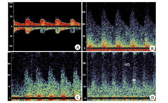
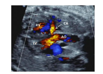
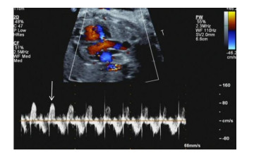
ObjectiveTo explore the value of coronary artery dilatation in prognosis of fetal growth restriction (FGR). MethodsSeventy-three fetuses with fetal growth restriction (FGR) confirmed by ultrasound in our center from January 2019 to December 2020 were collected. The blood flow spectrum characteristics included coronary artery (CA) dilatation, umbilical artery (UA), middle cerebral artery (MCA) and venous duct (DV) were observed by prenatal ultrasound. The fetuses were divided into two parts: poor prognosis group and good prognosis group according to fetal pregnancy outcome. After comparing the baseline data and blood flow parameters, a logistic regression model based on the characteristics of ultrasonic variableas was established. Receiver operating characteristic curve (ROC) was drawn to evaluate the predictive value of logistic regression model for fetal poor prognosis. ResultsThere were 46 cases in good prognosis group and 27 cases in poor prognosis group. The blood flow parameters including abnormal UA, abnormal DV, CA dilatation had significant differences (P < 0.05), whereas abnormal MCA did not (P>0.05) Logistic regression analysis show that CA dilatation, abnormal UA, and abnormal DV were independent risk factors for fetal poor prognosis(OR values=9.715, 4.956, 11.291, respectively). The Logistic regression model based on CA dilation, abnormal UA and abnormal DV predicted that the area under the curve, the sensitivity and specificity of poor prognosis of FGR were 0.874, 81.8% and 91.30%, respectively. ConclusionCoronary artery dilatation was an independent risk factor for poor prognosis in FGR fetuses. Logistic regression model based on three abnormal blood flow variables, including CA dilation, abnormal UA and abnormal DV could effectively predict the poor prognosis of FGR.
2021, 44(2): 286-290.
doi: 10.12122/j.issn.1674-4500.2021.02.14
Abstract:
ObjectiveTo analyze the developmental characteristics of follicles and endometrium in patients with infertility and provide precise dependence for clinical treatment. MethodsA total of 128 patients with infertility were selected and regarded as the observation group, and 128 women with successful pregnancy were regarded as the control group. The monitoring cycle was from the 8th day of the menstrual cycle to the ovulation period for 6 consecutive cycles. The differences in the monitoring results between the two groups were compared. ResultsCompared with the control group, the number of ovulation follicles, the maximum diameter of follicles, the daily growth diameter of follicles and the thickness of endometrium were all lower in the observation group, and the differences were statistically significant (P < 0.05). In the control group, there were 4 cases with normal ovulation type, which were significantly lower than 126 cases in the control group, and the differences were statistically significant (P < 0.05). In the observation group, there were 42 cases, 64 cases and 84 cases with luteinized unruptured follicles, flat follicles and non-dominant follicles respectively, which were significantly higher than 2 cases, 6 cases and 1 case in the control group, and the differences were statistically significant (P < 0.05). Among patients with type B endometrium in the observation group, there were 1 case and 18 cases with normal ovulation type and abnormal ovulation type, which were significantly lower than 125 cases and 3 cases in the control group, and the difference was statistically significant (P < 0.05). Among the patients with non-dominant follicles in the observation group, there were 76 cases and 7 cases with type A endometrium and type C endometrium, which were significantly higher than 0 cases and 0 cases in the control group, and the difference was statistically significant (P < 0.05); Among the patients with luteinized unruptured follicle type in the observation group, there were 11 cases and 15 cases with type A endometrium and type C endometrium, which were significantly higher than 0 cases and 0 cases in the control group, and the differences were statistically significant (P < 005). ConclusionThe developmental characteristics of follicles and endometrium in patients with infertility are abnormal. Realizing "early detection, early diagnosis, and early treatment" by transvaginal ultrasound dynamic monitoring for patients with infertility can effectively increase the pregnancy rate.
2021, 44(2): 291-294.
doi: 10.12122/j.issn.1674-4500.2021.02.15
Abstract:
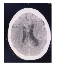
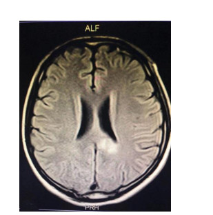
ObjectiveTo analyze the imaging manifestations and diagnostic value of MRI for corpus callosum injury caused by traumatic brain injury. MethodsThe general data of 103 patients with corpus callosum injury caused by traumatic brain injury who were diagnosed and treated in the hospital from February 2016 to November 2020 were retrospectively analyzed. All of the patients received MRI and CT examinations, and the imaging were analyzed. The diagnostic efficiencies of MRI and CT were compared, and MRI parameters of different types of corpus callosum injuries were analyzed. ResultsMRI examination showed no obvious enhancement but clear edges in 99 patients, and enhancement or unclear edges in 4 patients after enhanced scan. There were 45 patients with non-hemorrhagic injury, showing equal signal or low signal on T1WI, slightly higher signals on both T2WI and DWI. Besides, no bleeding signal was observed in each scan sequence. For 58 patients with hemorrhagic injury, there were mixed signals on each scan sequence, with bleeding signals. There were no significant differences between MRI and CT in the detection rates of genu injury, splenium injury, and body genu injury (P>0.05), but the detection rate of body injury by MRI was significantly higher than that by CT (P < 0.05). There was no significant difference between MRI and CT in the diagnostic accuracy for hemorrhagic injury (P>0.05), but the diagnostic accuracy rate of MRI was significantly higher than that of CT for non-hemorrhagic injury, corpus callosum atrophy, softening lesions and gliosis (P < 0.05). There were no significant differences in DWI sequence display and DAI scores between the two groups (P>0.05). The T2WIFLAIR and T2WI/T1WI display of hemorrhagic injury was significantly better than that of non-hemorrhagic injury (P < 0.05). ConclusionCompared with CT, MRI can show more lesions in the corpus callosum caused by traumatic brain injury, and it can be used to differentiate and diagnose hemorrhagic and non-hemorrhagic lesions, and accurately locate the specific location of corpus callosum injury.
2021, 44(2): 295-298.
doi: 10.12122/j.issn.1674-4500.2021.02.16
Abstract:
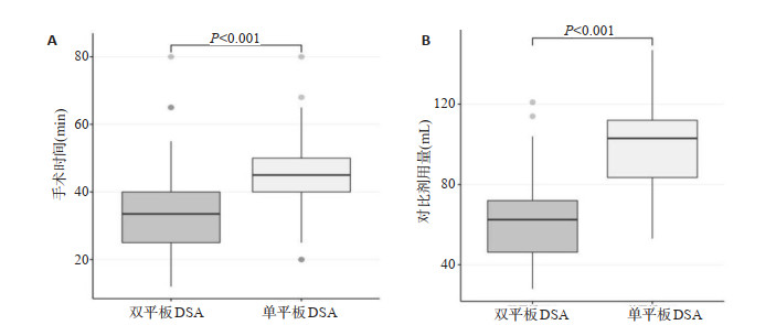
ObjectiveTo analyze the application values of bi- plane digital subtraction angiography (DSA) in whole cerebral angiography. MethodsThe patients who underwent cerebral angiography were randomly divided into the experimental group (n=50, using bi-plane DSA) and the control group (n=59, using single-plane DSA). And the operation time, the amount of contrast agent, the number of movie sequences, the unmber of photographic frames, the dose area product (DAP) and air kerma (AK) of the two groups were compared. ResultsThe operation time of the experimental group [33.50 (25.00, 40.50) min vs 45.00 (40.00, 50.00) min, P < 0.001], the amount of contrast agent (62.68 ± 22.40 mL vs 100.46 ± 20.91 mL, P < 0.001), and the number of movie sequences [10.00 (8.00, 13.25) vs 14.00 (12.00, 16.00), P < 0.001] were lower than the control group. The difference is statistically significant. The two groups of patients have difference in dose area product, air kerma, and number of photographic frames. The difference was not statistically significant (P>0.05). The number of movie sequences in both groups was positively correlated with operation time (r=0.637, P < 0.001) and intraoperative contrast agent dosage (r=0.586, P < 0.001). ConclusionThere is no significant difference between the radiation dose of bi- plane DSA angiography and conventional single-plate DSA. However, the use of bi-plane DSA angiography significantly reduces the operation time and the amount of contrast agent during the operation, which is beneficial to patients.
2021, 44(2): 299-303.
doi: 10.12122/j.issn.1674-4500.2021.02.17
Abstract:
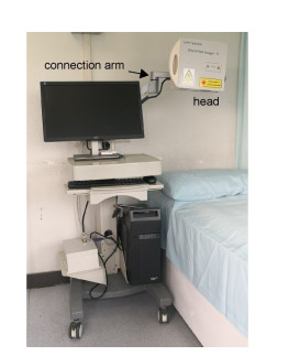
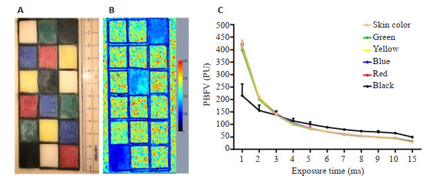
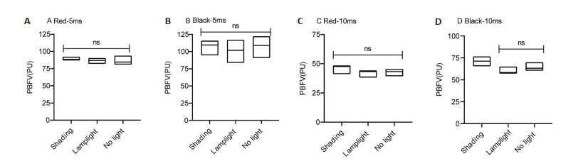
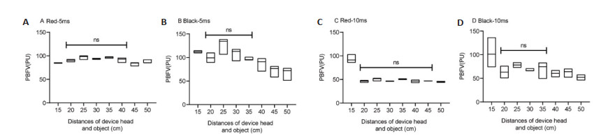
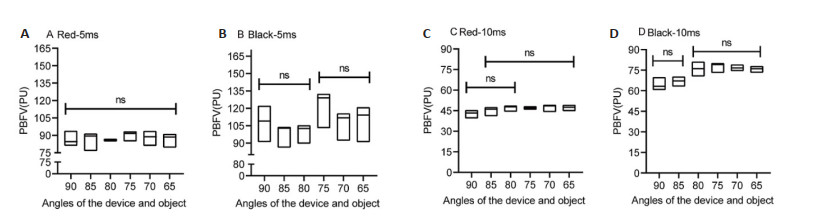
ObjectiveTo find out the factors that affect the measurement results of laser speckle contrast imaging (LSCI) by simulating using conditions. MethodsWe simulated using conditions in the pain clinic of The First Affiliated Hospital of Jinan University. The LSCI device we used is a commercial product from SIM Opto-Technology Co., Wuhan, China (Laser Speckle Blood Flow Imager Ⅱ). Using a silicon-imitated skin model, we collected the results changes of Laser Speckle Blood Flow Imager Ⅱ data in four using conditions, including different exposure times (1-15 ms), different lighting situations (shading condition, lamplight condition, and no light condition), different distances between the LSCI device and object (15, 20, 25, 30, 35, 40, 45, 50 cm), and different angles between the LSCI device laser beam and object (90°, 85°, 80°, 75°, 70°, 65°). ResultsThe influence of the exposure time: The measurement results of the six colors area of the object decreased when the setting of exposure time increased, and particularly showed different change pattern when the object color was black, compared to the other five colors. The influence of the light condition: When the detected area was red, the laser speckle results showed no significant difference in three light conditions; when the detected area was black, the laser speckle results showed no significant difference only in lamplight conditions and no light conditions. The influence of the distance of the device and object: When the detected area was red, and the distances were between 20-40 cm, the laser speckle results showed no significant difference; when the detected area was black, and the distances were between 20-35 cm, the laser speckle results showed no significant difference. The influence of the angle between the device laser beam and the object: When the detected area was red, and the exposure time was 5 ms, the laser speckle results showed no significant difference in all six angles between the device laser beam and object; and when the exposure time was 10ms, the laser speckle results showed no significant difference in two ranges of 80°-90° and 65°-85°. When the detected area was black, and the exposure time was 5 ms, the laser speckle results showed no significant difference in two ranges of 80°-90° and 65°-75°; when the exposure time was 10 ms, the laser speckle results showed no significant difference in two ranges of 85°-90° and 65°-80°. ConclusionExposure time, lighting condition, the distance between the LSCI device and object, the angle between the device laser beam and the object are all factors that affect the measurement results. In addition, compared to other colors, measurement results varied from the other results when the color of object is black, we should reconsider the comparability of LSCI blood flow value between different people.
2021, 44(2): 304-308.
doi: 10.12122/j.issn.1674-4500.2021.02.18
Abstract:
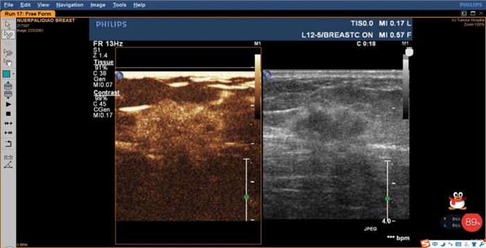
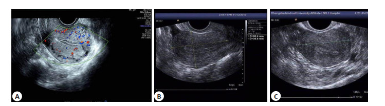
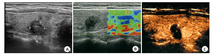
ObjectiveTo explore the application value of contrast-enhanced ultrasound combined with elastography in blood perfusion analysis and benign and malignant evaluation of breast nodules. MethodsThe study retrospectively analyzed clinical data of 102 patients with breast nodules treated in the hospital between January 2017 and June 2020. All patients were subjected to pathological examination, conventional ultrasound, contrast- enhanced ultrasound and ultrasound elastography. According to the pathological results, patients were divided into benign group and malignant group. Blood perfusion of the two groups was compared, and the diagnostic efficiencies of contrast-enhanced ultrasound and elastography for benign and malignant breast nodules were analyzed. ResultsIn the 102 patients with breast nodules, 69 cases with benign nodules (benign group) and 33 cases with malignant nodules (malignant group) were detected by surgical pathology. The maximum diameter of nodules in the malignant group was larger than that in the benign group (P < 0.05). The peak intensity (PI) of the malignant group was significantly lower than that of the benign group (P < 0.05), but no significant differences were found in time to peak (TP) and mean transit time (MTT) (P>0.05). In the malignant group, comparison of PI in different regions showed tissue around the nodule >nodule margin > nodule center (P < 0.05), but no significant differences were found in TP and MTT in different regions (P>0.05). In the benign group, no significant differences were found in PI, TP and MTT in different regions (P> 0.05). The ratio of 0-2 points was significantly lower and the ratio of 3-4 points was significantly higher in the malignant group than in the benign group (P < 0.05). The sensitivity, specificity and accuracy of contrast- enhanced ultrasound in diagnosing benign and malignant breast nodules were 83.78%, 84.62% and 84.31%, which of ultrasound elastography were 74.42%, 89.83 and 83.33%. The sensitivity, specificity and accuracy of combined diagnosis were 91.43%, 94.03%, and 93.14%, respectively. The accuracy of combined diagnosis was higher than that of contrast-enhanced ultrasound or ultrasound elastography alone (P < 0.05). ConclusionBoth contrast-enhanced ultrasound and ultrasound elastography can be used to diagnose breast nodules. The differences in quantitative parameters of contrast-enhanced ultrasound can reflect microvessel distribution characteristics of the nodule to a certain extent. Contrast-enhanced ultrasound combined with elastography can improve the diagnostic accuracy for benign and malignant breast nodules
2021, 44(2): 309-313.
doi: 10.12122/j.issn.1674-4500.2021.02.19
Abstract:
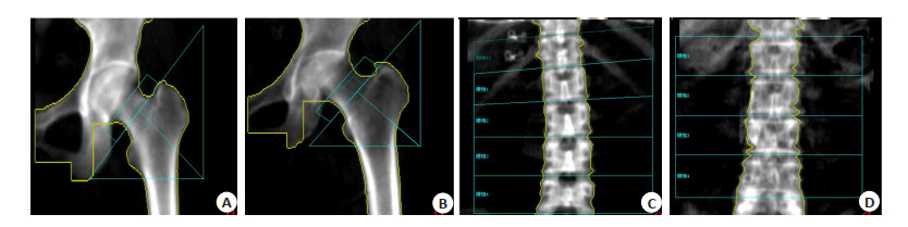
ObjectiveTo investigate the correlation between bone mineral density (BMD) and bone turnover rate in type 2 diabetes mellitus (T2DM) women. MethodsWe collected the clinical data of 201 women with T2DM in the Third Affiliated Hospital of South Medical University. Bone mineral density (BMD) at lumbar spine, the left femoral neck and the total hip were measured with dual energy X-ray absorptiometry. The selected subjects were divided into normal bone mass group (n=85, T>-1)、osteopenia group (n=87, -2.5 < T < -1) and osteoporosis group (n=29, T < -2.5). The T-score of bone formation and the T-score of bone resorption were calculated by testing osteocalcin and β-C-terminal telopeptide of typeⅠcollagen (β-CTX) respectively. The bone turnover rate and the bone remodeling rate in T2DM patients were evaluated by comparing the T-score of the bone turnover and bone remodeling between osteoporosis group and normal bone mass group, to investigate the correlation between the T-score of the bone turnover and bone remodeling and BMD in T2DM patients. ResultsThe T-score of the bone turnover rate showed significant differences between the osteoporosis group and the normal group in T2DM patients (P= 0.041). The T-score of the bone turnover rate was negatively correlated with hip BMD in T2DM patients (r=- 0.14, P=0.049). Controlling the glycosylated hemoglobin factors, the T-score of the bone turnover rate was negatively correlated with hip BMD in T2DM patients (r=-0.144, P=0.043). ConclusionWith the bone turnover rate increasing, BMD decreased and the risk of T2DM patients with hip osteoporosis would be increased.
2021, 44(2): 314-320.
doi: 10.12122/j.issn.1674-4500.2021.02.20
Abstract:
ObjectiveTo analyze the diagnostic value of dual-phase bronchial artery 64-slice CT for pulmonary nodules. MethodsA total of 120 patients with pulmonary nodules (under 3 cm in diameter) found by 64-slice spiral CT in our hospital between September 2016 and September 2020 were enrolled in the study. All of them were subjected to bronchial artery dualphase enhanced scan and targeted scan of the lesion. Pathology confirmed that there were 65 cases with malignant pulmonary nodules (malignant group), 30 cases with preinvasive nodules (carcinoma in situ) (preinvasive group), and 25 cases with benign pulmonary nodules (benign group). The detection rates of CT signs, bronchial artery morphology, image quality, CT values of plain scan and enhanced scan, and enhancement after dual-phase enhanced scan were compared between the two groups. The diagnostic value of bronchial artery dual-phase enhanced scan for malignant pulmonary nodules was analyzed. ResultsIn the malignant group, the proportions of deep lobulation sign, spicule sign, spiculate protuberance, blood vessel pack sign, vacuole sign, and pleural indentation sign were higher, the blood vessels and bronchi in pulmonary ground glass shadow mainly were type Ⅱ and Ⅲ. In the preinvasive group, the proportions of calcification and satellite lesions were higher, the blood vessels and bronchi in pulmonary ground glass shadow mainly were type Ⅳ. In the benign group, the proportion of square signs was higher, the blood vessels and bronchi in the pulmonary ground glass shadow mainly were type Ⅰ and Ⅱ (P < 0.05). The proportion of vascular lake was higher in the malignant group than in the preinvasive group and the benign group (P < 0.05). There were no statistically significant differences in the proportions of aggregation, tortuosity and interruption, image quality and CT value of plain scan among the 3 groups (P>0.05). The CT values of enhance scan at 30-45 s and 90-120 were higher in the malignant group and the benign group than in the preinvasive group (P < 0.05), and CT values of enhanced scan at 90~120 s were lower in the malignant group than in the benign group (P < 0.05). Statistically significant differences were found in enhancement after dual-phase enhanced scan among the 3 groups (P < 0.05). Besides, moderate and obvious enhancement was the majority in the malignant group, showing a fast-rising and fast-falling pattern. The sensitivity, specificity and accuracy of bronchial artery dual-phase 64-slice CT for diagnosing the malignancy of pulmonary nodules were 84.61%, 76.36%, and 80.83%, respectively. ConclusionThe bronchial artery dual-phase 64-slice CT has certain value in the diagnosis of benign and malignant pulmonary nodules. Accurate diagnosis can be made based on the CT signs, vascular and bronchial types in pulmonary ground glass shadow, bronchial morphology, and the degree of enhancement.
2021, 44(2): 321-326.
doi: 10.12122/j.issn.1674-4500.2021.02.21
Abstract:
ObjectiveTo explore the application value of ultrasound contrast quantitative analysis parameters in evaluating prognostic factors of invasive breast cancer. Methods110 cases of invasive ductal breast cancer were pre-operatively performed gray-scale ultrasound contrast. QLAB software was used to analyzed to generate time-intensity curve. The blood perfusion parameters of surrounding normal tissue and tumor tissue were compared. The relationship between quantitative parameters of ultrasound contrast perfusion of invasive breast cancer and prognostic factors were analyzed. Finally, ROC curve analysis was performed on the quantitative perfusion parameters of each ultrasound contrast. ResultsCompared with normal tissues, the peak intensity (PI) of tumor tissues was significantly increased, the peak time (TTP) and the mean transit time (MTT) were significantly shorter, the difference was statistically significant (P < 0.05). There is no statistically significant difference in the rise time (RT) (P>0.05). Compared with patients with high-to-medium differentiated invasive breast cancer, the RT and TTP of patients with poorly differentiated invasive breast cancer were significantly prolonged, and PI was significantly increased. The difference was statistically significant (P < 0.05). Compared with MTT, the difference was not statistically significant (P>0.05). The PI of invasive breast cancer patients with tumor diameter >20 mm was significantly higher than that of patients with tumor diameter ≤20 mm (P < 0.05), and there was no statistically significant difference between TTP, MTT and RT. Compared with patients with invasive breast cancer without lymph node metastasis, patients with lymph node metastasis had elevated PI and prolonged RT. The difference was statistically significant (P < 0.05), while the difference between TTP and MTT was not statistically significant (P>0.05). ER-negative and PR-negative invasive breast cancer patients were significantly higher PI than ER-positive and PR-positive patients, the difference was statistically significant (P < 0.05), but there was no statistically significant difference between TTP, MTT and RT (P>0.05). There is no statistically significant difference between PI-2, TTP, MTT, RT of patients with and without Her-2 expression (P>0.05). ROC curve analysis results show that PI has a medium-low diagnostic efficiency in the differential diagnosis of ER negative expression and PR negative expression. The cutoff point of PI is 12.975dB, and the standard errors are 0.073 (95%CI: 0.562-0.849), 0.074 (95%CI: 0.540-0.828), sensitivity and specificity were 77.40%, 72.20%, 65.20%, 66.70%, and the area under the curve was 0.705, 0.688, respectively. ConclusionPI, TTP, RT can reflect the blood perfusion in the lesion, which is helpful to the prognosis assessment of patients with invasive breast cancer.
2021, 44(2): 327-331.
doi: 10.12122/j.issn.1674-4500.2021.02.22
Abstract:
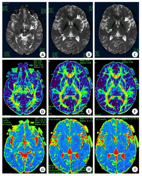
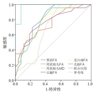
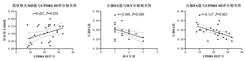
ObjectiveTo investigate the value of MR diffusion tensor imaging (DTI) of midbrain and basal ganglia nuclei in the diagnosis and severity assessment of Parkinson's disease (PD). MethodsThe clinical and MR data of 27 PD patients (PD group) and 23 normal controls (NC group) who visited our hospital from July 2017 to November 2020 were retrospectively analyzed. Routine MR scanning and DTI examination were performed on PD group and NC group. And DTI parameters, including the fractional anisotropy (FA) and mean diffusivity (MD), were measured for bilateral substantia nigra, red nucleus, caudate head, thalamus, globus pallidus and putamen of each subject. The FA and MD values of each nucleus were compared between PD and NC groups. This study analyzed the diagnostic effectiveness of single and combined application of parameters with inter-group differences and their correlation with course of disease, Hoehn-Yahr grade and exercise part of unified Parkinson's disease rating scale (UPDRS Ⅲ) score. ResultsThe FA values of substantia nigra, caudate head, thalamus, globus pallidus and putamen in PD group were all lower than those in NC group, while the MD values of caudate head were higher than that in NC group (all P < 0.05). The receiver operating characteristic curve analysis showed that the area under the curve (AUC) of combined application of FA values of substantia nigra, caudate head, thalamus, globus pallidus and putamen as well as MD value of caudate head for PD diagnosis was 0.981, which was significantly greater than the AUC of each parameter applied alone for PD diagnosis (all P < 0.05). The MD value of caudate head was positively correlated with UPDRS Ⅲ score of PD patients (r=0.402, P=0.038), and the FA value of thalamus was negatively correlated with the Hoehn-Yahr grade (r=-0.490, P=0.009) and UPDRS Ⅲ score (r=-0.547, P=0.003) of PD patients. ConclusionThe DTI parameters of midbrain and basal ganglia nuclei are helpful for the diagnosis and severity assessment of PD. The combined application of DTI parameters of multiple nuclei has better diagnostic efficacy.
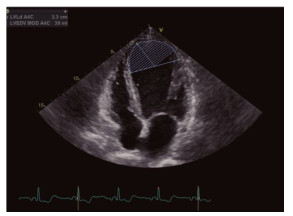
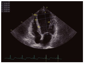
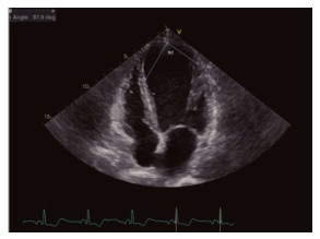
2021, 44(2): 332-335.
doi: 10.12122/j.issn.1674-4500.2021.02.23
Abstract:
ObjectiveTo explore the correlation between the apical morphology and cardiac function of left ventricular and plaque vulnerability in patients with coronary heart disease. Methods53 patients with coronary heart disease in our hospital from January 2018 to January 2020 were enrolled. Echocardiography and coronary CT angiography (CTA) were performed in all patients. According to CTA results, patients were divided into stable plaque group (n=28) and vulnerable plaque group (n= 25). Various indexes were compared between groups, including left ventricular end systolic diameter (LVESD), left ventricular end diastolic diameter (LVEDD), left ventricular ejection fraction (LVEF), anterior to posterior diameter (DAP), apical spherical index (Siap), end diastolic apex angle and end systolic apex angle. Correlation between the apical morphology and cardiac function of left ventricular and plaque vulnerability was analyzed using Logistic regression analysis. ResultsLVEDD and LVESD in stable plaque group were significantly lower than those in vulnerable plaque group (P < 0.05), and LVEF was significantly higher than that in vulnerable plaque group (P < 0.05). The left ventricular Dap, end-diastolic apex Angle and endsystolic apex Angle in stable plaque group were significantly lower than those in vulnerable plaque group (P < 0.05), and Siap was significantly higher than that in vulnerable plaque group (P < 0.05). Plaque vulnerability was an independent risk factor affecting left ventricular Dap, end-diastolic apex angle, end-systolic apex angle, LVEDD, LVESD, and LVEF (P < 0.05). ConclusionThe morphology and function of left ventricular apex in patients with coronary heart disease have significant correlation with plaque vulnerability.
2021, 44(2): 336-340.
doi: 10.12122/j.issn.1674-4500.2021.02.24
Abstract:
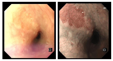
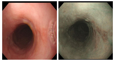
ObjectiveTo explore the value of high-definition intelligent electronic staining endoscopy (i-Scan) combined with magnifying endoscopy in the diagnosis of early cancer and precancerous lesions of the esophagus. MethodsWe selected the endoscopy data of 100 patients with early esophageal cancer (early cancer group) and 200 patients with esophageal precancerous lesions (precancerous lesions group) confirmed by pathological examination in our hospital. The pathological results was treated as the gold standard. The detection rate and diagnostic indicators for the diagnosis of early esophageal cancer and precancerous lesions under i-Scan combined with magnifying endoscopy and white light endoscopy alone and in combination were calculated. Resultsi-Scan had a diagnostic detection rate of 88.00% for early esophageal cancer, 82.50% for precancerous lesions, 28.00% for mild dysplasia, 29.00% for moderate dysplasia, white light endoscopy. The diagnosis and detection rate of early cancer of the esophagus was 57.00%, the detection rate of precancerous lesions was 56.00%, the detection rate of mild dysplasia was 16.00%, and the detection rate of moderate dysplasia was 17.00%. The detection rate of I-Scan was higher than that of white light endoscopy and the difference was statistically significant (P < 0.05). The detection rate of I-Scan for severe esophageal mucosal dysplasia was 25.50% compared with 22.50% of white light endoscopy, with no significant difference (P>0.05). 100 patients with early esophageal cancer Twelve cases were diagnosed as type A, 2 of patients with moderate dysplasia were diagnosed as type B1, and 4 of patients with severe dysplasia were diagnosed as type B1. White light endoscopy differential diagnosis of early esophageal cancer, esophageal cancer. The sensitivity of precancerous lesions was 57.00%, and the specificity was 56.00%. The sensitivity of I-Scan for differential diagnosis of early esophageal cancer and precancerous lesions of esophagus was 88.00%, and the specificity was 82.50%. The sensitivity for diagnosing early esophageal cancer and precancerous lesions of esophagus was 88.00%, and the specificity was 97.00%. Conclusioni-Scan combined with magnifying endoscopy has high sensitivity and specificity in the differential diagnosis of early cancer and precancerous lesions of the esophagus.
2021, 44(2): 341-345.
doi: 10.12122/j.issn.1674-4500.2021.02.25
Abstract:
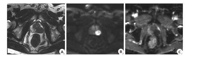
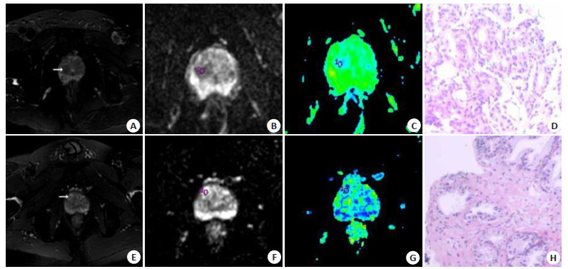
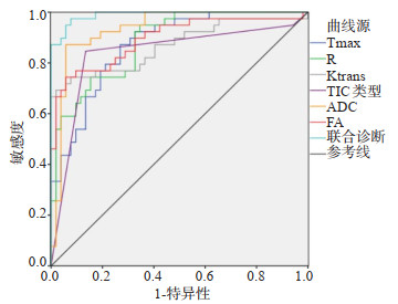
ObjectiveTo investigate the value of dynamic contrast-enhanced MRI (DCE-MRI), diffusion tensor imaging (DTI) and their combination in the differential diagnosis of central prostate nodules. MethodsFrom January 2019 to June 2020, 79 patients with central prostate nodules in our hospital, 91 nodules, were examined by DCE-MRI and DTI, and the differences of parameters between benign and malignant nodules were analyzed. ResultsThe pathological diagnosis was made, among the 91 nodules; 39 were malignant and 52 were benign. The Tmax of DCE-MRI parameters of malignant nodules was 103.36 ± 32.50s, which was significantly lower than that of benign nodules (P < 0.05), while the enhancement rate and Ktrans were (4.90± 1.10)% and 12.20±4.11 min-1, which were significantly higher than those of benign nodules (P < 0.05). The proportion of TIC type Ⅲ in malignant nodules was 84.62%, which was significantly higher than that in benign nodules (P < 0.05). The proportion of TIC type Ⅱ in benign nodules was 80.77%, which was significantly higher than that in malignant nodules (P < 0.05). There was no significant difference in the proportion of TIC type Ⅰ between benign and malignant nodules (P>0.05). The ADC value of malignant nodules was (1.03±0.22)×10-3 mm2/s, which was significantly lower than that of benign nodules (P < 0.05), while the FA was 0.32 ± 0.10, which was significantly higher than that of benign nodules (P < 0.05). The area under ROC curve of Tmax, enhancement rate, Ktrans, TIC type, ADC, FA and combined diagnosis of central prostate malignant nodules were 0.870, 0.883, 0.868, 0.838, 0.903, 0.885 and 0.933, respectively (P < 0.05). ConclusionDCE-MRI, DTI and combined diagnosis have good application value in the differential diagnosis of central prostate nodules and is worthy of clinical use.
2021, 44(2): 346-349.
doi: 10.12122/j.issn.1674-4500.2021.02.26
Abstract:
ObjectiveTo explore the clinical value of CT target reconstruction combined with MRI scan in the diagnosis of solitary pulmonary nodules of early and mid-term peripheral lung cancer. MethodsOne hundred and eighteen patients with solitary pulmonary nodules peripheral lung cancer admitted to our hospital from August 2017 to August 2020 were selected. The patients received CT target reconstruction and MRI scan for staging examinations. The pathological characteristics of the two examination methods were analyzed. Finally, postoperative pathological examination was used as the gold standard to analyze the accuracy and diagnostic value of the two examination results. ResultsCT target reconstruction indicated that there were 38 patients in T1 stage, 35 in T2 stage, 30 in T3 stage, 15 in T4 stage, and 73 early and mid-term patients, with an accuracy rate of 91.21%. MRI scan diagnosis indicated there were 39 in T1 stage, 33 in T2, 32 in T3, 14 in T4, and 72 in early and mid-term, with an accuracy rate of 90.00%. CT target reconstruction combined with MRI scan diagnosis indicated that there were 42 patients in T1, 37 in T2 stage, 29 in T3 stage, 10 in T4 stage, and 79 in early and mid-term, with an accuracy rate of 98.75%. The detection rate of CT target reconstruction of the internal tumor with in the cavity characteristics and the peripheral characteristics of the tumor was significantly higher than that of MRI scanning mode (P < 0.05). The detection rate of MRI examination for patients' lymph nodes, vascular cross-sections, invasion, effusion, and pleural indentation signs was significantly higher than that of CT target reconstruction examination. CT target reconstruction examination had a significantly higher detection rate of lesion calcification for patients than MRI examination (P < 0.05). The specificity and the sensitivity of CT target reconstruction combined with MRI scan for early-stage peripheral lung cancer in solitary pulmonary nodules were higher than those of individual examinations. ConclusionCT target reconstruction combined with MRI scan has a higher diagnostic value for solitary pulmonary nodules of early and mid-term stage peripheral lung cancer. The specificity and sensitivity are higher than those of individual diagnosis.
2021, 44(2): 350-354.
doi: 10.12122/j.issn.1674-4500.2021.02.27
Abstract:
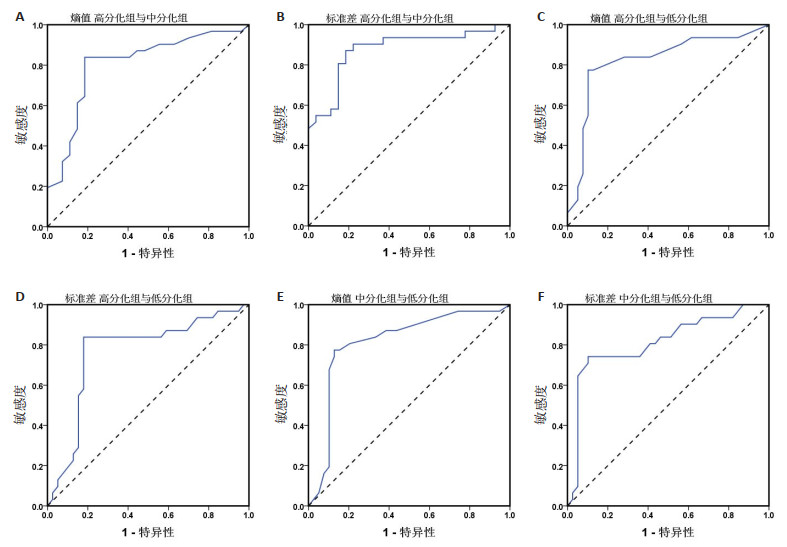
ObjectiveTo discuss application value of texture analysis (TA) based on MRI in distinguishing high and low differentiation of cervical cancer. MethodsThe clinical records of 97 patients who were pathologically confirmed cervical cancer and admitted to our hospital from January 2019 to January 2020 were retrospectively analyzed. Based on MRI, patients were divided into three groups: high differentiation group (n=27), medium differentiation group (n=31) and low differentiation group (n=39). Parameters including skewness, kurtosis, entropy and standard deviation were compared. Spearman correlation was used to analyze the correlation between cervical cancer differentiation degree and texture parameters. ROC curve was used to analyze the value of TA in differentiation of cervical cancer. ResultsAmong high, medium and low differentiation groups, there was an increase in entropy, and a decrease in standard deviation, with statistic significance (P < 0.05). The degree of differentiation of cervical cancer was negatively correlated with entropy (r=-0.269, P < 0.05) and positively correlated with standard deviation (r=0.288, P < 0.05). ROC curve analysis showed that when the entropy value was set at 5.34, the AUC of entropy in differentiating high and medium differentiation was 0.805. AUC of entropy in differentiating high and low differentiation was 0.821 when value was set at 5.18. Moreover, AUC of entropy in differentiating medium and low differentiation was 0.813 when value was set at 5.08. AUC of standard deviation in differentiating high and medium differentiation was 0.875 when value was set at 67.35. AUC of standard deviation in differentiating high and low differentiation was 0.764 when value was set at 59.97. Further, AUC of standard deviation in differentiating medium and low differentiation was 0.811 when value was set at 58.25. ConclusionThe entropy value and standard deviation of MRI texture parameters are significantly correlated with differentiation degree of cervical cancer, and it has good efficiency in identifying the differentiation degree of cervical cancer.
2021, 44(2): 355-358.
doi: 10.12122/j.issn.1674-4500.2021.02.28
Abstract:
ObjectiveTo evaluate the value of HR-MRI detection of the core types of vulnerable carotid plaques to provide valuable clues for further clinical prevention and diagnosis of cerebral infarction. MethodsA total of 150 ACI patients who were treated in our hospital from June 2017 to June 2020 were selected as the observation group. Thirty volunteers who received physical examinations during the same period were randomly selected as the healthy control group. MRI examination adoptted GE Discovery MR750 3.0 T superconducting magnetic resonance instrument, -channel phased array surface coil imaging method, scanning slice thickness 4 mm, slice spacing 1mm inspection. All patients were performed with conventional transverse T1WI, T2WI and T2 fluid attenuation inversion recovery (FLAIR), sagittal T1WI examination. ResultsThe differrences of gender, age, TC and TG levels of the observation group and the healthy control group were not significant (P>0.05). The observation group had higher LDL-C and FIB than the healthy control group (P < 0.05). HDL-C was lower in the healthy control group (P < 0.05). Serum FFA and oxLDL-C levels in the observation group were significantly higher than those in the healthy control group (P < 0.05). Patients in the observation group were divided into non-plaque group (n=35), stable plaque group (n=50) and unstable plate group (n=35) based on the stability of carotid atherosclerotic plaque determined by MRI examination (n=65). The differrences of serum FFA and oxLDL-C levels between the three groups were significant (P < 0.05). According to Spearman correlation analysis, the stability of carotid atherosclerotic plaque in ACI patients was positively correlated with serum FFA and oxLDL-C levels (P < 0.05). ConclusionThe evaluation value of HR-MRI for the core type of carotid artery vulnerable plaque is good.
2021, 44(2): 391-395.
doi: 10.12122/j.issn.1674-4500.2021.02.35
Abstract:
As the fMRI molecular imaging technology maturing, the research on the central mechanism for acupuncture effect has also entered the visualization phase, and it is widely used in the field of stroke. At present, fMRI research mainly bases on task-state and resting-state, and analysis methods include brain region activation, functional connection, brain network analysis, etc. Based on the existing literature on acupuncture-fMRI, this review summarizes the progress of central mechanism research on acupuncture in the treatment of stroke, proposes possible problems in the aspects of its experimental design and analysis methods and looks forward to its application prospect. Results show that acupuncture could activate the cerebral cortex motor area, sensory area and cerebellum, basal ganglia to different degrees, and improve the functional connection of some brain regions, regulate brain motor, sensory, language networks. However, there are still many problems that need to be improved. In the future, traditional Chinese medicine theory should be proposed as the outline combine with modern technology through multidisciplinary partnerships to build a specific brain effect network for acupuncture treatment of stroke.
As the fMRI molecular imaging technology maturing, the research on the central mechanism for acupuncture effect has also entered the visualization phase, and it is widely used in the field of stroke. At present, fMRI research mainly bases on task-state and resting-state, and analysis methods include brain region activation, functional connection, brain network analysis, etc. Based on the existing literature on acupuncture-fMRI, this review summarizes the progress of central mechanism research on acupuncture in the treatment of stroke, proposes possible problems in the aspects of its experimental design and analysis methods and looks forward to its application prospect. Results show that acupuncture could activate the cerebral cortex motor area, sensory area and cerebellum, basal ganglia to different degrees, and improve the functional connection of some brain regions, regulate brain motor, sensory, language networks. However, there are still many problems that need to be improved. In the future, traditional Chinese medicine theory should be proposed as the outline combine with modern technology through multidisciplinary partnerships to build a specific brain effect network for acupuncture treatment of stroke.
2021, 44(2): 396-399.
doi: 10.12122/j.issn.1674-4500.2021.02.36
Abstract:
Prostate cancer is a serious threat to men's health. In recent years, the incidence of prostate cancer in my country has also increased rapidly. Early detection of prostate cancer has a very positive significance for improving the survival rate. At present, the prostate cancer guidelines still need to be diagnosed by ultrasound-guided prostate biopsy, and then treated by means of active monitoring, radical resection, radiotherapy, and local radiotherapy. However, prostate biopsy increases unnecessary risks such as urinary retention, hematuria, and misses up to one-third of cancer tissues. The current treatment methods lack specificity and over-or under-treatment of patients. In recent years, the diagnosis and treatment of prostate cancer have entered the molecular level, and the non-invasive and accurate diagnosis and treatment of prostate cancer through molecular imaging has shown great development prospects. This article reviews and summarizes the application progress in prostate cancer of four common molecular imaging technologies, nuclear medicine molecular imaging, MR molecular imaging, optical molecular imaging, and ultrasound molecular imaging.
Prostate cancer is a serious threat to men's health. In recent years, the incidence of prostate cancer in my country has also increased rapidly. Early detection of prostate cancer has a very positive significance for improving the survival rate. At present, the prostate cancer guidelines still need to be diagnosed by ultrasound-guided prostate biopsy, and then treated by means of active monitoring, radical resection, radiotherapy, and local radiotherapy. However, prostate biopsy increases unnecessary risks such as urinary retention, hematuria, and misses up to one-third of cancer tissues. The current treatment methods lack specificity and over-or under-treatment of patients. In recent years, the diagnosis and treatment of prostate cancer have entered the molecular level, and the non-invasive and accurate diagnosis and treatment of prostate cancer through molecular imaging has shown great development prospects. This article reviews and summarizes the application progress in prostate cancer of four common molecular imaging technologies, nuclear medicine molecular imaging, MR molecular imaging, optical molecular imaging, and ultrasound molecular imaging.
2021, 44(2): 400-404.
doi: 10.12122/j.issn.1674-4500.2021.02.37
Abstract:
Cancer has always been a difficult problem for human beings, and conventional imaging methods have some limitations in the diagnosis of tumors. Photoacoustic imaging (PAI)is one of flourishing fields of medical imaging methods. Compared with conventional radiology techniques, it can use endogenous or exogenous contrast agents such as melanin and hemoglobin to monitor the concentration of subst-ances related to tumor angiogenesis in real time and noninvasively, or through the molecular targeting exogenous contrast agent to combine with antibodies or peptides to provide information about the structure of the tumor and its molecular information, so as to achieve morphological and functional imaging. In recent years, PAI has made a valuable contribution to the early diagnosis of cancer, the study of tumor angiogenesis, the detection of tumor microenvironment, and the monitoring of cancer progression and treatment response. According to the unique advantages of PAI in tumor imaging. The application progress of photoacoustic imaging in tumor diagnosis, management and treatment guidance were reviewed.
Cancer has always been a difficult problem for human beings, and conventional imaging methods have some limitations in the diagnosis of tumors. Photoacoustic imaging (PAI)is one of flourishing fields of medical imaging methods. Compared with conventional radiology techniques, it can use endogenous or exogenous contrast agents such as melanin and hemoglobin to monitor the concentration of subst-ances related to tumor angiogenesis in real time and noninvasively, or through the molecular targeting exogenous contrast agent to combine with antibodies or peptides to provide information about the structure of the tumor and its molecular information, so as to achieve morphological and functional imaging. In recent years, PAI has made a valuable contribution to the early diagnosis of cancer, the study of tumor angiogenesis, the detection of tumor microenvironment, and the monitoring of cancer progression and treatment response. According to the unique advantages of PAI in tumor imaging. The application progress of photoacoustic imaging in tumor diagnosis, management and treatment guidance were reviewed.