Find Duplicates
 Check Document
 Submission(new)
 Experts Office
 Editorial Office
2021 Vol. 44, No. 3
Display Method:
2021, 44(3): 417-421.
doi: 10.12122/j.issn.1674-4500.2021.03.01
Abstract:
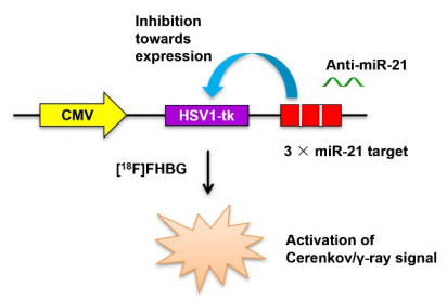
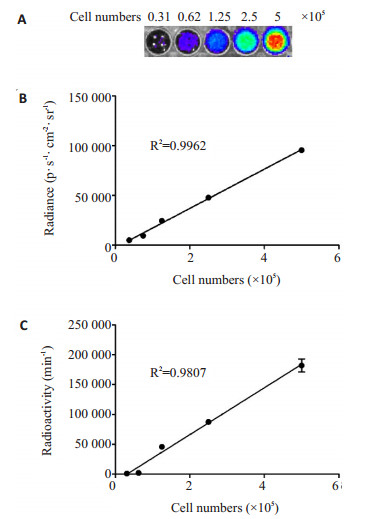
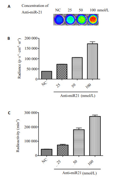
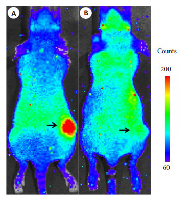
ObjectiveTo establish a novel reporter gene system based on the herpes simplex virus type-1 thymidine kinase (HSV1-tk) gene for the Cerenkov optical imaging of microRNA (miR-21) expression inside tumors. MethodsThe cytomegalovirus (CMV) promoter gene, HSV1-tk gene, and the triple complementary target sequences for miR-21 were constructed in series to form the MV-HSV1-tk-3×miR-21t reporter gene. The CMV-HSV1-tk-3×miR-21t gene was inserted into the pcDNA3.1 vector and transfected to the A549 cell line. The transfected cells which can stably express the reporter gene aforementioned (A549T cells) were incubated with 9-[4-[18F]fluoro-3-(hydroxymethyl)butyl)guanine ([18F]FHBG), or treated with antisense miR-21 ribonucleic acid (Anti-miR-21) in gratitude dose and then incubated with [18F]FHBG. The Cerenkov optical signal and γ radioactive counts per minute were acquired, respectively. Except that, we set up subcutaneous A549T xenograft models using athymic nude mice and divide them into two groups: one group was intratumorally injected with Anti-miR-21, and another group was the control injected with the control of RNA mixture only. Then these two groups were administrated with [18F]FHBG and in vivo scanned in Cerenkov modality. ResultsAfter uptaking [18F]FHBG, the A549T cells can transmit marked optical signal and γ radioactivity in a linear positive correlation with the number of cells(R2=0.9962, 0.9807). The intensity of the optical signal and γ radioactivity is dose-dependently correlated with the Anti-miR-21 added into the A549T cell culture(P < 0.05). The in vivo Cerenkov images of the subcutaneous xenograft model show that compared to the group intratumorally injected with control RNA, the signal from the xenograft of the group injected with Anti-miR-21 is visually higher with a sharp contrast. ConclusionA novel reporter gene system employing the regulation on tracer uptake via microRNA for the Cerenkov imaging of endogenous miR-21 expression has been established successively.
2021, 44(3): 422-426.
doi: 10.12122/j.issn.1674-4500.2021.03.02
Abstract:
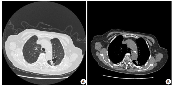
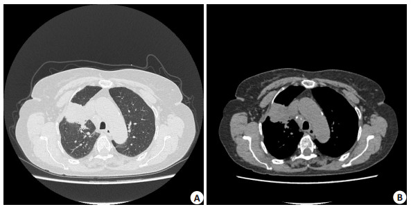
ObjectiveTo evaluate the difference of clinical and imaging findings in sinonasal NK/T cell lymphoma (NKTCL) and diffuse large B cell lymphoma (DLBCL) and to improve the diagnostic accuracy. MethodsPathology-proven fifteen patients with NKTCL and nine patients with DLBCL were collected in this study. Patient's age, gender distribution, clinical symptoms, mass location, volume, morphology, T1WI and T2WI signal intensity, enhancement degree and homogeneity, surrounding tissue invasion of the two groups were retrospectively analyzed. ResultsThe mean age at diagnosis for DLBCL was significantly higher than NKTCL(P < 0.001). Gender distribution did not have significant difference between the two groups(P= 0.351). The most common symptom for patients with NKTCL was nasal congestion (93.3%) whereas ocular signs more often occur in patients with DLBCL (55.5%). The most common region of NKTCL was nasal cavity whereas DLBCL more often located in paranasal sinus(P=0.000). NKTCL was commonly diffuse infiltration growth and DLBCL commonly in mass, but there was no significant difference between the two groups in this study(P=0.099). The lesion of DLBCL was larger than NKTCL (P=0.019). There was no difference in T1WI, T2WI signal intensity and enhancement degree (P values were 0.669, 0.118 and 0.617, respectively). NKTCL was characterized by heterogeneous enhancement and DLBCL was homogeneous enhancement (P=0.009). NKTCL often involved nasal dorsum(70.0%), and the region invasion of orbital cavity was more often in DLBCL (62.5%). The bone destruction degree was more serious in DLBCL than NKTCL. ConclusionThere are valuable differentiating clinical and imaging features between sinonasal NKTCL and DLBCL. Nasal obstruction is the most common presenting symptom in NKTCL patients, NKTCL is predominant in nasal cavity, with small volume, MR heterogeneous enhancement, and bone destruction mildly. The most common presenting symptoms in DLBCL patients are proptosis and epiphora, DLBCL often located in paranasal sinus, with large volume, MR homogeneous enhancement, and osteolytic bone destruction.
2021, 44(3): 427-434.
doi: 10.12122/j.issn.1674-4500.2021.03.03
Abstract:
ObjectiveTo construct an effective and reliable three categories prediction model to distinguish RCC subtypes based on 3D multi-phase enhanced computed tomography (CT) radiomic features (RFs). MethodsA total of 210 RCC were included in this study (143 clear cell, 25 papillary, 29 chromophobe, and 13 other RCC). The 3D multi-phase enhanced CT-based RFs were used to construct a prediction model. CT included a non-contrast phase (NCP), cortico-medullary phase (CMP), parenchyma phase (PP), excretory phase (EP), and all-phase (ALL-P), which contains all the single-phase information. The ensemble learning stratified bagging method was used to predict the RCC subtype by using LASSO regression and a 1 vs. rest logistic regression algorithm. Five-fold and external stratified cross-validation was used to assess the performance of the different prediction models. ResultsThere were 105 RFs extracted from each single phase of the CT scan. The 4 CMP, 3 PP, 1 EP, and 1 NCP RF were selected in the ALL-P model, and these RFs was no overlap with the other 4 single-phase models. The prediction efficiency of ALL-P was the best, with a diagnostic accuracy of 0.81 [area under the receiver operating characteristic curve (AUC)=0.853, precision=0.717, sensitivity=0.799, kappa=0.679). Among the four single phase models, the PP model had the best performance, with accuracy of 78.3% (AUC=0.811, precision=0.689, sensitivity=0.735, kappa=0.532). The performance of the CMP model was similar to that of the EP model, but the kappa value of the EP model was 0.285, which was significantly lower than that of the CMP model (0.446). The performance of the model in NCP was the worst, AUC was 0.693. ConclusionThe ALL-P prediction model based on 3D CT RFs is an effective and reliable method for distinguishing RCC subtypes.
2021, 44(3): 435-440.
doi: 10.12122/j.issn.1674-4500.2021.03.04
Abstract:
ObjectiveTo investigate the differential diagnosis value and incremental value of the maximum standard uptake value (SUVmax) of quantitative SPECT/CT imaging in benign and malignant bone lesions. MethodsFrom April to December in 2019, 124 patients with extraskeletal malignant tumors who underwent whole body bone scan (WBS) and quantitative SPECT/ CT were enrolled. A total of 294 abnormal tracer-positive concentration lesions was detected, and 92 normal vertebras were included as the control group. The differences of SUVmax among the benign, malignant, and control groups were analyzed. All lesions were further divided into two groups: The first group collected 46 cases totaling 108 lesions from April to July in 2019 and plotted ROC curve to obtain cut-off values of SUVmax. The second group prospectively collected 78 cases with 186 lesions from August to December 2019 and compared the diagnostic efficacy between SPECT/CT qualitative analysis and SPECT/CT qualitative+SUVmax quantitative analysis. ResultsUsing pathology and/or follow-up imaging (WBS, CT, MRI and/or PET/CT) for at least 12 months as the golden standard, 137 malignant, and 157 benign lesions were determined. The SUVmax of malignant lesions (27.29±14.44 g/mL) was significantly higher than benign lesions (16.28±10.21 g/mL, P=0.00) and the controls (6.91±1.41 g/mL, P=0.00). Using the cut-off value of SUVmax≥18.2 g/mL as the diagnostic criterion for malignant lesions obtained by the ROC curve in the first group, the sensitivities of SPECT/CT qualitative analysis and SPECT/CT qualitative + SUVmax quantitative analysis were 88.5% and 94.9%; specificities were 93.5%, and 93.5%; accuracies were 91.4% and 94.1%, respectively. The area under curve of ROC curve were 0.91(95% CI: 0.86-0.95) and 0.96(95% CI: 0.94-0.98), respectively, the differences between them were significant (P=0.01). ConclusionQuantitative SPECT/CT SUVmax has important clinical application and incremental value in the differential diagnosis of benign and malignant bone lesions.
2021, 44(3): 441-446.
doi: 10.12122/j.issn.1674-4500.2021.03.05
Abstract:
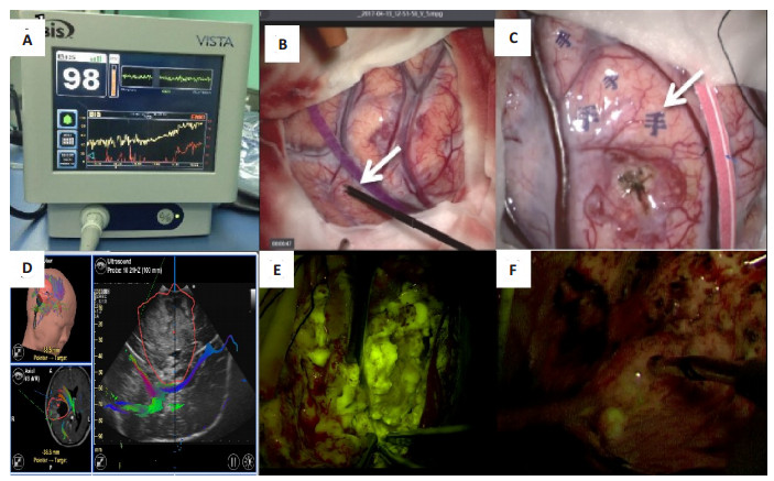
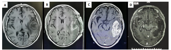
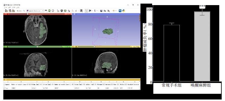
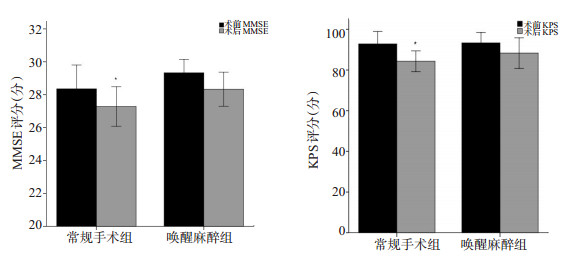
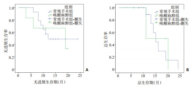
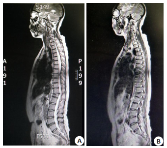
ObjectiveTo observe the effect of intraoperative awake anesthesia combined with multimodal images in the resection of recurrent glioma. MethodsThe clinical data of 20 patients with recurrent glioma who underwent reoperation in our hospital from January 2017 to December 2020 were retrospectively analyzed. They were divided into awake anesthesia group (n=6) and conventional operation group (n=14). Under the guidance of preoperative fMRI neuronavigation, the language or motor cortex was located by cortical electrical stimulation technology, and the tumor was removed under fluorescence microscope. 3D-slicer software was used to evaluate the extent of tumor resection. Mini mental state examination and Karnofsky functional state score were used to compare the cognitive function and life status of patients before and 1 week after surgery. Follow-up was conducted every 3 months. ResultsTwenty patients with recurrent gliomas were selected for reoperation, the average recurrence time was 14.65±7.64 months. There was no significant difference in preoperative baseline data between the two groups (P > 0.05). All the operations were carried out smoothly. In the awake anesthesia group, the patients fully cooperated with the operator's instructions. During the operation, 1 case of language functional area and 1 case of motor functional area were confirmed by electrical stimulation. Postoperative pathology was glioblastoma, WHO grade IV. According to 3D-slicer software measurement, the tumor burden reduction rate of awake anesthesia group was significantly higher than that of conventional operation group (P < 0.05). There were no postoperative complications and death cases in the awake anesthesia group, but 1 case of recent neurological dysfunction occurred in the conventional operation group. The Mini mental state examination and Karnofsky functional state score in the conventional operation group were significantly lower than those before operation (P < 0.05), but there was no significant difference in the awake anesthesia group (P > 0.05). Follow-up time was 3~24 months, there was no significant difference in progression free survival and overall survival between the two groups (P > 0.05). ConclusionIntraoperative awake anesthesia combined with multimodal images is helpful to maximize the resection of brain recurrent gliomas on the premise of protecting patients' neurological function, and provides a safe surgical strategy for ensuring the quality of life of patients after reoperation.
2021, 44(3): 447-451.
doi: 10.12122/j.issn.1674-4500.2021.03.06
Abstract:
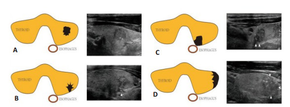
ObjectiveTo explore the risk factors of central and lateral cervical lymph node metastasis in papillary thyroid carcinoma (PTC). MethodsA total of 247 patients with PTC were retrospectively analyzed. According to the metastatic region, the patients were divided into three groups: non metastatic group (NLNM, n=152), simple central region metastasis group (CLNM, n=47) and lateral cervical region metastasis group (LLNM, n=48). Univariate analysis was used to analyze the risk factors of cervical lymph node metastasis, and multivariate analysis was used to analyze the independent risk factors of central and lateral cervical lymph node metastasis. ResultsUnivariate analysis showed that age, maximum diameter of lesion, aspect ratio and capsular contact were significantly in predicting cervical lymph node metastasis (P < 0.05). Multivariate analysis showed that age and the maximum diameter of lesion were independent predictors of clnm and llnm (P < 0.05). The capsule contact area was only independent predictor of llnm (P < 0.001, OR=2.090). Age was negatively correlated with clnm and llnm (β=-1.596, -1.285). ConclusionAge < 45 years old, maximum diameter of lesion > 10 mm and capsule contact are important predictors of cervical lymph node metastasis. The larger the contact area, the greater the possibility of metastasis.
2021, 44(3): 452-456.
doi: 10.12122/j.issn.1674-4500.2021.03.07
Abstract:
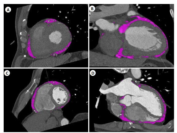
ObjectiveTo investigate whether the volume of Epicardial Adipose Tissue (EAT) measured by CT is a risk factor of Cardiac Troponin I (cTnI) elevation in patients with acute Paroxysmal Supraventricular Tachycardia (PSVT). MethodsThe objecters were patients with acute PSVT received treatment in Peking University Shenzhen Hospital from June 2014 to August 2020(n=30). The cTnI and CT Scan of the Heart were detected during the hospitalization. All the patients were divided into the cTnI positive group and the cTnI negative group. Their clinical and demographic characteristics, EAT volume, the incidence of CHD were compared. PSVT patients without CHD were divided into cTnI positive group and cTnI negative group, and their EAT volume was compared. ResultsCompared with the patients with cTnI negative at the onset of PSVT, the patients with cTnI elevation had a larger EAT volume (P=0.032). EAT volume was a related risk factor for cTnI elevation in patients with acute PSVT(r=0.351, P=0.028). In PSVT patients without diagnosed CHD, the EAT volume of cTnI positive group made no difference with that of cTnI negative group (P=0.062). ConclusionThough patients with cTnI elevation during the attack of PSVT had lager EAT volume, they should be on the alert. In addition, EAT volume is a related risk factor for cTnI elevation in PSVT patients, which provides reference information for improving the prognosis. PSVT patients with cTnI positive need more frequent follow-up to object the occurrence of cardiovascular diseases, even if CHD has not been confirmed for the time being.
2021, 44(3): 457-461.
doi: 10.12122/j.issn.1674-4500.2021.03.08
Abstract:
ObjectiveTo investigate the clinical value of MRI combined with cerebrospinal fluid (CSF) in the diagnosis and differential diagnosis of central nervous system infection. MethodsOne hundred and fifty-one patients with central nervous system infectious diseases treated in our hospital from January 2019 to may 2020 were selected, including 84 cases of viral meningitis, 35 cases of purulent meningitis and 32 cases of tuberculous meningitis. All patients were examined by MRI. The lactate dehydrogenase (LDH), protein, lactic acid (LA) and white blood cell count (WBC) in CSF were examined. ResultsThe abnormal detection rate of MRI in tuberculous meningitis was significantly higher than that in viral meningitis and purulent meningitis (P < 0.05). There was no significant difference in abnormal detection rate of MRI between viral meningitis and purulent meningitis (P > 0.05). The detection rate of meningeal enhancement in viral meningitis was significantly higher than that in purulent meningitis and tuberculous meningitis (P < 0.05). The detection rate of ring enhancement in purulent meningitis was significantly higher than that of viral meningitis (P < 0.05). The detection rate of meningeal enhancement in tuberculous meningitis was significantly higher than that in viral meningitis and purulent meningitis (P < 0.05). The LDH, protein, LA and WBC in CSF of viral meningitis were significantly lower than those in purulent meningitis and tuberculous meningitis (P < 0.05). The LDH in CSF of tuberculous meningitis was significantly higher than that in purulent meningitis (P < 0.05), while the protein and WBC were significantly lower than those in purulent meningitis (P < 0.05). Three indicators, LDH (X1), protein (X2) and WBC (X3), were screened out to establish discriminant functions, namely: Yviral meningitis=-0.056X1-0.065X2-0.062X3+ 1.168, Ypurulent meningitis=-0.041X1 + 0.102X2 + 0.089X3-1.102, Ytuberculous meningitis =0.112X1-0.057X2-0.078 + 1.032. The sensitivity, specificity and accuracy of MRI combined with CSF parameter function in the diagnosis of viral meningitis were 79.76%, 74.63% and 77.48%, the sensitivity, specificity and accuracy of diagnosing purulent meningitis were 80.00%, 81.03% and 80.79%, the sensitivity, specificity and accuracy rate of diagnosing tuberculous meningitis were 78.13%, 84.87% and 83.44%. ConclusionMRI combined with CSF detection has high application value in the diagnosis and differential diagnosis of central nervous system infection.
2021, 44(3): 462-466.
doi: 10.12122/j.issn.1674-4500.2021.03.09
Abstract:
ObjectiveTo evaluate the diagnostic value of three different molecular methods, GeneXpert MTB/RIF, TaqMan probe fluorescence quantitative PCR, and RNA constant temperature amplification, combined with imaging findings in the diagnosis of tuberculosis. MethodsWe collected 38 patient of suspected Mycobacterium tuberculosis infection on chest X-ray. The sensitivity, specificity, positive predictive value and negative predictive value of the three different molecular methods were compared. GeneXpert MTB/RIF, TaqMan probe fluorescence quantitative PCR, and RNA constant temperature amplification, when the clinical diagnosis of tuberculosis was taken as the gold standard. ResultsAmong the patients whose imaging findings were suspected of Mycobacterium tuberculosis infection, 23 were clinically diagnosed with tuberculosis, and 15 were clinically excluded from tuberculosis. The sensitivity of GeneXpert MTB/RIF, TaqMan probe fluorescence quantitative PCR, and RNA constant temperature amplification were 87.0%, 52.2% and 13.0%, respectively. The negative predictive value were 83.3%, 57.7% and 43.2%, respectively (P < 0.05). The positive predictive value of the three methods is 100%.GeneXpert MTB/RIF method had the highest sensitivity and negative predictive value, followed by TaqMan probe fluorescence quantitative PCR, and RNA isothermal amplification had the lowest sensitivity and negative predictive value. ConclusionFor patients with suspected imaging findings of Mycobacterium tuberculosis infection, GeneXpert MTB/RIF has higher sensitivity in detecting Mycobacterium tuberculosis, and good auxiliary diagnostic value in the diagnosis of tuberculosis. It should be recommended as a test for Mycobacterium tuberculosis infection.
2021, 44(3): 467-471.
doi: 10.12122/j.issn.1674-4500.2021.03.10
Abstract:
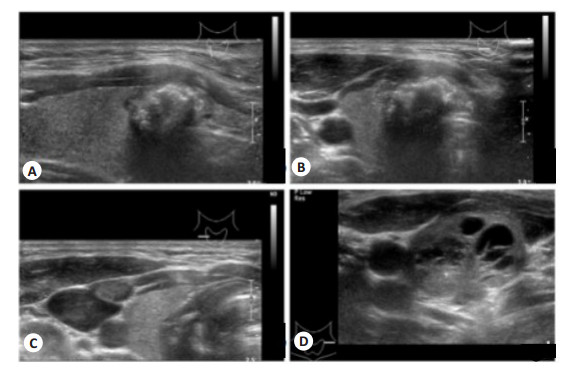
ObjectiveTo explore the blood flow resistance index (RI), blood flow characteristics, sound image characteristics and diagnostic value of high-frequency color Doppler ultrasound in breast cancer patients. MethodsFrom January 2019 to January 2021, 96 breast cancer patients (case group) diagnosed by pathology in the Department of Thyroid Breast Surgery of Anhui Cancer Hospital and 100 patients with benign breast disease diagnosed by pathological examination during the same period (benign group). The high-frequency color Doppler ultrasound examination data of the patients in the two groups were analyzed. The differences in RI, blood flow characteristics, and sound image characteristics of the two groups were compared. The pathological results were used as the gold standard to calculate the value of various indicators in the diagnosis of breast cancer. ResultsThe detection rates of irregular shape, edge burr sign, microcalcification lesions, and blurred borders in the case group were higher than those in the benign group(P < 0.05). The breast was diagnosed by drawing the ROC curve and the shape of the tumor. The sensitivity of cancer and benign breast diseases was 66.67%, the specificity was 60.00%, and the area under the ROC curve was AUC value of 0.633. The marginal burr sign for the diagnosis of breast cancer and benign breast diseases had a sensitivity of 73.96%, specificity of 58.00%.The area under the AUC value was 0.660. The sensitivity of microcalcified lesions in the diagnosis of breast cancer and benign breast diseases was 31.25%, the specificity was 91.00%, and the AUC value of the area under the ROC curve was 0.611. The sensitivity of the fuzzy boundary in the diagnosis of breast cancer and benign breast diseases was 27.08%, specificity 89.00%, area under the ROC curve AUC value of 0.580. The blood flow classification of the case group were mainly grade II (48.96%) and grade III (27.08%). The blood flow classification of the benign group was mainly grade 0 (53.00%), grade I (24.00%). The difference between the two groups was significant (P < 0.05). The sensitivity of blood flow classification in the diagnosis of breast cancer and benign breast diseases was 76.04%, the specificity was 77.00%, and the ROC curve was below. The area AUC value was 0.765. The RI value of the case group was lower than that of the benign group (P < 0.05). The sensitivity of the RI value of the lesion to diagnose breast cancer and benign breast diseases was 77.03%, and the specificity was 55.17%, the AUC value of the area under the ROC curve was 0.681. ConclusionAccording to the characteristics of high-frequency color Doppler ultrasonography, blood flow classification, and RI parameters, the differential diagnosis of breast cancer and benign breast diseases has certain clinical value. Several indicators can be analyzed clinically to improve clinical diagnosis efficiency.
2021, 44(3): 472-477.
doi: 10.12122/j.issn.1674-4500.2021.03.11
Abstract:
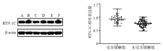
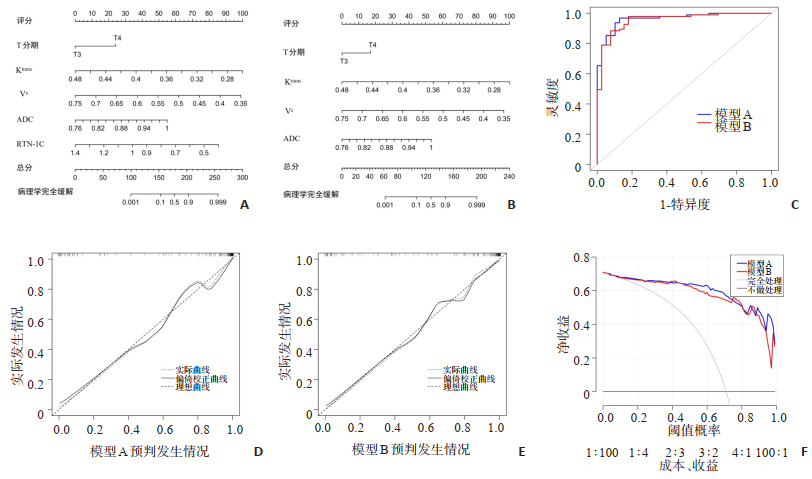
ObjectiveTo establish a model for evaluating the efficacy of neoadjuvant radiotherapy and chemotherapy for advanced rectal cancer based on MRI and reticulin 1C (RTN-1C) and evaluate the efficacy of the model. MethodsA total of 134 patients with advanced rectal cancer treated in Linxi Hospital of Kailuan General Hospital from August 2018 to November 2020 were selected as subjects. According to whether they achieved pathological complete response, they were divided into two groups : complete remission group (n=39) and incomplete remission group (n=95). The mononuclear cells in peripheral blood were obtained by Ficoll density gradient separation method, and the expression of RTN-1C in mononuclear cells was detected by Western blot. Logistic regression was used to analyze the risk factors of neoadjuvant radiotherapy and chemotherapy efficacy in advanced rectal cancer, and the nomogram regression model of these risk factors was established. Receiver operating characteristic (ROC) curve, calibration curve and decision curve analysis were used to evaluate the value of the prediction model. ResultsThe volume transfer constant (Ktrans), extravascular extracellular space volume ratio (Ve), reflux rate constant (Kep) and relative expression of RTN-1C in the complete remission group were all higher than those in the incomplete remission group; apparent diffusion coefficient (ADC) in the complete remission group was lower than that in the incomplete remission group (P < 0.05). The results of logistic regression analysis showed that T4 stage and high ADC value were independent risk factors for the efficacy of neoadjuvant radiotherapy and chemotherapy in advanced rectal cancer (P < 0.05). High Ktrans, high Ve value and high relative expression of RTN-1C were independent protective factors for the efficacy of neoadjuvant radiotherapy and chemotherapy in advanced rectal cancer (P < 0.05). When the threshold probability was 0.39%-0.42%, 0.75%-0.80% and 0.86%-0.88%, the net benefit of model B evaluating the efficacy of neoadjuvant radiotherapy and chemotherapy in advanced rectal cancer was higher than that of model A. When the threshold probability was in other ranges, the net benefit of model A evaluating the efficacy of neoadjuvant radiotherapy and chemotherapy in advanced rectal cancer was higher than that of model B. ConclusionModel A based on MRI and RTN-1C have a high degree of discrimination, calibration and clinical application value for the efficacy of neoadjuvant radiotherapy and chemotherapy in advanced rectal cancer, which can assist clinical decision-making.
2021, 44(3): 478-481.
doi: 10.12122/j.issn.1674-4500.2021.03.12
Abstract:
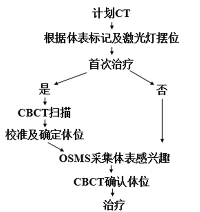
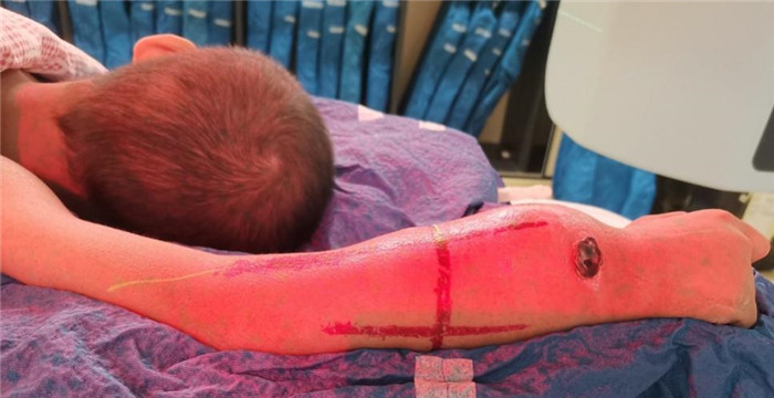
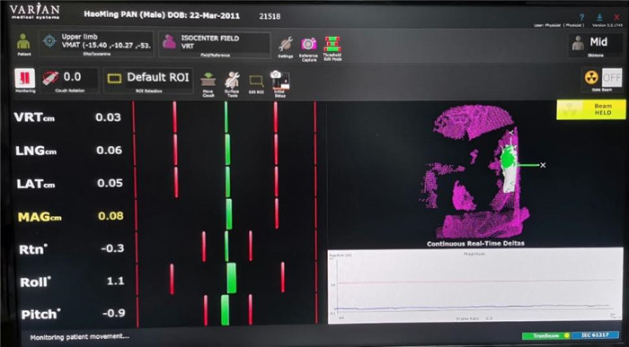
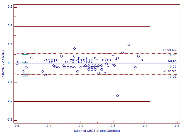
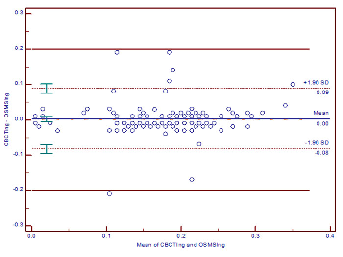
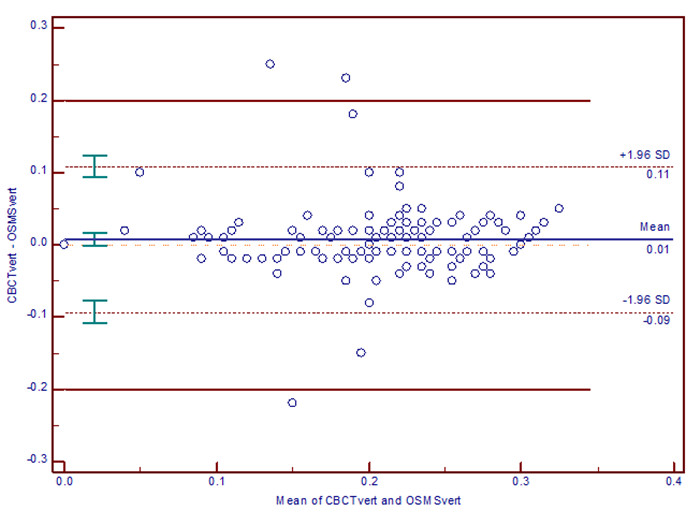
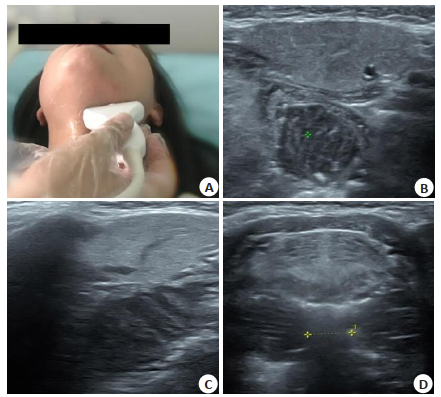
ObjectiveTo explore the positioning accuracy and application value of optical body surface tracking technology (OSMS) in precision radiotherapy for children's tumors. MethodsSix pediatric tumor patients were included. The body surface marking, laser light and optical surface tracking technology (OSMS) were applied to verified the patients pretreatment position, and perform CBCT scan before treatment after each positioning. The error of the left and right, head and foot, up and down directions of OSMS and CBCT were record. The χ2 test was used to compare the two groups to analyze the set-up error relationship between the two groups, and the Bland-Altman method was used to evaluate the consistency of the two systems. During treatment, the patient's posture changes were observed by OSMS monitors. ResultsThe movement errors of CBCT and OSMS in the left and right, head and foot, and up and down directions were: 0.207±0.076 cm, 0.207±0.073 cm; 0.186±0.072 cm, 0.183±0.069 cm; 0.206±0.068 cm, 0.198±0.071 cm, which had no significant difference (P>0.05). There were 4.65% (6/129), 5.43% (7/129), 3.88% (5/129) of movement differences between CBCT and OSMS in the left and right, head and foot, up and down directions Outside of 95% LoA. In the consistency range, the maximum absolute values of the differences were 0.06, 0.09, 0.11 cm, respectively. ConclusionOSMS is a high-precision and high-efficiency image guidance method, which can accurately reduce positioning errors and improve positioning efficiency. It is suitable for precise radiotherapy of childhood tumors.
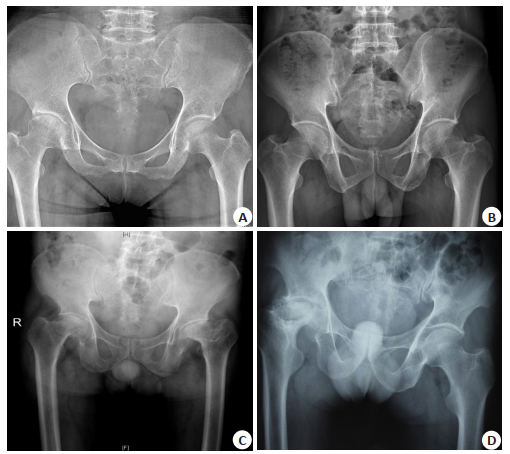
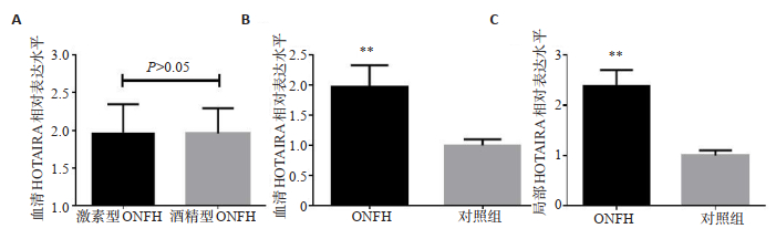
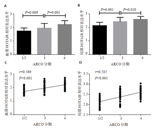
Correlation of local and serum LncRNA HOTAIR expressions with disease severity of non-traumatic ONFH
2021, 44(3): 482-487.
doi: 10.12122/j.issn.1674-4500.2021.03.13
Abstract:
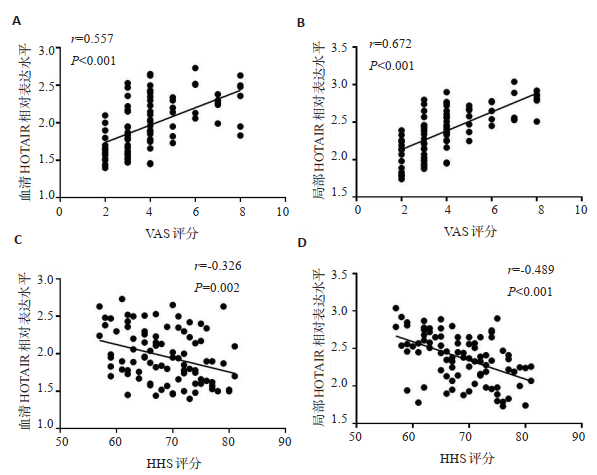
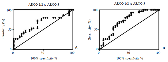
ObjectiveTo investigate the relationship between the serum and local expressions of LncRNA HOTAIR and the disease severity in patients with non- traumatic ONFH. MethodsNinety non- traumatic ONFH patients and 84 healthy controls were enrolled. RT-PCR was used to detect the expression of serum and local HOTAIR. The radiographic progression was determined by ARCO stage. The clinical severity of ONFH was evaluated by VAS and Harris hip score. ROC curve was used to determine the diagnostic value of serum HOTAIR with regard to radiographic progression. Resultsthe expression of serum HOTAIR in patients with non-traunatic ONFH was significantly higher than that in healthy controls (1.97±0.32 vs 1.00± 0.10, P < 0.001), and the local HOTAIR expression in the necrotic area was significantly higher compared with the non-necrotic area (2.37±0.25 vs 1.00±0.10, P < 0.001). The expressions of serum and local HOTAIR in ARCO 4 were significantly higher than ARCO 3(2.22±0.28 vs 1.94±0.36, P=0.001;2.58±0.22 vs 2.42±0.32, P=0.020), and the expression of serum and local HOTAIR in ARCO 3 was significantly higher than ARCO 1/2(1.94±0.36 vs 1.73±0.23, P=0.009;2.42±0.32 vs 2.13±0.24, P < 0.001). Expressions of HOTAIR in serum and local area were positively associated with ARCO grade(r=0.569, P < 0.001; r=0.585, P < 0.001), VAS score (r=0.557, P < 0.001; r=0.672, P < 0.001)and negatively related to Harris hip score(r=-0.326, P=0.002; r=-0.489, P < 0.001). Further ROC curve analysis showed that serum HOTAIR could be used as a decent indicator for the radiograohic progression of nontraumatic ONFH (AUC=0.663, P=0.030; AUC=0.726, P=0.003). ConclusionThe increase of serum and local HOTAIR expression in patients with non-traumatic ONFH may reflect the severity of the disease.
2021, 44(3): 488-491.
doi: 10.12122/j.issn.1674-4500.2021.03.14
Abstract:
ObjectiveTo explore the consistency between ultrasound-guided transperineal and transrectal prostate biopsy in the diagnosis of prostate cancer. MethodsThe clinical data of 121 patients undergoing prostate biopsy who were admitted to the hospital from February 2017 to February 2020 were collected. According to different examination methods, they were divided into rectum examination group (n=60) and perineum examination group (n=61). The consistency between the two groups in the diagnosis of prostate cancer was analyzed. The diagnostic efficiency of prostate cancer, detection rate of prostate cancer in PSA gray zone, examination time and occurrence of complications were compared between the two groups. ResultsKappa value in rectum examination group was close to that in perineum examination group for diagnosis of prostate cancer (0.699 vs 0.668). The sensitivity, specificity and accuracy for diagnosis of prostate cancer in rectum examination group were 87.1%, 82.8% and 85.0%, without significant difference compared with those in perineum examination group (85.3%, 81.5%, 83.6%) (P>0.05). The detection rate of prostate cancer in PSA gray area in perineum examination group was significantly higher than that in rectum examination group (P < 0.05). The examination time in rectum examination group was significantly shorter than that in perineum examination group, while incidence of bloody stool and fever was significantly higher than that in perineum examination group (P < 0.05). ConclusionConsistency between ultrasound- guided transperineal and transrectal prostate biopsy is comparable in the diagnosis of prostate cancer. The detection rate of prostate cancer in PSA gray area is higher by the former, with higher safety. However, its examination time is longer. In clinical practice, the most reasonable puncture method can be selected according to the specific conditions of patients.
2021, 44(3): 492-495.
doi: 10.12122/j.issn.1674-4500.2021.03.15
Abstract:
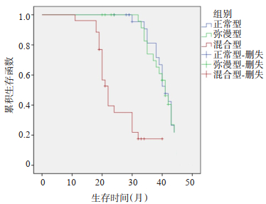
ObjectiveTo explore the whole body diffusion-weighted imaging(WB-DWI)findings of multiple myeloma patientswith different international staging system(ISS)stages and their correlation with prognosis. MethodsEighty patients withmultiple myeloma treated in our hospital from January 2014 to January 2020 were selected, including 19 patients with ISS stageI, 28 patients with ISS stage Ⅱ, and 33 patients with ISS stage Ⅲ.The WB-DWI was performed, the differences of WB-DWIclassification in patients with different ISS stages were analyzed, and the prognosis of patients with different WB-DWIclassification were analyzed. ResultsThe diffuse type and mixed type total proportion of WB-DWI in patients with ISS stage IIand Ⅲ were significantly higher than that in patients with ISS stage I(P < 0.05).The difference in the total proportion of diffusetype and mixed type of WB-DWI in patients with ISS stage Ⅱ and Ⅲ was not significant(P>0.05).There was no significantdifference in apparent diffusion coefficient between patients with different ISS stages(P>0.05).The difference in ISS stagesbetween patients with diffuse and mixed types and normal type of WB-DWI was significant(P < 0.05).The serum β2microglobulin of B-DWI patients with diffuse type and mixed type was significantly higher than that of normal type(P < 0.05).The median overall survival of mixed WB-DWI patients(95%CI: 19.73-24.27)was significantly shorter than that of normal anddiffuse types(95%CI: 38.39-43.62, P < 0.05). ConclusionThe WB-DWI findings of multiple myeloma patients with different ISSstages are different.There is a certain relationship between WB-DWI findings and prognosis.
2021, 44(3): 496-501.
doi: 10.12122/j.issn.1674-4500.2021.03.16
Abstract:
ObjectiveTo explore the effect of ultrasound-guided low-temperature plasma technology in the treatment of tonsil hypertrophy in children with obstructive sleep apnea-hypopnea syndrome (OSAHS). MethodsA total of 120 children with OSAHS caused by tonsillar hypertrophy were selected in Xinhua Hospital of Huainan Xinhua Medical Group to receive ultrasound-guided low-temperature plasma surgery. The 60 cases received total tonsillectomy (totalectomy group) and other 60 cases received partial tonsillectomy as non-totalectomy group. The operation time, bleeding, the immune function, quality of life, and sleep monitoring index of the children before and after the operation in the two groups were compared. ResultsThe operation time and blood loss of the total resection group were greater than those of the non-total resection group(P < 0.05). Before surgery, the difference of peripheral blood CD3+, CD4+, CD8+, CD4+/CD8+ between the total resection group and the non-total resection group was not significant (P>0.05). One month after operation, the peripheral blood CD3+, CD4+, CD4+/ CD8+ values of the non-total resection group were greater than those of the total resection group (P < 0.05). Before and 1 month after the operation, there was no significant difference in sleep disturbance, physical symptoms, emotional effects, daytime problems, and caregiver care scores between the total resection group and the non-full resection group (P>0.05). One month after the operation, the sleep disturbance, physical symptoms, emotional impact, daytime problems, and caregiver care scores of the patients in the group were significantly lower than those in the preoperative group (P < 0.05). Before and 1 month after the operation, there was no significant difference of AHI, minimum blood oxygen saturation, and average blood oxygen saturation between the total and non-total resection group (P>0.05). One month after surgery, the AHI of the two groups of patients were significantly lower than that of the preoperative group (P < 0.05). The blood oxygen saturation and average blood oxygen saturation were significantly higher than those of this group before operation (P < 0.05). ConclusionUltrasound-guided low-temperature plasma technology has little difference in effect between partial tonsillectomy and total tonsillectomy, but the former has the advantages of short operation time, less trauma, and less impact on the immune function of children after surgery.
2021, 44(3): 502-506.
doi: 10.12122/j.issn.1674-4500.2021.03.17
Abstract:
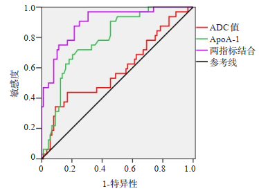
ObjectiveTo explore the value of magnetic resonance diffusion weighted imaging(MR DWI)combined with level ofserum apolipoprotein A-1(ApoA-1)in predicting the curative effect of chemoradiotherapy on patients with non-small celllung cancer(NSCLC). MethodsA total of 120 NSCLC patients who underwent chemoradiotherapy and admitted fromDecember 2019 to December 2020 were enrolled.All patients underwent DWI examination and were tested for serum ApoA-1before and after treatment.DWI indexes and ApoA-1 level before and after treatment were compared.The correlation betweenDWI indexes and ApoA-1 level were analyzed.The curative effect on patients was evaluated.DWI imaging findings beforeand after treatment were analyzed.DWI indexes and level of serum ApoA-1 were compared among patients with differentcurative effect after 2 courses.Predictive value DWI indexes and serum ApoA-1 for curative effects were analyzed. ResultsAfter treatment, ADC and ApoA-1 level were significantly increased(P < 0.05).As the increase of treatment cycle, ADC valueand ApoA-1 level were gradually increased, and the differences were significant(P < 0.05).AFC value was positively correlatedwith ApoA-1(r=0.633, P < 0.05).ADC value and ApoA-1 level in effective group were significantly higher than those inineffective group(P < 0.05).ROC curves showed that AUC values of ADC value, ApoA-1 and their combination for predictingcurative effect were 0.587, 0.786 and 0.899, respectively. ConclusionThe application of MR DWI combined with level of serumApoA-1 can effectively improve the predictive value for curative effect of chemoradiotherapy on NSCLC patients.
2021, 44(3): 507-511.
doi: 10.12122/j.issn.1674-4500.2021.03.18
Abstract:
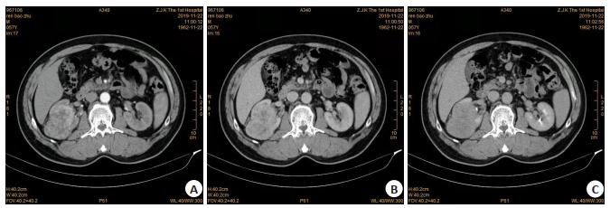
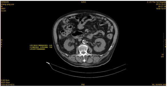
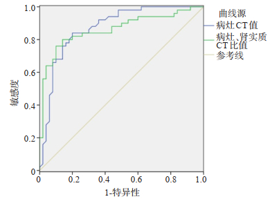
ObjectiveTo evaluate the value of multidetector CT(MDCT)in the diagnosis of angiomyolipomas with minimal fat. MethodsA total of 117 patients with angiomyolipomas with minimal fat who were treated in our hospital from January 2012to February 2020 were selected as the observation group, 210 patients with renal clear cell carcinoma were control group.Bothgroups were given MDCT examination.The differences of CT signs and CT values between the two groups were analyzed.The receiver operating characteristic(ROC)curve was used to analyze the CT value in the diagnosis of angiomyolipomas withminimal fat. ResultsThe female proportion of the observation group was higher than that of the control group(P < 0.05).Theage of the observation group was lower than that of the control group(P < 0.05).In the observation group, the proportion of thedensity was uniform, the boundary was clear, and vascular shadow in the tumor were 80.34%, 100.00% and 16.24%, whichwere significantly higher than that in the control group(P < 0.05).The proportion of pseudocapsule in the observation groupwas significantly lower than that in the control group(P < 0.05).In CT signs, there were no significant difference in morphologyand enhancement mode between the observation group and the control group(P>0.05).The lesion CT value and CT ratio oflesion/renal parenchyma in the observation group were 46.10±7.82 Hu and 1.37±0.32, which were significantly higher thanthose in the control group(P < 0.05).There was no significant difference in CT value of renal parenchyma between theobservation group and the control group(P>0.05).The area under ROC curve of lesion CT value and lesion/renal parenchymaCT ratio in the diagnosis of angiomyolipomas with minimal fat were 0.872 and 0.862, cut-off values were 41.50 Hu and 1.20, the sensitivity were 78.80% and 76.50%, and the specificity were 75.50% and 74.40%. ConclusionMDCT has a good value inthe diagnosis of angiomyolipomas with minimal fat, which is worthy of clinical use.
2021, 44(3): 512-515.
doi: 10.12122/j.issn.1674-4500.2021.03.19
Abstract:
ObjectiveTo explore the value of ultrasound combined with thoracoscopy in the diagnosis of benign and malignantpleural effusion. MethodsA total of 102 patients with unexplained pleural effusion in our hospital from January 2020 to March2021 were selected.The differences of thoracoscopic morphology and ultrasound characteristics between benign andmalignant pleural effusion patients were analyzed. ResultsIn the 102 patients, 76 were benign and 26 were malignant.Theetiology of benign patients was tuberculous pleurisy, while the main cause of malignant was metastatic adenocarcinoma ofpleura, accounting for 88.46%.The proportions of pleural mass and porcelain like thickening in malignant group were higherthan those in benign group(P < 0.05).The proportions of pleural congestion and edema and pleural adhesions were lower thanthose in benign group(P < 0.05).In the malignant group, the pleural thickness and the proportion of non-uniform pleuralthickening were significantly higher than those in benign group(P < 0.05).The proportion of hyperechoic fiber septum wassignificantly lower than that in benign group(P < 0.05).The area under the ROC curve of membrane thickness in differentiatingmalignant pleural effusion was 0.736(P < 0.05), and the cut-off value was 10.73 mm, the sensitivity and specificity were 53.80%and 99.70%, respectively.The sensitivity, specificity, accuracy, positive predictive value and negative predictive value ofultrasonography combined with thoracoscopic morphological diagnosis of malignant pleural effusion were 76.92%, 81.58%, 80.39%, 58.82% and 91.18%, respectively. ConclusionUltrasound combined with thoracoscopy has a certain value in thediagnosis of benign and malignant pleural effusion.
2021, 44(3): 516-520.
doi: 10.12122/j.issn.1674-4500.2021.03.20
Abstract:
ObjectiveTo evaluate the value of transthoracic echocardiography(TTE)and multi-slice spiral CT(MSCT)in thediagnosis of aortic dissection in emergency. MethodsA total of 141 patients with suspected aortic dissection who were treatedin our hospital from January 2016 to April 2020 were selected.The TTE and MSCT were examined, the difference of diagnosticvalue between them was analyzed by χ2 test, the differences of MSCT signs between aortic dissection and non-aortic dissection, as well as the differences of MSCT signs between different types of aortic dissection were analyzed. ResultsAmong 141suspected aortic dissection patients, 112 patients were diagnosed as aortic dissection by surgery or CT angiography, and 29patients were non aortic dissection patients.The sensitivity, accuracy and negative predictive values of MSCT in the diagnosisof aortic dissection were 95.54%, 94.33% and 83.87%, which were significantly higher than that of TTE(P < 0.05).There were nosignificant difference in specificity and positive predictive value between MSCT and TTE in the diagnosis of aortic dissection(P>0.05).Among the MSCT signs, the proportions of calcification metastasis, aortic area density, linear pattern and aorticwidening in aortic dissection were significantly higher than those in non-aortic dissection(P < 0.05).There was no significantdifference in the proportion of central effusion and pleural effusion between aortic dissection and non-aortic dissection(P>0.05).In MSCT findings, the high density ratio of aortic area in type A active aortic dissection was significantly higher than thatin type B active aortic dissection(P < 0.05).There was no significant difference in the proportion of calcification metastasis, linear sign, pericardial effusion, pleural effusion and aortic widening between type A and type B aortic dissection(P>0.05). ConclusionCompared with TTE, MSCT has better application value in the diagnosis of emergency aortic dissection and isworthy of clinical use.
2021, 44(3): 521-525.
doi: 10.12122/j.issn.1674-4500.2021.03.21
Abstract:
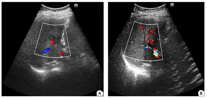
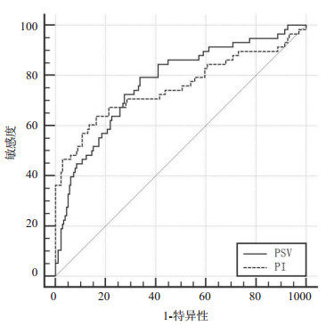
ObjectiveTo explore the value of color Doppler ultrasound combined with hepatic artery related index in predicting the efficacy and prognosis of interventional chemotherapy for elderly patients with primary liver cancer (PLC). MethodsThe clinical data of 236 elderly patients with PLC admitted to our hospital from March 2017 to December 2018 were retrospectively analyzed. Statistics of elderly patients with PLC were analyzed. The clinical data of patients in the invalid group and the effective group were compared. Logistic regression analysis was used to analyze the factors of TACE treatment in elderly patients with PLC. ROC was used to analyze the predictive value of hepatic artery correlation index for the treatment effect of elderly patients with PLC. After 1 year follow-up, the hepatic artery related index was used to analyze the prognosis of elderly patients with PLC. ResultsThe incidence of treatment failure in elderly PLC patients was 24.58% after TACE treatment. The pathological classification was poorly differentiated. Peak hepatic artery systole (PSV) and Hepatic artery pulsation index (PI) were all factors affecting the treatment effect of TACE in elderly patients with PLC (OR=4.011, 3.340, 2.807, all P < 0.05). ROC analysis showed that the best cut-off points for PSV and PI to predict treatment failure in elderly PLC patients were 82.42 cm/s and 1.73, respectively. The AUC values were 0.772 and 0.753, respectively. The PSV and PI of the poor prognosis group were significantly higher than those of the poor prognosis group (P < 0.05). ConclusionColor doppler ultrasound combined with PSV and PI has a higher efficacy in predicting the efficacy and prognosis of PLC interventional chemotherapy in the elderly. It can be used as an important indicator for evaluating the efficacy and prognosis.
2021, 44(3): 526-530.
doi: 10.12122/j.issn.1674-4500.2021.03.22
Abstract:
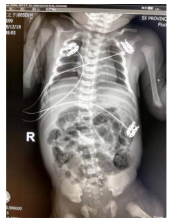
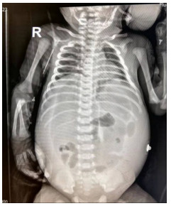
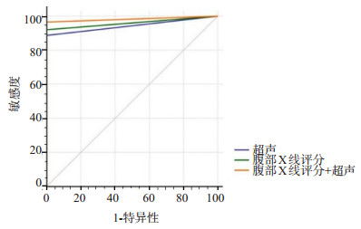
ObjectiveTo evaluate the value of duck abdominal assessment scale (DAAS) combined with abdominal ultrasound in the diagnosis of neonatal necrotizing enterocolitis (NEC). MethodsA total of 97 children with suspected NEC who were diagnosed and treated in our hospital from January 2017 to December 2019 were prospectively included. Among them, 59 were males with an average of 6.4±3.5 d and 38 females with an average of 6.9±2.8 d. Abdominal X-ray examination and abdominal ultrasound were performed on all patients. Regarding the surgical pathological results as the gold standard, the agreement rate, sensitivity, specificity, positive predictive value and negative predictive value by DAAS, abdominal ultrasound and DAAS combined with abdominal ultrasound were used to assess the diagnostic ability. The ROC curve was used to analyze the diagnostic performance of the three methods. The DAAS of children with different pathological stages were compared, and the DAAS of children with different intestinal diseases were compared. ResultsEighty-nine cases were diagnosed as NEC by surgical pathology. Comparing the surgical pathological results, the agreement rate, sensitivity, and specificity of abdominal X-ray examination, abdominal ultrasound, and abdominal X-ray combined with abdominal ultrasound for the diagnosis of NEC were all higher than 85%. The area under the curve of the three methods for the diagnostic efficiency of NEC were 0.96, 0.94 and 0.98, respectively. The diagnostic efficiency of DAAS combined with abdominal ultrasound was the highest. As the severity of the disease increased, the DAAS of children was significantly increased (P < 0.05). DAAS of children in the intestinal perforation group were significantly higher than those in the intestinal necrosis group(P < 0.05). ConclusionThe combination of DAAS and abdominal ultrasound can identify NEC better, which has important clinical value.
2021, 44(3): 531-535.
doi: 10.12122/j.issn.1674-4500.2021.03.23
Abstract:
ObjectiveTo evaluate the value of ultrasonography in the diagnosis of ALN metastasis of breast cancer and ovarian function after chemotherapy. MethodsSixty-two breast cancer patients diagnosed and treated in our hospital from June 2019 to March 2021were selected. Among them, 28 patients with ALN metastasis were compared in cortical morphology, blood flow and ultrasonic measurement indexes. The diagnostic efficacy of ultrasound indicators on ALN metastasis was analyzed. The correlation between ovarian ultrasound index and ovarian function was analyzed. ResultThe stenosis type, concentric thickening type, eccentric thickening type and non hilar type between the two groups were significantly different (χ2=27.871, P < 0.001). The hilar type, scattered type, peripheral type and mixed type between the two groups were significantly different(χ2= 28.802, P < 0.001). The longitudinal diameter (t=17.037, P < 0.001), cortical thickness (t=15.491, P < 0.001) and D/W (t=7.243, P < 0.001) of ALN metastasis group were significantly higher than those of non ALN metastasis group. The diagnostic specificity of combined detection for ALN metastasis was significantly higher than that of single detection. The AMH and FSH between the two groups before chemotherapy were not significantly different(P>0.05). After treatment, AMH of the two groups decreased significantly, FSH increased significantly, and AMH (t=6.003, P < 0.001) and FSH (t=4.307, P < 0.001) of patients without ALN metastasis were significantly higher than those of patients with ALN metastasis. The RI and Pi of two groups were significantly decreased. The RI (t=6.887, P < 0.001) and PI (t=26.430, P < 0.001) of patients without ALN metastasis were significantly lower than those of patients with ALN metastasis. AMH, FSH and RI, PI were positively correlated. ConclusionUltrasound imaging has a good diagnostic effect on ALN metastasis of breast cancer. It has great significance for the evaluation of ovarian function after chemotherapy by ultrasound analysis of local ovarian lesions.
2021, 44(3): 536-540.
doi: 10.12122/j.issn.1674-4500.2021.03.24
Abstract:
ObjectiveTo explore the ability of contrast- enhanced ultrasound (CEUS) in the differential diagnosis of primary hepatocellular carcinoma (PHC) and hepatic hemangioma (HCH) by blood flow signals inside and around the lesion, and analyze the risk factors of tumor metastasis in PHC patients. MethodsSixty-five patients with liver tumors from February 2019 to December 2020 were selected as the study subjects, including 35 patients with PHC and 30 patients with HCH. CEUS examinations were performed on the two groups of patients, and the blood flow signals inside and around the tumor lesions of the two groups were compared, as well as related ultrasound indicators including the increase time, peak time, peak enhancement intensity, enhancement rate and 50% tilt rate. The differences in the clinical data, the blood flow signals inside and around the tumor lesions, and related ultrasound indexes of patients in different metastasis groups in PHC patients were compared. In addition, the risk factors of tumor metastasis were analyzed by multivariate logistic. ResultsThe differences in blood flow signals inside and around the tumor lesion in the PHC group and the HCH group were significant (P < 0.05). The increase time, peak time and enhancement rate of PHC patients were significantly higher than those of HCH patients(P < 0.05). The peak enhancement intensity and 50% tilt rate were significantly lower than those of HCH patients(P < 0.05). There were significant differences in terms of age, number of tumors and tumor stage between the metastatic group and the nonmetastatic group (P < 0.05), but the differences in gender, body mass index and tumor diameter were no significant (P > 0.05). The difference in blood flow signals inside and around the tumor lesion between the metastatic group and the non- metastatic group was significant (P < 0.05). The increase time, peak time and enhancement rate of patients in the metastasis group were significantly higher than those in the non-metastasis group, and the peak enhancement intensity and 50% tilt rate of metastasis group were significantly lower than those of non-metastasis group, and the difference was statistically significant (P < 0.05). The age, number of tumors, stage of tumors, blood flow signal, 50% tilt rate, peak enhancement intensity, increase time, peak time and enhancement rate were all independent risk factors for tumor metastasis in PHC patients. ConclusionCEUS can effectively differentiate PHC and HCH. The age, number of tumors, stage of tumors and related ultrasound indicators can affect the risk of tumor metastasis.
2021, 44(3): 541-545.
doi: 10.12122/j.issn.1674-4500.2021.03.25
Abstract:
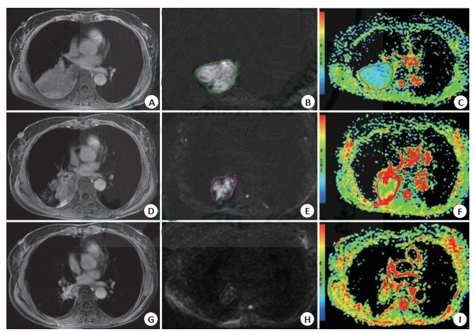
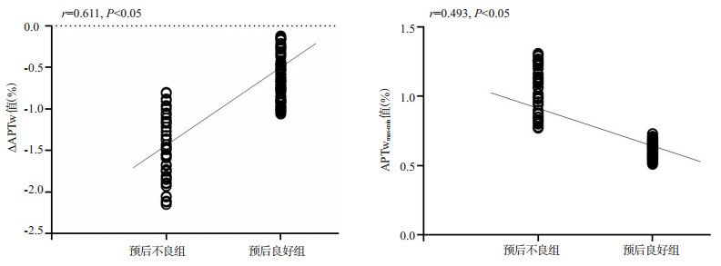
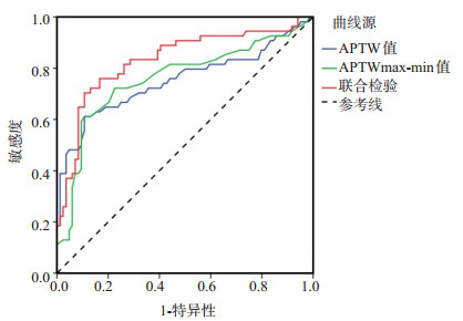
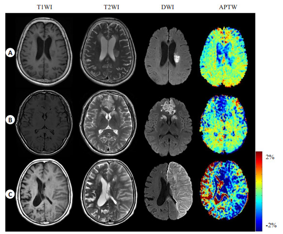
ObjectiveTo investigate value of amide proton chemical exchange saturation transfer MRI in predicting the prognosis of neurological function in cerebral infarction. MethodsA total of 138 patients with cerebral infarction in our hospital from July 2017 to July 2019 were enrolled. Based on modified Rankin scale score (mRS) at post-admission 3 months, they were divided into two groups: good prognosis group (n=84, mRS < 2) and poor prognosis group (n=54, mRS≥2). All patients received amide proton chemical exchange saturation transfer MRI examinations. The amide proton transfer-weighted (APTw) signal intensity was quantitatively analyzed in both groups. The correlation of ∆APTw value and APTwmax-min value with neurological function prognosis of cerebral infarction was analyzed, and the predictive value of two indicators on neurological function prognosis was also verified. ResultsThe ∆APTw value of poor prognosis group was lower than that of good prognosis group (P < 0.05). APTwmax-min value of poor prognosis group was higher than that of good prognosis group (P < 0.05). ∆APTw value was positively correlated with prognosis of cerebral infarction neurological function (P < 0.05). APTwmax-min value was negatively correlated with neurological function prognosis of cerebral infarction (P < 0.05). Decrease in ∆APTw value and increase in APTwmax-min value were risk factors for poor neurological prognosis of cerebral infarction (P < 0.05). The AUC, sensitivity, specificity and accuracy of ∆APTw value combined with APTwmax-min in predicting the neurological prognosis of cerebral infarction were 0.832, 75.91%, 83.32%, and 88.32%, respectively. The critical values of ∆APTw and APTwmax-min were-0.92 and 0.75, respectively. ConclusionQuantitative analysis of APTW in amide proton chemical exchange saturation transfer MRI has high value in predicting the neurological function prognosis of cerebral infarction, moreover, clinical intervention measures are of vital importance when there is a decrease in ∆APTw value and increase in APTwmax-min value.
2021, 44(3): 546-551.
doi: 10.12122/j.issn.1674-4500.2021.03.26
Abstract:
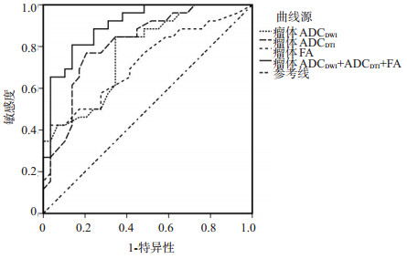
ObjectiveTo investigate the diagnostic value of diffusion weighted imaging (DWI), diffusion tensor imaging (DTI) and conventional MRI in high-grade glioma. MethodsSixty-five patients with glioma in our hospital from October 2018 to July 2020 were selected. According to the pathological results, they were divided into high-grade glioma group and low-grade glioma group. MRI was performed before operation, and the scanning sequences were T1WI, T2WI, DWI and DTI. The MRI images of different grades of gliomas were analyzed, and the apparent dispersion coefficient (ADC) and fractional anisotropy (FA) values of different grades of gliomas were compared. The diagnostic threshold, sensitivity and specificity of ADCDWI, ADCDTI and FA were analyzed. The specificity of ADCDWI, ADCDTI and FA in the diagnosis of high-grade glioma were analyzed. ResultsAll 65 patients were diagnosed as glioma by postoperative pathological examination, including 29 cases of low-grade glioma and 36 cases of high-grade glioma. The difference in ADCDWI, ADCDTI and FA values among different grades of gliomas were not significant(P > 0.05). The difference in FA between different grades of glioma was not significant(P > 0.05). The values of ADCDWI, ADCDTI in high-grade gliomas were significantly lower than those in low-grade gliomas(P < 0.05). The AUC of ADCDWI, ADCDTI and FA were 0.775, 0.817 and 0.716, respectively.The sensitivity and specificity of DWI and DTI in the diagnosis of high-grade gliomas were higher, AUC was 0.903(P < 0.05). ConclusionConventional MRI combined with DWI and DTI can improve the accuracy of diagnosis and pathological grading of glioma.
2021, 44(3): 552-555.
doi: 10.12122/j.issn.1674-4500.2021.03.27
Abstract:
ObjectiveTo explore the relationship between radiation-induced lung injury after three dimensional adaptive radiology and lung low-dose volume in patients with esophageal cancer. MethodsIn this retrospective study, 96 patients with esophageal cancer diagnosed in our hospital from January 2015 to January 2020 were selected. All patients were treated with Three dimensional adaptive radiology. According to the diagnostic criteria of the American cancer radiotherapy collaboration, the general information, V5, V10, V20, V30, average lung dose and minimum lung dose of patients with radiation-induced lung injury and non radiation-induced lung injury were compared to analyze the correlation between radiation-induced lung injury and the exposure volume in low dose area. ResultsThere was no significant difference in gender, age, body mass index, TNM stage and pathological type between radiation-induced and non radiation-induced lung injury patients (P > 0.05). V5 (P < 0.001), V10 (P < 0.001), V20 (P < 0.001), V30 (P < 0.001) and average lung dose (P < 0.001) of radiation-induced lung injury patients occurred. There was no significant difference in the minimum lung dose and maximum lung dose between the two groups(P > 0.05). Through the regression analysis of lung dose of lung injury severity, in the treatment of patients, V5, V10 and V20 were the only high-risk factors of radiation-induced lung injury above grade 2, while the patients were more likely to suffer from radiation-induced lung injury. There was no correlation between lung dose and radiation-induced pulmonary fibrosis(P > 0.05). ConclusionV5, V10, V20 are effective predictors of radiation pneumonitis. In the treatment of patients, we can adjust the radiation volume of low-dose area of lung to reduce the occurrence of radiation-induced lung injury in time with the premise of ensuring the treatment effect. It has positive significance for the prognosis of patients.
2021, 44(3): 556-558.
doi: 10.12122/j.issn.1674-4500.2021.03.28
Abstract:
ObjectiveTo investigate the influence of BMI on ultrasound diagnosis of ectopic pregnancy. MethodsA total of 62 patients with ectopic pregnancy who were admitted to our hospital and diagnosed by ultrasound from November 2015 to November 2019 were included and retrospectively analyzed according to the screening criteria. According to the value of BMI, the patients were divided into the normal BMI group (18.5 kg/m2 < BMI < 25 kg/m2) and the high BMI group (BMI≥25 kg/m2). The clinical baseline data of all patients were analyzed, and the baseline data of patients in different groups were compared. The imaging findings of ectopic pregnancy were compared between different BMI groups. ResultsThe difference in baseline data between the two groups was not significant (P > 0.05). Compared with the normal BMI group, the positive rate of ultrasound diagnosis in the high BMI group was significantly lower(P < 0.05). Compared with the normal BMI group, the positive rate of ultrasound diagnosis of phantom pregnancy bursa, pelvic effusion and mixed mass was lower in the high BMI group (P < 0.05). However, the difference in the diagnosis rate of germ and fetal heart between the two groups was not significant(P > 0.05). ConclusionWhen using ultrasound to diagnose patients with ectopic pregnancy, the influence of BMI on the positive rate of ultrasound diagnosis should be considered, and other methods are recommended to diagnose ectopic pregnancy in obese patients.
2021, 44(3): 559-562.
doi: 10.12122/j.issn.1674-4500.2021.03.29
Abstract:
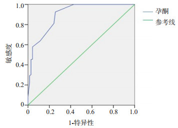
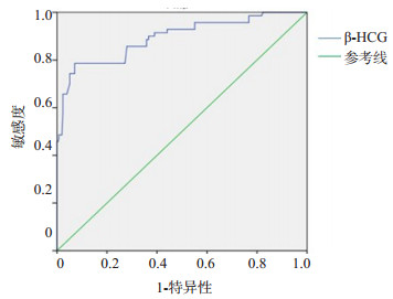
ObjectiveTo explore the clinical value of subchorionic hemorrhage(SCH)combined with progesterone and β-HCG for predicting early pregnancy outcome in threatened abortion patients. MethodsA total of 324 patients who came to our hospital due to threatened abortion from June 2020 to December 2020 were selected for serum progesterone, β-HCG and ultrasound examination. They were followed up for 14 weeks of gestation to observe the pregnancy outcome. The patients who continued pregnancy after treatment were defined threatened abortion group, and those who developed inevitable abortion were defined abortion group. The differences of progesterone, β-hCG and the positive rate of SCH between two groups were compared. The prediction value of progesterone and β-hCG for abortion were analyzed by receiver operating characteristic (ROC) curve. ResultsThe 254 cases were in threatened abortion group and 70 cases were in abortion group. The levels of serum progesterone and β-hCG in abortion group were significantly lower than those in threatened abortion group, and the positive rate of SCH in abortion group was significantly higher than those in threatened abortion group (P < 0.05). The area under ROC curve of serum progesterone was 0.90 (95% CI: 0.85-0.94). When cut off value was 18.5, the sensitivity and specificity of predicting abortion were 92.5% and 74.3%, respectively. The area under ROC curve of serum β-hCG was 0.89 (95% CI: 0.84-0.94). When cut off value was 6926, the sensitivity and specificity of predicting abortion were 78.6% and 92.9%, respectively. The sensitivity, specificity, positive predictive value and negative predictive value of serum progesterone, β-hCG and SCH were 70.8%, 98.3%, 97.1% and 80.8% respectively. ConclusionsThe SCH, Serum progesterone and β-HCG level have certain value for predicting the early pregnancy outcome in threatened abortion patients. The combined detection of the three indicators can improve the specificity and positive predictive value.
2021, 44(3): 563-566,封3.
doi: 10.12122/j.issn.1674-4500.2021.03.30
Abstract:
ObjectiveTo explore the correlation between MSCT imaging findings of lung adenocarcinoma and CEA and CA125. MethodsA total of 94 patients with lung adenocarcinoma admitted to our hospital from June 2018 to February 2021 were selected. All patients acceptted multislice CT scanning (MSCT). The imaging features of patients with package including the tumor edge (burr), Ye Zheng, tumor signs, air bronchogram, cavitation and cystic cavity change, the structure of the tumor around sign, pleural indentation, blood vessels, cluster were analyzed. The serum CEA and CA125 level were analyzed by using chemiluminescence detection, and its correlation with MSCT imaging findings was analyzed. ResultsAll the 94 patients underwent MSCT examination. We found that 58 patients had burr sign, 69 patients had lobulation sign, 9 patients had air bronchial sign, 8 patients had vacuolation sign, 13 patients had vascular cluster sign, 60 patients had pleural depression sign, and 3 patients had cystic cavity like change. CEA was significantly different with MSCT imaging burr sign, lobulation sign, vacuole sign, cystoid change and pleural depression sign (P < 0.05). CA125 was significantly different with MSCT imaging burr sign, lobulation sign, vacuole sign, cystoid change and pleural depression sign (P < 0.05). ConclusionMSCT imaging manifestations of lung adenocarcinoma include burr sign, lobulation sign, vacuole sign, cystoid change and pleural depression, which are correlated with CEA and CA125 levels.