


Value of MR diffusion tensor imaging of midbrain and basal ganglia nuclei in the diagnosis and severity assessment of Parkinson's disease
-
摘要:
目的探讨中脑和基底节神经核团的磁共振扩散张量成像(DTI)在帕金森病(PD)诊断及病情评估中的应用价值。 方法回顾性分析2017年7月~2020年11月在我院就诊的27例PD患者(PD组)及23例正常人群(对照组)的临床及MR资料。对两组进行常规MR平扫及DTI检查,测量各受试者双侧黑质、红核、尾状核头、丘脑、苍白球、壳核的DTI参数,包括各项异性分数(FA)、平均扩散系数(MD)。比较各核团FA和MD值在PD组和对照组间的差异。分析存在组间差异的参数单独及联合应用的诊断效能,以及和病程、Hoehn-Yahr分级及统一帕金森病评定量表运动部分(UPDRS Ⅲ)评分的相关性。 结果PD组黑质、尾状核头、丘脑、苍白球及壳核的FA值均低于对照组,尾状核头MD高于对照组(P < 0.05)。受试者工作特征曲线分析示联合应用黑质、尾状核头、丘脑、苍白球及壳核的FA值和尾状核头的MD值诊断PD的曲线下面积(AUC)为0.981,均明显大于各参数单独应用于诊断PD的AUC(P < 0.05)。尾状核头MD值与PD患者UPDRS Ⅲ评分呈正相关(r=0.402, P=0.038),丘脑FA值与PD患者Hoehn-Yahr分级(r=-0.490, P=0.009)、UPDRS Ⅲ评分(r=-0.547, P=0.003)呈负相关。 结论中脑、基底节神经核团DTI参数有助于PD的诊断及病情严重程度的评估,联合应用多个核团DTI参数具有较好的诊断效能。 Abstract:ObjectiveTo investigate the value of MR diffusion tensor imaging (DTI) of midbrain and basal ganglia nuclei in the diagnosis and severity assessment of Parkinson's disease (PD). MethodsThe clinical and MR data of 27 PD patients (PD group) and 23 normal controls (NC group) who visited our hospital from July 2017 to November 2020 were retrospectively analyzed. Routine MR scanning and DTI examination were performed on PD group and NC group. And DTI parameters, including the fractional anisotropy (FA) and mean diffusivity (MD), were measured for bilateral substantia nigra, red nucleus, caudate head, thalamus, globus pallidus and putamen of each subject. The FA and MD values of each nucleus were compared between PD and NC groups. This study analyzed the diagnostic effectiveness of single and combined application of parameters with inter-group differences and their correlation with course of disease, Hoehn-Yahr grade and exercise part of unified Parkinson's disease rating scale (UPDRS Ⅲ) score. ResultsThe FA values of substantia nigra, caudate head, thalamus, globus pallidus and putamen in PD group were all lower than those in NC group, while the MD values of caudate head were higher than that in NC group (all P < 0.05). The receiver operating characteristic curve analysis showed that the area under the curve (AUC) of combined application of FA values of substantia nigra, caudate head, thalamus, globus pallidus and putamen as well as MD value of caudate head for PD diagnosis was 0.981, which was significantly greater than the AUC of each parameter applied alone for PD diagnosis (all P < 0.05). The MD value of caudate head was positively correlated with UPDRS Ⅲ score of PD patients (r=0.402, P=0.038), and the FA value of thalamus was negatively correlated with the Hoehn-Yahr grade (r=-0.490, P=0.009) and UPDRS Ⅲ score (r=-0.547, P=0.003) of PD patients. ConclusionThe DTI parameters of midbrain and basal ganglia nuclei are helpful for the diagnosis and severity assessment of PD. The combined application of DTI parameters of multiple nuclei has better diagnostic efficacy. -
Key words:
- magnetic resonance imaging /
- diffusion tensor imaging /
- Parkinson's disease /
- midbrain /
- basal ganglia
-
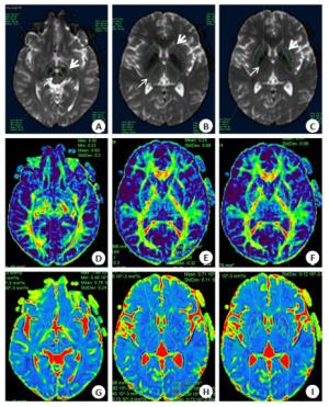
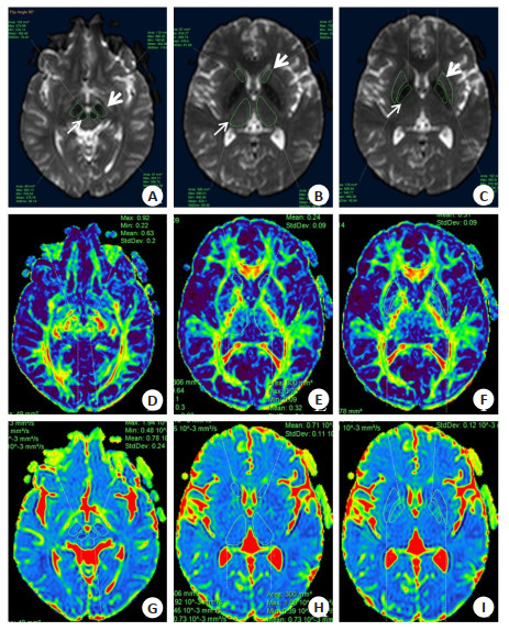
图 1 手动勾画中脑和基底节神经核团的ROI
A: 黑质(粗箭头)和红核(细箭头)ROI; B: 尾状核头(粗箭头)和丘脑(细箭头)ROI; C: 苍白球(细箭头)和壳核(粗箭头)ROI; D~F: 对应的FA图; G~I: 对应的MD图.
Figure 1. Manually delineate the ROI of the midbrain and basal ganglia nuclei
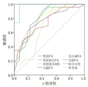
图 2 扩散张量成像参数诊断帕金森病的受试者工作特征曲线分析
Figure 2. Receiver operating characteristic curve analysis for diagnosis of Parkinson's disease by diffusion tensor imaging parameters.
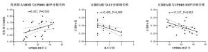
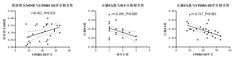
图 3 尾状核头及丘脑扩散张量成像参数与H-Y分级和UPDRS Ⅲ评分相关性
H-Y: Hoehn-Yahr grade, UPDRS Ⅲ: Unified Parkinson's disease rating scale; FA: 各项异性分数, MD: 平均扩散系数.
Figure 3. The correlations of diffusion tensor imaging parameters of caudate head and thalamus with H-Y grading and UPDRS Ⅲ score
表 1 两组中脑和基底节神经核团扩散张量成像参数比较
Table 1. Comparison of diffusion tensor imaging parameters of midbrain and basal ganglia nuclei between PD and NC groups (Mean±SD)
参数 黑质 红核 尾状核头 丘脑 苍白球 壳核 FA PD组 0.53±0.09 0.45±0.05 0.21±0.04 0.29±0.03 0.26±0.04 0.19±0.03 对照组 0.61±0.04 0.47±0.05 0.25±0.03 0.33±0.05 0.31±0.05 0.23±0.04 t -4.626 -0.931 -4.016 -3.418 -3.901 -3.648 P < 0.001 0.361 < 0.001 0.002 < 0.001 0.001 MD(×10-3 mm2/s) PD组 0.67±0.04 0.62±0.04 0.70±0.06 0.71±0.02 0.67±0.09 0.70±0.07 对照组 0.72±0.09 0.64±0.06 0.67±0.04 0.72±0.03 0.67±0.10 0.68±0.05 t -1.336 -0.846 2.606 -0.194 -0.071 0.881 P 0.208 0.406 0.012 0.848 0.944 0.387  下载: 导出CSV
表 2 扩散张量成像参数对帕金森病的诊断效能
Table 2. Diagnostic efficacy of diffusion tensor imaging parameters in Parkinson's disease
参数 AUC P 最佳界值 敏感度(%) 特异性(%) 约登指数 黑质FA 0.814 < 0.001 0.565 91.3 59.3 0.506 尾状核头FA 0.786 0.001 0.205 95.7 51.9 0.475 尾状核头MD 0.686 0.025 0.730 100.0 40.7 0.407 丘脑FA 0.761 0.002 0.305 65.2 77.8 0.430 苍白球FA 0.788 < 0.001 0.265 82.6 63.0 0.456 壳核FA 0.754 0.002 0.225 52.2 85.2 0.374 联合应用 0.981 < 0.001 - 100.0 92.6 0.926 AUC: 曲线下面积.
下载: 导出CSV
表 3 扩散张量成像参数与病程及临床量表的相关性
Table 3. Correlation of diffusion tensor imaging parameters with course of disease and clinical scale
参数 病程a H-Y分级b UPDRS Ⅲ评分a 黑质FA r=-0.171,P=0.394 r=0.069,P=0.732 r=-0.009,P=0.966 尾状核头FA r=-0.155, P=0.440 r=-0.112,P=0.579 r=-0.337,P=0.086 尾状核头MD r=-0.296,P=0.134 r=0.304, P=0.123 r=0.402,P=0.038 丘脑FA r=-0.149, P=0.459 r=-0.490,P=0.009 r=-0.547,P=0.003 苍白球FA r=-0.346,P=0.077 r=-0.186,P=0.353 r=-0.240,P=0.227 壳核FA r=0.088,P=0.663 r=-0.164,P=0.412 r=-0.082,P=0.684 a呈正态分布的计量资料, 行Pearson相关性分析; b等级资料, 行Spearman相关性分析.
下载: 导出CSV
-
[1] Reich SG, Savitt JM. Parkinson's Disease[J]. Med Clin North Am, 2019, 103(2): 337-50. doi: 10.1016/j.mcna.2018.10.014 [2] Hayes MT. Parkinson's disease and Parkinsonism[J]. Am J Med, 2019, 132(7): 802-7. doi: 10.1016/j.amjmed.2019.03.001 [3] 姚佳琪, 陆鹏, 姜磊, 等. 扩散峰度成像技术在帕金森患者黑质纹状体区的临床价值[J]. 分子影像学杂志, 2020, 43(2): 230-4. doi: 10.12122/j.issn.1674-4500.2020.02.11 [4] Braak H, Del Tredici K. Neuropathological staging of brain pathology in sporadic Parkinson's disease: separating the wheat from the chaff [J]. J Parkinsons Dis, 2017, 7(s1): S71-85. doi: 10.3233/JPD-179001 [5] Le HB, Zeng WK, Zhang HH, et al. Mean apparent propagator MRI is better than conventional diffusion tensor imaging for the evaluation of Parkinson's disease: a prospective pilot study[J]. Front Aging Neurosci, 2020, 12: 563595. doi: 10.3389/fnagi.2020.563595 [6] 滕佳岐, 李坤成. 磁共振弥散张量成像在帕金森病诊断中的价值[J]. 中国CT和MRI杂志, 2018, 16(3): 1-3, 7. doi: 10.3969/j.issn.1672-5131.2018.03.001 [7] 李东升, 马建军, 李学, 等. 磁共振弥散张量及磁敏感加权成像对早期原发性单侧症状帕金森病的诊断价值[J]. 中国实用神经疾病杂志, 2018, 21(19): 2143-8. doi: 10.12083/SYSJ.2018.19.466 [8] 刘莹, 王红, 马景旭. 磁共振扩散张量成像及扩散峰度成像在帕金森病诊断中的价值[J]. 医学影像学杂志, 2020, 30(3): 358-62. https://www.cnki.com.cn/Article/CJFDTOTAL-XYXZ202003005.htm [9] Atkinson-Clement C, Pinto S, Eusebio A, et al. Diffusion tensor imaging in Parkinson's disease: Review and meta-analysis[J]. Neuroimage: Clin, 2017, 16: . doi: 10.1016/j.nicl.2017.07.011 [10] 司海娜, 田玉玲, 王效春, 等. 不同运动亚型帕金森病患者脑深部核团扩散峰度成像的比较研究[J]. 中华神经科杂志, 2019, 52(5): 379-86. doi: 10.3760/cma.j.issn.1006-7876.2019.05.004 [11] Ji GJ, Hu PP, Liu TT, et al. Functional connectivity of the corticobasal Ganglia-thalamocortical network in parkinson disease: a systematic review and meta-analysis with cross-validation[J]. Radiology, 2018, 287(3): 973-82. doi: 10.1148/radiol.2018172183 [12] Luo C, Song W, Chen Q, et al. White matter microstructure damage in tremor-dominant Parkinson's disease patients[J]. Neuroradiology, 2017, 59(7): 691-8. doi: 10.1007/s00234-017-1846-7 [13] Postuma RB, Berg D, Stern M, et al. MDS clinical diagnostic criteria for Parkinson's disease[J]. Mov Disord, 2015, 30(12): 1591-601. doi: 10.1002/mds.26424 [14] 李伟, 肖卫民, 符小丽, 等. 帕金森病伴很可能快速眼动相睡眠行为障碍患者的临床影像特征及影响因素[J]. 分子影像学杂志, 2020, 43(1): 88-93. doi: 10.12122/j.issn.1674-4500.2020.01.18 [15] Mentis AFA, Dardiotis E, Efthymiou V, et al. Non-genetic risk and protective factors and biomarkers for neurological disorders: a metaumbrella systematic review of umbrella reviews[J]. BMC Med, 2021, 19(1): 1-28. doi: 10.1186/s12916-020-01826-0 [16] Onyango IG, Bennett JP, Stokin GB. Regulation of neuronal bioenergetics as a therapeutic strategy in neurodegenerative diseases [J]. Neural Regen Res, 2021, 16(8): 1467-82. doi: 10.4103/1673-5374.303007 [17] Cui L, Hou NN, Wu HM, et al. Prevalence of Alzheimer's disease and Parkinson's disease in China: an updated systematical analysis[J]. Front Aging Neurosci, 2020, 12: 603854. DOI:10.3389/ fnagi.2020.603854. [18] Dunn AR, O'Connell KMS, Kaczorowski CC. Gene-by-environment interactions in Alzheimer's disease and Parkinson's disease[J]. Neurosci Biobehav Rev, 2019, 103: 73-80. doi: 10.1016/j.neubiorev.2019.06.018 [19] Lo Buono V, Palmeri R, Corallo F, et al. Diffusion tensor imaging of white matter degeneration in early stage of Alzheimer's disease: a review[J]. Int J Neurosci, 2020, 130(3): 243-50. doi: 10.1080/00207454.2019.1667798 [20] Rektorova I. Imaging Parkinson's disease using functional and diffusion MRI [J]. Parkinsonism Relat Disord, 2019, 62: 1-2. doi: 10.1016/j.parkreldis.2019.06.006 [21] 李燕, 司海娜, 田玉玲, 等. 扩散峰度成像在帕金森病早期诊断中的应用价值探讨[J]. 磁共振成像, 2019, 10(7): 486-90. https://www.cnki.com.cn/Article/CJFDTOTAL-CGZC201907003.htm -

 点击查看大图
计量
- 文章访问数: 668
- HTML全文浏览量: 285
- PDF下载量: 6
- 被引次数: 0