


Application value of texture analysis based on MRI in distinguishing high and low differentiation of cervical cancer
-
摘要:
目的探讨基于MRI纹理分析(TA)鉴别宫颈癌高低分化的应用价值。 方法回顾性分析我院2019年1月~2020年1月收治97例经手术病理证实的宫颈癌患者临床病案资料,所有患者均经磁共振成像检查,根据肿瘤分化水平不同分为高分化组(n=27)、中分化组(n=31)和低分化组(n=39)。比较各组病灶纹理参数偏度、峰度、熵值、标准差。采用Spearman相关性分析宫颈癌分化程度与纹理参数的相关性,采用ROC曲线分析TA对鉴别宫颈癌高低分化的价值。 结果高分化组、中分化组、低分化组熵值依次升高,标准差依次降低,比较差异均有统计学意义(P < 0.05);宫颈癌分化程度与熵值呈负相关(r=-0.269,P<0.05),与标准差呈正相关(r=0.288,P < 0.05);经ROC曲线分析,熵值取5.34时,鉴别高分化与中分化的AUC为0.805,取5.18时,鉴别高分化与低分化的AUC为0.821,取5.08时,鉴别中分化与低分化的AUC为0.813;标准差取67.35时,鉴别高分化与中分化的AUC为0.875,取59.97时,鉴别高分化与低分化的AUC为0.764,取58.25时,鉴别中分化与低分化的AUC为0.811。 结论MRI纹理参数熵值及标准差与宫颈癌分化程度具有显著相关性,用于鉴别宫颈癌分化程度具有较好效能。 Abstract:ObjectiveTo discuss application value of texture analysis (TA) based on MRI in distinguishing high and low differentiation of cervical cancer. MethodsThe clinical records of 97 patients who were pathologically confirmed cervical cancer and admitted to our hospital from January 2019 to January 2020 were retrospectively analyzed. Based on MRI, patients were divided into three groups: high differentiation group (n=27), medium differentiation group (n=31) and low differentiation group (n=39). Parameters including skewness, kurtosis, entropy and standard deviation were compared. Spearman correlation was used to analyze the correlation between cervical cancer differentiation degree and texture parameters. ROC curve was used to analyze the value of TA in differentiation of cervical cancer. ResultsAmong high, medium and low differentiation groups, there was an increase in entropy, and a decrease in standard deviation, with statistic significance (P < 0.05). The degree of differentiation of cervical cancer was negatively correlated with entropy (r=-0.269, P < 0.05) and positively correlated with standard deviation (r=0.288, P < 0.05). ROC curve analysis showed that when the entropy value was set at 5.34, the AUC of entropy in differentiating high and medium differentiation was 0.805. AUC of entropy in differentiating high and low differentiation was 0.821 when value was set at 5.18. Moreover, AUC of entropy in differentiating medium and low differentiation was 0.813 when value was set at 5.08. AUC of standard deviation in differentiating high and medium differentiation was 0.875 when value was set at 67.35. AUC of standard deviation in differentiating high and low differentiation was 0.764 when value was set at 59.97. Further, AUC of standard deviation in differentiating medium and low differentiation was 0.811 when value was set at 58.25. ConclusionThe entropy value and standard deviation of MRI texture parameters are significantly correlated with differentiation degree of cervical cancer, and it has good efficiency in identifying the differentiation degree of cervical cancer. -

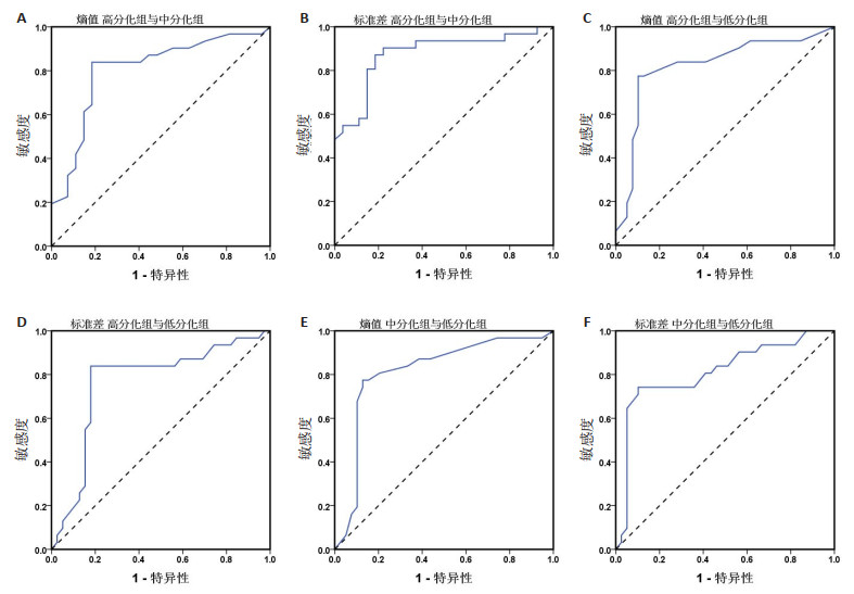
图 1 纹理参数鉴别宫颈癌分化程度的ROC分析
A、C、E: 分别为熵值对宫颈癌高分化与中分化、高分化与低分化、中分化与低分化的ROC曲线分析; B、D、F: 分别为标准差对宫颈癌高分化与中分化、高分化与低分化、中分化与低分化的ROC曲线分析.
Figure 1. ROC analysis of texture parameters in diagnosing differentiation degree of cervical cancer
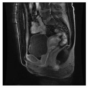
图 2 子宫颈中分化鳞状细胞癌MRI图
患者年龄55岁, 宫内膜菲薄, 内膜下见大小3.5 cm×3 cm×2.5cm灰白结节, 宫颈管长2.5 cm, 外口直径2.3 cm, 宫颈外口见大小2 cm×1.3 cm糜烂区域, 诊断为子宫颈中分化鳞状细胞癌.
Figure 2. MRI image of moderately differentiated squamous cell carcinoma
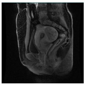
图 3 子宫非角化型颈鳞状细胞癌MRI图
患者年龄48岁, 宫颈糜烂, 1点、3点处水肿、充血、出血明显, 穹隆光滑,病理诊断为宫颈1点、3点非角化型鳞状细胞癌, 宫颈9点局灶高度鳞状上皮内病变.
Figure 3. MRI image of non keratinized cervical squamous cell carcinoma
表 1 纹理参数比较
Table 1. Comparison of texture parameters(Mean±SD)
组别 偏度 峰度 熵值 标准差 高分化组(n=27) 0.23±0.04 3.28±0.69 5.04±0.05 73.29±6.24 中分化组(n=31) 0.25±0.03 3.12±0.63 5.70±0.14a 65.23±5.03a 低分化组(n=39) 0.24±0.04 2.96±0.60 6.38±0.27 ab 54.69±3.78 ab F 2.113 2.042 398.362 115.772 P 0.128 0.135 < 0.001 < 0.001 aP < 0.05 vs高分化组; bP < 0.05 vs中分化组.  下载: 导出CSV
表 2 宫颈癌分化程度与纹理参数的Spearman相关性分析
Table 2. Spearman correlation analysis of cervical cancer differentiation and texture parameters
指标 宫颈癌分化程度 r P 偏度 -0.097 0.347 峰度 0.161 0.115 熵值 -0.269 0.008 标准差 0.288 0.004
下载: 导出CSV
表 3 MRI纹理参数鉴别宫颈癌分化程度的ROC分析
Table 3. ROC analysis of texture parameters in differentiation of cervical cancer
指标 阈值 AUC 特异度 敏感度 熵值 高分化组与中分化组 5.34 0.805 83.9 71.5 高分化组与低分化组 5.18 0.821 77.4 89.7 中分化组与低分化组 5.08 0.813 77.4 77.2 标准差 高分化组与中分化组 67.35 0.875 87.1 71.5 高分化组与低分化组 59.97 0.764 83.9 82.1 中分化组与低分化组 58.25 0.811 74.2 89.7
下载: 导出CSV
-
[1] 胡艳, 常晓斌, 王国庆, 等. 宫颈癌538例预后影响因素分析[J]. 陕西医学杂志, 2017, 46(11): 1531-4. doi: 10.3969/j.issn.1000-7377.2017.11.013 [2] Balcacer P, Shergill A, Litkouhi B. MRI of cervical cancer with a surgical perspective: staging, prognostic implications and pitfalls [J]. Abdom Radiol (NY), 2019, 44(7): 2557-71. doi: 10.1007/s00261-019-01984-7 [3] 胡莎莎, 陈小莉, 刘海峰, 等. 磁共振DWI、IVIM与宫颈癌病理学特征的相关性研究[J]. 磁共振成像, 2017, 8(10): 780-4. doi: 10.12015/issn.1674-8034.2017.10.011 [4] 谢元亮, 杜丹, 谢伟, 等. DCE-MRI纹理分析鉴别宫颈鳞癌与腺癌及预测分级的价值[J]. 放射学实践, 2019, 34(8): 835-40. https://www.cnki.com.cn/Article/CJFDTOTAL-FSXS201908002.htm [5] 郑明雪, 董江宁, 李翠平, 等. 表观扩散系数联合纹理特征评估宫颈鳞癌分化程度的价值[J]. 实用放射学杂志, 2020, 36(4): 592-5, 614. doi: 10.3969/j.issn.1002-1671.2020.04.021 [6] 中国抗癌协会妇科肿瘤专业委员会. 宫颈癌诊断与治疗指南(第四版[) J]. 中国实用妇科与产科杂志, 2018, 34(6): 613-22. https://www.cnki.com.cn/Article/CJFDTOTAL-ZGSF201806011.htm [7] Balcacer P, Shergill A, Litkouhi B. MRI of cervical cancer with a surgical perspective: staging, prognostic implications and pitfalls [J]. Abdom Radiol (NY), 2019, 44(7): 2557-71. doi: 10.1007/s00261-019-01984-7 [8] 张秀芳, 杨静, 郑薇薇. 宫颈癌淋巴结转移情况的超声与CT检查对比分析[J]. 癌症进展, 2020, 18(1): 92-5. https://www.cnki.com.cn/Article/CJFDTOTAL-AZJZ202001026.htm [9] 田兆荣, 郭玉林, 蔡磊, 等. MRI在宫颈癌分期诊断中的应用[J]. 中国医学影像学杂志, 2016, 24(2): 138-40, 147. doi: 10.3969/j.issn.1005-5185.2016.02.014 [10] Théodore C, Levaillant JM, Capmas P, et al. MRI and ultrasound fusion imaging for cervical cancer [J]. Anticancer Res, 2017, 37(9): 5079-85. http://www.ncbi.nlm.nih.gov/pubmed/28870937 [11] Mao JJ, Fang J, Duan XH, 等. 原发鼻咽癌病人治疗前MRI纹理分析的预测价值[J]. 国际医学放射学杂志, 2019, 42(5): 628. http://www.cnki.com.cn/Article/CJFDTotal-GWLC201905089.htm [12] 刘辉, 吴琼雅, 宫晓梅, 等. 基于增强MRI诊断并行全脑放疗的非小细胞肺癌脑转移预后分析[J]. 中国肺癌杂志, 2011, 14(9): 719-22. doi: 10.3779/j.issn.1009-3419.2011.09.05 [13] 祁红琳, 胡先玲, 李传明, 等. 基于MRI纹理特征的早期肝癌术后复发预测[J]. 中国医学物理学杂志, 2017, 34(9): 908-11. doi: 10.3969/j.issn.1005-202X.2017.09.010 [14] 李翠平, 李信响, 董江宁, 等. IVIM-DWI参数及纹理特征术前鉴别宫颈癌亚型的价值[J]. 临床放射学杂志, 2020, 39(6): 1127-32. https://www.cnki.com.cn/Article/CJFDTOTAL-LCFS202006022.htm [15] 陈文林, 胥明婧, 李绍东. 磁共振扩散加权成像纹理分析在对宫颈癌术后早期复发的预测价值[J]. 广西医学, 2018, 40(13): 1440-3. https://www.cnki.com.cn/Article/CJFDTOTAL-GYYX201813016.htm [16] Hernández VJDD, Mejía-Rosales S, Gama GA. Fractal properties of biophysical models of pericellular brushes can be used to differentiate between cancerous and normal cervical epithelial cells [J]. Colloid Surfaces B, 2018, 170: 572-7. doi: 10.1016/j.colsurfb.2018.06.059 [17] 何陈伟, 陈亚君, 邹靖, 等. 动态对比增强磁共振成像在局部晚期宫颈癌同步放化疗效果评估中的价值[J]. 分子影像学杂志, 2021, 44(1): 74-7. doi: 10.12122/j.issn.1674-4500.2021.01.14 [18] 武科, 孙洪赞. 磁共振成像在宫颈癌治疗疗效评价中的应用现状及展望[J]. 磁共振成像, 2019, 10(10): 792-6. doi: 10.12015/issn.1674-8034.2019.10.016 [19] 刘颖, 叶兆祥. 基于磁共振扩散加权成像的表观扩散系数直方图分析在宫颈癌中的应用[J]. 临床放射学杂志, 2018, 37(11): 1940-3. https://www.cnki.com.cn/Article/CJFDTOTAL-LCFS201811045.htm [20] Wang BT, Fan WP, Xu H, et al. Value of magnetic resonance imaging texture analysis in the differential diagnosis of benign and malignant breast tumors [J]. Chin Med Scis J, 2019, 34(1): 33-7. doi: 10.24920/003516 [21] Xie L, Chu R, Wang K, et al. Prognostic assessment of cervical cancer patients by clinical staging and surgical-pathological factor: a support vector machine-based approach[J]. Front Oncol, 2020, 10: 1353. doi: 10.3389/fonc.2020.01353 [22] 叶瑞婷, 邹玉坚, 郑晓林, 等. IVIM-DWI参数分析宫颈癌的组织学特征及其临床诊断价值[J]. 放射学实践, 2020, 35(6): 750-5. https://www.cnki.com.cn/Article/CJFDTOTAL-FSXS202006013.htm [23] 李海蛟, 曹崑, 郑虹, 等. 多序列MRI纹理分析预测宫颈癌新辅助化学治疗疗效[J]. 中国医学影像技术, 2020, 36(8): 1215-9. https://www.cnki.com.cn/Article/CJFDTOTAL-ZYXX202008031.htm [24] 陈浩, 袁子龙, 郑丽丽, 等. 宫颈癌体素不相干运动的直方图和纹理特征的可重复性研究[J]. 临床放射学杂志, 2020, 39(8): 1577-82. https://www.cnki.com.cn/Article/CJFDTOTAL-LCFS202008027.htm -

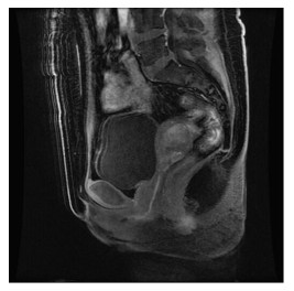
 点击查看大图
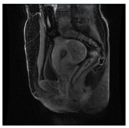
计量
- 文章访问数: 684
- HTML全文浏览量: 229
- PDF下载量: 4
- 被引次数: 0