

 Find Duplicates
 Check Document
 Submission(new)
 Experts Office
 Editorial Office
2021 Vol. 44, No. 1
Display Method:
2021, 44(1): 1-7.
doi: 10.12122/j.issn.1674-4500.2021.01.01
Abstract:
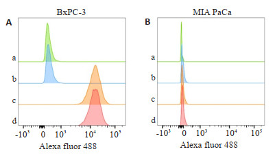
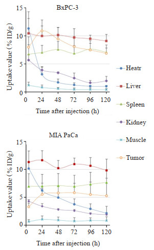
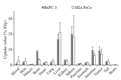
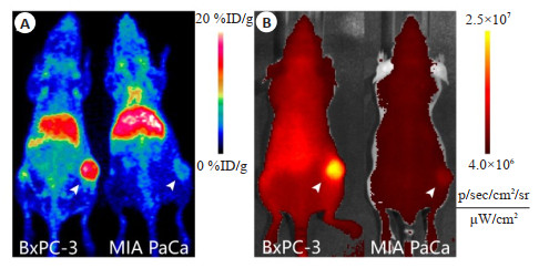
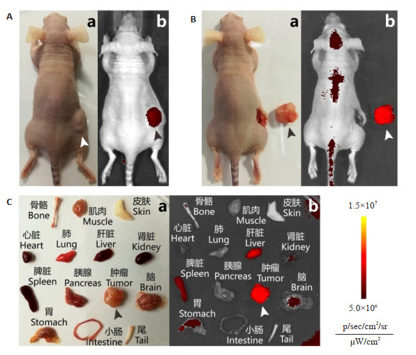
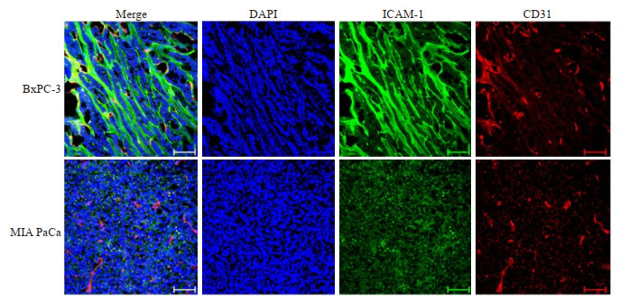
ObjectiveTo validate the feasibility of intermodal imaging of pancreatic cancer tissue employing positron emission tomography (PET) and near-infrared fluorescent (NIRF) imaging, based on a dual-labeled monoclonal ICAM-1 antibody tracer. MethodsICAM-1 expression in pancreatic cancer cell lines (BxPC-3 and MIA PaCa) was determined via flow cytometry. NIRF fluorophore and zirconium-89 Dual-labeled tracer was synthesized via bio- conjugation reaction and coordination chemistry. In the mouse models bearing subcutaneous tumors established using the aforementioned cell lines, we assessed the specificity, performance of intermodal imaging, and radioactive bio-distribution of the tracer. The in situ ex vivo NIRF imaging of resected tumor and major organs at pre-/post-dissection timepoints in BxPC-3 subcutaneous mouse model was also performed. The expression of ICAM-1 in the tissue of transplanted tumors was validated via pathohistochemistry. ResultsSignificant discrepancy in the expression level of ICAM-1 was found between BxPC-3 and MIA PaCa cell lines. The results of PET/NIRF intermodal imaging and radioactive bio-distribution indicated that peak values of tracer uptake in tumors were notably different between the two mouse models of pancreatic cancer (P < 0.05). There was strong evidence for the optimal avidity and specificity of the tracer against ICAM-1. PET and NIRF imaging colocalized the tumor, and the NIRF signal translocated with resected tumor nodules, leaving negligible residual signal in remaining surrounding tissues. Immunohistochemical staining showed that the difference in the expression of ICAM-1 in tumor tissues derived from the two cell lines was positively correlated with the contrast of tracer deposition in tumors between the two mouse models. ConclusionWe confirmed that the preclinical intermodal imaging of malignant tissues in pancreatic cancer using dual-labeled monoclonal antibody targeting ICAM-1 is feasible. This attempt exemplifies the simultaneous implementation of whole-body in vivo focal imaging and in situ visualization of tumor tissue. It implies the potential of ICAM-1-targeted imaging in clinical applications involving lesion detection and intraoperative navigation.
2021, 44(1): 8-12.
doi: 10.12122/j.issn.1674-4500.2021.01.02
Abstract:
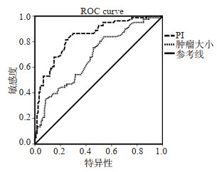
ObjectiveTo explore the ultrasonic diagnosis factors influencing the 5-year recurrence of postoperative hepatocellular carcinoma (HCC) and establish Predict Index (PI) model. MethodsKaplan-Meier and Log- rank univariate analysis and multivariate Cox regression analysis were used to analyze all related ultrasonic diagnosis factors, and the predictive factors and regression coefficients were obtained. According to the predictive factors and regression coefficients, a PI model was established to predict the 5-year recurrence of postoperative HCC by ultrasonic diagnosis. The validity of the PI model was tested and multivariate adjusted to draw ROC curve of PI model to predict the 5-year recurrence of postoperative HCC and evaluate the predictive value of this model for the 5-year recurrence of postoperative HCC. ResultsMultivariate Cox regression analysis showed that tumor boundary (β=0.14, OR=1.23), tumor site (β=0.36, OR=1.58), tumor size (β=0.59, OR=2.42), tumor number (β=0.41, OR=1.79) were independent predictors of the 5-year recurrence of postoperative HCC. According to the predictors and regression coefficient, PI prediction model was established as: PI=0.14×X1 (clear boundary=0, unclear boundary= 1)+0.36×X2 (surrounding type=0, central type=1)+0.59×X3 (cm) +0.41×X4 (single engine=0, multiple engine=1), the cut off of PI was 1.66, the validity of PI was 12.43 by χ2 test (P < 0.05). PI model was an independent predictor after multivariate factor correction (β =1.08, OR=2.91), the area under ROC curve of PI model was 0.812 (0.765-0.957), and the accuracy was 78.7%. ConclusionWe put forward PI model of ultrasonic diagnosis predicting the 5-year recurrence of postoperative HCC and the critical value at the first time. This model had a high predictive value, which provided a strong basis with clinical accurate assessment of the recurrence of postoperative HCC.
2021, 44(1): 13-21.
doi: 10.12122/j.issn.1674-4500.2021.01.03
Abstract:
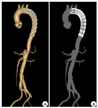
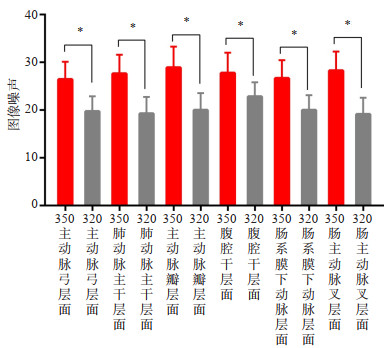
ObjectiveTo explore the value of low tube voltage, low iodine concentration contrast medium (CM) and adaptive statistical iterative reconstruction-V (ASIR-V) protocol in the follow-up of Stanford type B aortic dissection after endovascular repair. Methods128 subjects were included and were randomly divided into the test (100 kVp, 320 mgI/mL, FBP and ASIR-V reconstruction, n=64) and control (120 kVp, 350 mgI/mL, FBP reconstruction, n=64) groups. Image quality of the aorta was evaluated subjectively by a 5 points grading scale and objectively by calculating the signal- and contrast-to-noise ratios (SNR and CNR, respectively). Radiation and CM doses were also evaluated. ResultsThe CT attenuation, SNR, CNR, and subjective image quality assessment of different levels of aorta didn't exhibit significant differences between the groups (P>0.05). In the test group, images reconstructed with FBP and ASIR-V showed significant differences in image noise, SNR and CNR (P < 0.05). The test group resulted in 31.69% less radiation (P < 0.05) and 24.14% less iodine weight than the control group (P < 0.05). Conclusions Protocol of low tube voltage, low iodine concentration CM and ASIR-V algorithm can be used in the follow-up of Stanford type B aortic dissection after endovascular repair. It can decrease the radiation and iodine doses while maintaining good image quality.
2021, 44(1): 22-26.
doi: 10.12122/j.issn.1674-4500.2021.01.04
Abstract:
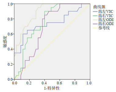
ObjectiveTo explore the clinical value of Neurite orientation dispersion and density imaging (NODDI) in cerebellar microstructural changes in patients with Parkinson's disease (PD). MethodsTwenty healthy persons and 35 patients with Parkinson's disease (PD) were selected as case group and control group respectively. According to the relevant parameters of NODDI scanning results, orientation dispersion index (ODI), intracellular volume fraction (VIC) and isotropic volume fraction (VISO) value, ROC curve was used to evaluate whether the related parameters had certain clinical diagnostic value for Parkinson's disease. ResultsThe VIC and ODI values of dentate nucleus, cerebellar white matter and dentate nucleus in PD patients were significantly lower than those in healthy controls (P < 0.05). The ODI values of cerebellar white matter in PD patients were higher than that in the controls (P < 0.05). At the same time, ROC curve showed that the AUC of patients with PD diagnosed by VIC on the left and right side of dentate nucleus were 0.743 and 0.767, and the AUC of patients with PD diagnosed by ODI on the left and right side of dentate nucleus were 0.891 and 0.694, respectively. ConclusionNODDI can reflect the changes of cerebellar microstructure in patients with PD, and its parameters can be used as an evaluation index for patients with PD.
2021, 44(1): 27-30.
doi: 10.12122/j.issn.1674-4500.2021.01.05
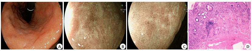
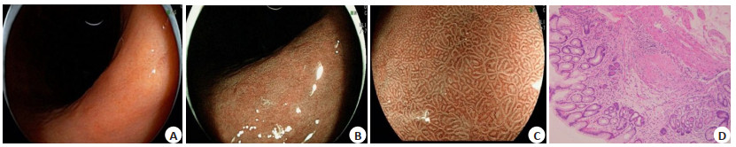
Abstract:
ObjectiveTo evaluate the clinical value of blue light endoscopy in the diagnosis of histological changes and grading of gastric mucosa. MethodsA total of 160 patients with intestinal metaplasia and atrophy of gastric mucosa confirmed by pathological examination from July 2019 to September 2020 were selected. They were all performed the blue light imaging magnifying endoscopy in Shenzhen Hospital of Southern Medical University for physical examination or for dyspepsia and other symptoms. In the process of blue light imaging endoscopy, the appearance of the whole gastric mucosa was observed in the normal white light mode, the bright blue light imaging mode and the blue light imaging mode in turn. Under the white light mode, the samples were taken according to the pathological diagnosis consensus of chronic gastritis biopsy. Under the bright blue light imaging mode and blue light imaging mode, the samples were taken at the abnormal part of mucosal color. Paired chi- square test was used to analyze the diagnostic rate of biopsy pathology for intestinal metaplasia and atrophy of gastric mucosa under different observation model. Wilcoxon signed rank test was used to analyze the auxiliary recognition ability of CG histological change grades (0, +, + +, + + +) under different observation modes. ResultsThe diagnostic accuracy of gastric mucosal atrophy were 6.9% (11/160), 49.4% (79/160) and 20.6% (33/160) in white light mode, bright blue light mode and blue light mode, respectively. The diagnostic rates of intestinal metaplasia were 3.75% (6/160), 22.5% (36/160), 55.0% (88/160), respectively. Compared with the white light mode, the diagnosis rate of gastric mucosal atrophy was the highest under bright blue light imaging mode (P < 0.01). The diagnosis rate of intestinal metaplasia of gastric mucosa was the highest in blue light imaging mode (P < 0.01). The diagnosis rate of intestinal metaplasia of gastric mucosa was the highest in the blue light imaging mode (P < 0.01). In that aspect of the auxiliary identification ability in the CG histological grade, compared with the white light mode, bright blue light imaging mode was the best auxiliary recognition ability for grading gastric mucosal atrophy (Z=-7.685, P < 0.01), blue light imaging was the best auxiliary recognition ability for grading intestinal metaplasia (Z=-8.272, P < 0.01). ConclusionBlue light imaging endoscopy has a good clinical value in the diagnosis of intestinal metaplasia and atrophy of gastric mucosa in CG. Bright blue light imaging mode is more suitable for biopsy pathology and auxiliary histological grading of gastric mucosa atrophy. Blue light imaging mode is more suitable for biopsy pathology and auxiliary histological grading of intestinal metaplasia of gastric mucosa.
2021, 44(1): 31-35.
doi: 10.12122/j.issn.1674-4500.2021.01.06
Abstract:
ObjectiveTo investigate the effect of breast lesion type, benign or malignant lesion and lesion size on the average glandular dose (AGD) in low-energy, high-energy and subtraction images of contrast-enhanced mammography. MethodsA total of 123 female patients who underwent CESM examination in our hospital from February 2018 to October 2018 and confirmed by histopathology were enrolled as subjects. According to benign and malignant lesions, major lesion types, and mass lesion size, patients were divided and the total AGD of low energy imaging, high energy imaging and subtracted imaging (the subtraction imaging AGD is the sum of low energy imaging and high energy imaging) were compared in different groups. ResultsThere were 65 cases of malignant lesions and 58 cases of benign lesions. There were 86 cases of mass lesions and 37 cases of non-mass lesions. The tumor size was ≥2 cm in 44 cases, < 2 cm in 42 cases, including malignant mass ≥ 2 cm in 32 cases and < 2 cm in 25 cases. The AGD of the malignant lesion group was higher than the benign lesion group regardless of low energy, high energy and subtraction, and the results were statistically significant (P < 0.05). Both benign and malignant lesions were higher on the affected side than on the healthy side, and the results were statistically significant (P < 0.05). The AGD of the low energy, high energy and subtraction in the mass lesion group were higher than those in the nontumor lesion group, but the results were not statistically significant (P>0.05). The AGD of high energy, low energy and subtraction in the group with a long diameter ≥2 cm was higher than that in a group with a long diameter < 2 cm, and the results were statistically significant (P < 0.05). The AGD was higher than the group with a diameter < 2 cm, and the results were statistically significant (P < 0.05). ConclusionThe AGD of malignant lesions and ≥2 cm masses were higher than that of benign lesions and < 2 cm masses. The high energy AGD of the affected side was higher than that of the healthy side, no matter whether the lesions were benign or malignant.
2021, 44(1): 36-40.
doi: 10.12122/j.issn.1674-4500.2021.01.07
Abstract:
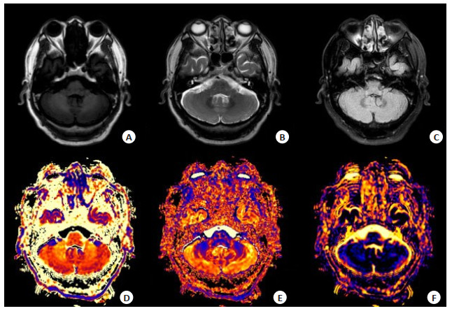
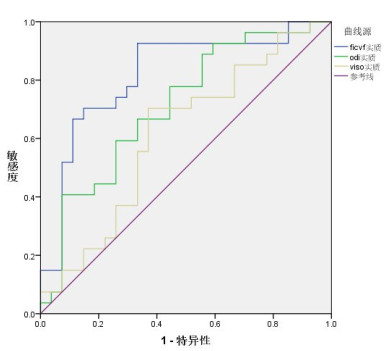
ObjectiveTo explore the application value of neuronal orientation dispersion and density imaging (NODDI) technology in preoperative diagnosis and classification of glioma disease, by using NODDI parameters to quantitatively evaluate the difference between high grade glioma and low grade glioma. MethodsTwenty-seven postoperative patients diagnosed with glioma were selected as the research subjects. Preoperative routine plain scan, enhanced scan, and advanced diffusion scan were performed. Images of neuronal orientation dispersion index (ODI), intracellular volume fraction (FICVF), volume fraction of the isotropic compartment (VISO) were obtained by the poster processing soft. The average values of various parameters of tumor parenchymal area, peritumor edema area and contralateral normal brain white matter area were measured respectively. Comparative analysis of parameters was performed using the receiver operating characteristic (ROC) curve. ResultsFICVF and VISO values in tumor parenchymal area of the high grade glioma group were higher than that of low grade glioma group, and the differences were statistically significant. FICVF, ODI and VISO values in peritumor edema area of the high grade glioma group were higher than that of low grade glioma group, and the differences were statistically significant. The FICVF, ODI and VISO values under the ROC curve of the tumor parenchymal area were 0.82 (P < 0.05), 0.71 (P < 0.05) and 0.61 (P>0.05), respectively. ConclusionNODDI parameters including FICVF, ODI and VISO can all be used to identify high and low grade glioma, and FICVF has the highest diagnostic performance, while VISO has the lowest performance.
2021, 44(1): 41-46.
doi: 10.12122/j.issn.1674-4500.2021.01.08
Abstract:
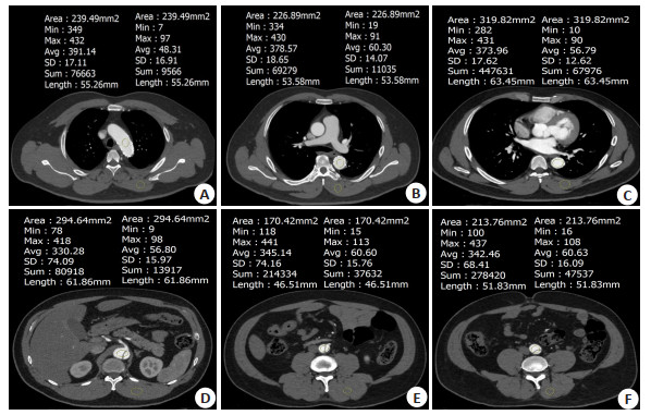
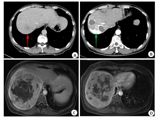
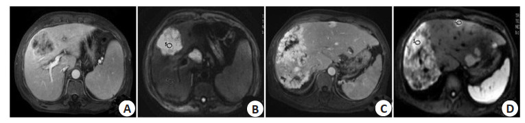
ObjectiveTo explore the value of CT and MRI in evaluating the effects of intervention and molecular targeted therapy on patients with primary hepatocellular carcinoma (HCC). MethodsA total of 80 patients with advanced HCC admitted to the hospital from January 2017 to January 2019 were retrospectively enrolled as the research objects. All patients underwent hepatic artery chemoembolization (TACE) and sorafenib therapy, and CT, MRI and digital subtraction angiography (DSA) were performed after 3 months. The number of clinically effective cases and response rate were compared after 3 months treatment by CT combined MRI and CT alone. Taking DSA results as the golden standard, the sensitivity, specificity, accuracy and kappa values of the two methods were calculated. The detection situations of residual and recurrence lesions were compared after treatment by the two methods. ResultsAfter 3 months of TACE and sorafenib treatment, DSA showed that the total number of target lesions decreased from 98 to 86, the lesions significantly reduced There were 64 clinical disease control patients and 16 ineffective patients, with 80.00% disease control rate. There were 82 residual and recurrence lesions. Among them, there were 9, 18, 42 and 17 residual and recurrence lesions of type I, II, III and IV iodized oil deposition, respectively. The detected number of type II iodized oil deposition and recurrence lesions by CT combined with MRI was more than that by CT (P < 0.05). The sensitivity of CT combined with MRI for evaluating the curative effect of TACE and sorafenib on HCC patients was higher than that of CT examination alone (95.31% vs 84.38%, P < 0.05). Kappa value of CT combined with MRI for evaluating clinical curative effect and DSA examination was 0.809, which was greater than that of CT alone (0.605). ConclusionThe clinical curative effect of TACE and sorafenib is good on HCC patients 3 months later. Compared with CT alone, CT combined with MRI is more conducive to detecting residual and recurrent lesions after treatment, which has of higher sensitivity and application value in evaluating curative effect after 3 months of treatment.
2021, 44(1): 47-52.
doi: 10.12122/j.issn.1674-4500.2021.01.09
Abstract:
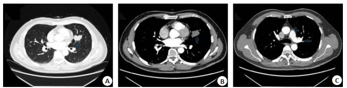
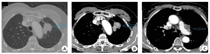
ObjectiveTo explore the relationship between the preoperative CT indexes and conversion to thoracotomy during video assisted thoracoscopic surgery (VATS) lobectomy. MethodsA total of 871 patients with lung space-occupying lesions in our hospital from December 2016 to December 2019 were selected. The patients completed the CT examination and underwent VATS lobectomy. Among them, 28 cases were converted to thoracotomy, accounting for 3.21%. 100 cases from patients who successfully completed VATS surgery were extracted as VATS group by the random number table method. The general data, CT examination results and surgical results were compared between the two groups. The influence and predictive value of preoperative CT examination on conversion of VATS to thoracotomy were analyzed. ResultsThe surgical time, intraoperative blood loss, drainage time and hospitalization timem in VATS group were significantly lower than those in conversion group(P < 0.05). There were no significant differences in the complications such as postoperative infection, pneumothorax and atelectasis between the two groups (P>0.05). The CT signs of short diameter of the largest lymph node and incidence rates of tumor infiltration and pleural indentation in VATS group were significantly lower than those in conversion group (P < 0.05). There were no significant differences in the location of lung field lesions, the largest diameter of lesions and lymph node calcification between the two groups (P>0.05). Logistics regression analysis showed that tumor infiltration was an independent risk factor affecting the conversion to thoracotomy in VATS (P < 0.05). ROC curve analysis showed that the sensitivities of tumor infiltration, short diameter of the largest lymph node, pleural indentation and the combination of the three CT indexes in predicting VATS conversion to thoracotomy were 82.14%, 67.86%, 67.86% and 78.57%, and the specificities were 89.00%, 69.00%, 93.00% and 93.00% respectively. ConclusionVATS conversion to thoracotomy can adversely affect postoperative rehabilitation of patients. The main reasons are that tumor infiltration or lymph node adhesion leads to increase difficulty in separating blood vessels and other anatomical structures, and preoperative CT indexes such as tumor infiltration, short diameter of the largest lymph node and pleural indentation have a certain reference value in assessing the risk of conversion to thoracotomy.
2021, 44(1): 53-58.
doi: 10.12122/j.issn.1674-4500.2021.01.10
Abstract:
ObjectiveTo investigate the correlation between O6- methylgua-nine-DNA methyltransferase (MGMT) expression and CT imaging, clinicopathological features of esophageal squamous cell carcinoma (ESCC). MethodsNon-enhanced and enhanced thoracic CT images of 142 cases for ESCC were retrospectively analyzed preoperatively. CT values, maximum axial thickness and vertical diameter of lesions were measured, and clinicopathological features were recorded. Categorical variable was tested using the χ2 test or Fisher's exact test. Independent-samples t test or Mann-Whitney U test was applied to compare the differences in CT parameters between different MGMT expression status. Receiver operating characteristic (ROC) curve was used to analyze the effectiveness of CT parameters in predicting different expression status of MGMT and Spearman was used to analyze the correlation between MGMT expression and CT parameters. ResultsThere were significant differences in total pathological stage, enhanced CT, ∆ CT and maximum thickness among different MGMT expression groups (all P < 0.05). The diagnostic efficiency of enhanced CT and ∆CT values in differentiating different expression states of MGMT was higher, with AUC of 0.810, 0.817, respectively. There was a high correlation between the enhanced CT, ∆ CT values and MGMT expression (r=- 0.444, - 0.473, P < 0.05). ConclusionAbnormal expression of MGMT may be involved in the development and progression of ESCC. Preoperative tumor enhanced CT values can better predict the expression status of MGMT, which has auxiliary value for evaluating development of tumor and selecting appropriate treatment scheme.
2021, 44(1): 59-62.
doi: 10.12122/j.issn.1674-4500.2021.01.11
Abstract:
ObjectiveTo investigate the clinical value of 3D ASL in assessing the perfusion status of cerebral watershed after different degrees of carotid stenosis. MethodsSixty patients with ischemic cerebrovascular disease who were initially diagnosed in the Department of encephalopathy from January 2018 to March 2020 were collected. The degree of bilateral carotid artery stenosis was evaluated by CDFI. Meanwhile, 3D ASL imaging was performed, and the original data were automatically generated into a pseudo color map of cerebral blood flow (CBF) using functool software, and three-dimensional arterial spin labeling (3D ASL) imaging examination. The interest areas (ROI=200±20 mm2) were selected from the front and back corners of the cortical watershed, the radial crown and the center of the semicircular circle. The CBF values of the cerebral watershed in patients with different degrees of carotid stenosis were measured and analyzed. ResultsThere were 120 cases of carotid artery, 30 cases of no stenosis, 49 cases of mild stenosis, 26 cases of moderate stenosis, 15 cases of severe stenosis. There was no significant difference in CBF between the patients with mild stenosis and the patients without stenosis (P > 0.05). The CBF value in the inner watershed was lower than that in the patients without stenosis (P < 0.05), The CBF values of cortical watershed and internal watershed in patients with moderate and severe carotid stenosis were significantly lower than those in patients without stenosis (P < 0.05). Conclusion3D ASL imaging technology can sensitively evaluate the cerebral perfusion in the watershed area of patients with different degrees of carotid stenosis. It has important application value for the selection of treatment plan and prognosis evaluation of patients.
2021, 44(1): 63-67.
doi: 10.12122/j.issn.1674-4500.2021.01.12
Abstract:
ObjectiveTo explore the value of color Doppler ultrasound (CDFI) in the detection of blood flow parameters of carotid artery and retro-ocular vascular in patients with non-arteritic anterior ischemic optic neuropathy (NAION). MethodsEighty NAION patients admitted to our hospital were selected as the NAION group and 80 volunteers without eye-related diseases as the control group. CDFI was used to detect the internal carotid artery blood flow parameters, internal carotid artery vascular elasticity parameters, and eyeballs of the two groups of subjects. The posterior arterial blood flow parameters were measured by coherent optical tomography to detect the optic disc diameter ratio, macular ganglion cell complex thickness, and optic disc retinal nerve fiber layer thickness in the two groups. Simple linear correlation Pearson correlation analysis method was used Analyze the relationship between internal carotid artery blood flow parameters, internal carotid artery vascular elastic parameters, posterior arterial blood flow parameters and NAION lesions. ResultsThe Vd value of the internal carotid artery in the NAION group was lower than that of the control group (P < 0.05). The RI and IMT values of the internal carotid artery in the NAION group were higher than those in the control group (P < 0.05). The dilatation, arterial tension, and pressure of the internal carotid artery in the NAION group-The strain coefficient value was lower than the control group (P < 0.05). The measured values of Vs and Vd of the OA artery in the NAION group were lower than the control group (P < 0.05). The measured value of arterial RI was higher than the control group (P < 0.05). The horizontal and vertical optic disc diameter ratio and the thickness of the macular ganglion cell complex in the NAION group were lower than those of the control group (P < 0.05). EDV value was significantly positively correlated with the ratio of the optic disc diameter of the horizontal and vertical optic cups, the thickness of the macular ganglion cell complex (P < 0.05). The RI value was significantly correlated with the ratio of the optic disc diameter of the horizontal and vertical optic cups, and the macular nerve The thickness of the ganglion cell complex was significantly negatively correlated (P < 0.05). ConclusionThe blood flow parameters of the carotid artery and posterior blood vessels of NAION patients often have significant changes. There is a certain relationship with the degree of ocular lesions in NAION patients.
2021, 44(1): 68-73.
doi: 10.12122/j.issn.1674-4500.2021.01.13
Abstract:
ObjectiveTo explore the effect of total hip arthroplasty between Super PATH approach and posterolateral approach on clinical efficacy, fracture healing and joint function in patients with traumatic femoral neck fracture. MethodsThe patients with surgey for traumatic femoral neck fracture in this hospital from June 2017 to June 2019 were analyzed and classified into two groups: total hip arthroplasty through posterolateral approach (referred to as control group) (n=42), and total hip arthroplasty through SuperPATH approach (referred to as observation group)(n=54). The Harris score of hip function was used to evaluate the joint function of patients in the two groups after surgey. The clinical effects between the two groups [operation time, intraoperative blood loss, total blood loss, length of surgical incision, postoperative hospitalization time, time required for fracture healing, pain, hemoglobin (Hb), hematocrit, C-reactive protein (CRP), erythrocyte sedimentation rate (ESR), and creatine kinase] were statistically analyzed. The t test was used for comparison between groups. ResultsThere was no difference in intraoperative blood loss, total blood loss and postoperative hospitalization time between two groups. However, the operation time of the observation group was longer than that of the control group, and the length of the surgical incision and the time required for fracture healing were significantly shorter than those of the control group, and the differences were statistically significant (P < 0.05). There was no significant difference in preoperative Hb, hematocrit, CRP, ESR and creatine kinase of patients between the two groups (P > 0.05). However, after operation, Hb and hematocrit decreased in both groups, while CRP, ESR and creatine kinase increased. There was no difference of Hb, hematocrit, CRP and ESR between the two groups after operation, but the level of creatine kinase in the observation group was significantly lower than that in the control group (P < 0.05). There were no significant differences of preoperative VAS and Harris scores between the two groups (P > 0.05). 3 days and 14 days after operation, the average VAS of two groups was lower than that before surgery, and the Harris score was higher than that before surgery. The VAS of the observation group 3 days after surgery was significantly lower than that of the control group, and the Harris score was significantly higher than that of the control group (P < 0.05). There was no significant difference in the VAS and Harris scores 14 days after surgery between two groups (P > 0.05). There were no complications and the sinking and loosening of the prosthesis in both groups. In the final follow-up, the scores in vitality, physical function, social function and overall health of the observation group were significantly higher than those of the control group (P < 0.05). ConclusionCompared with the posterolateral approach, SuperPATH approach of total hip arthroplasty is more effective in the treatment of patients with traumatic femoral neck fractures, which can reduce early postoperative pain and muscle damage. It can significantly improve early hip function and recovery of prognosis.
2021, 44(1): 74-77.
doi: 10.12122/j.issn.1674-4500.2021.01.14
Abstract:
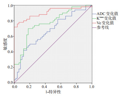
ObjectiveTo investigate the value of dynamic contrast- enhanced magnetic resonance imaging (DCE-MRI) in the evaluation of concurrent chemoradiotherapy for locally advanced cervical cancer. MethodsA total of 200 patients with locally advanced cervical cancer treated in our hospital from January 2019 to January 2020 were selected. All patients were treated with concurrent chemoradiotherapy, the changes of DCE-MRI parameters before and after treatment were observed, and the differences of DCE-MRI parameters between effective and ineffective patients were analyzed. ResultsAll patients received concurrent chemoradiotherapy. There were 21 patients with complete remission, 67 patients with partial remission, 72 patients with stable disease and 40 patients with disease progression, the effective rate was 44.00%. The proportion of FIGO stage ⅣA and Karnofsky score of patients with ineffective treatment were significantly higher than those of patients with effective treatment (P < 0.05). The apparent diffusion coefficient (ADC), volume metastasis constant (Ktrans) and volume of extracellular space (Ve) were significantly higher than those before treatment (P < 0.05). The change values of ADC, Ktrans and Ve in the effective patients were significantly higher than those in the ineffective patients (P < 0.05). The area under the ROC curve of ADC change, Ktrans change and Ve change to predict the treatment effectiveness were 0.675, 0.770 and 0.905 (P < 0.05), and the cutoff values were 0.35×10⁻³ mm2/s, 0.54 min-1 and 0.22, the sensitivity were 66.60%, 78.80% and 85.50%, and the specificity were 62.00%, 75.50% and 82.00%, respectively. ConclusionDCE-MRI has certain application value in evaluating the effect of concurrent chemoradiotherapy for local advanced cervical cancer and is worthy of clinical use.
2021, 44(1): 78-82.
doi: 10.12122/j.issn.1674-4500.2021.01.15
Abstract:
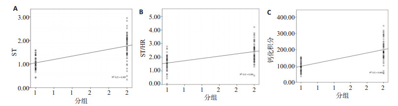
ObjectiveTo explore the diagnostic performance of synchronous 12-lead ambulatory electrocardiogram (AECG) combined with spiral CT in coronary vascular lesions. MethodsBetween April 2018 and August 2020, 90 patients with coronary heart disease who were subjected to spiral CT and AECG examinations were enrolled. According to the degree of coronary vascular lesions, the patients were divided into group A and B. Taking coronary angiography as the golden standard, the diagnostic value of the combination of the two methods for coronary vascular lesions was evaluated. ResultsThe differences in HR, atrioventricular premature beats, atrial tachycardia, and ventricular tachycardia between the two groups were not significant (P > 0.05), but the ST and ST/HR of group A were significantly lower than those of group B (P < 0.05). Group A had significantly lower calcification scores than group B (P < 0.05). ST, ST/HR and calcification scores were positively correlated with the degree of coronary artery lesions (P < 0.05). The sensitivity, specificity, accuracy rate, positive and negative predictive values of AECG combined with spiral CT in the diagnosis of coronary vascular lesions were higher than those of single diagnosis (P < 0.05). ConclusionThe changes of ST, ST/HR and calcification scores are closely related to coronary vascular lesions. Combining synchronous 12-lead AECG with spiral CT can improve the diagnostic efficiency.
2021, 44(1): 83-87.
doi: 10.12122/j.issn.1674-4500.2021.01.16
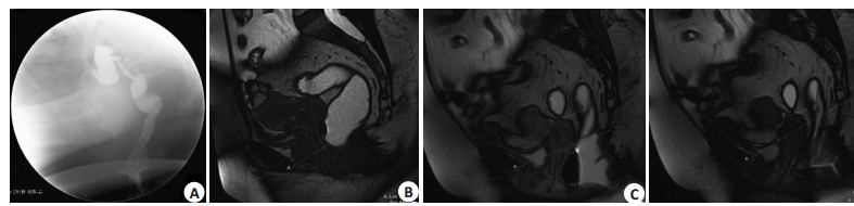
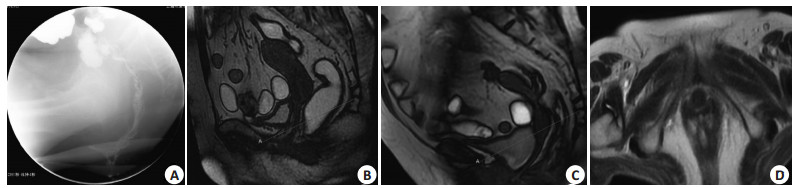
Abstract:
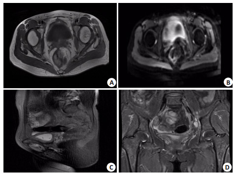
ObjectiveTo explore the clinical value of MR defecography in diagnosing female patients with outlet obstructive constipation. MethodsWe selected 80 patients with constipation of outlet obstruction who were clearly diagnosed in our hospital's anorectal department. The data of X-ray defecography and MR defecography were retrospectively analyzed. The clinical diagnosis results were used as the diagnostic criteria to calculate the two angiography. The coincidence rate of the method was calculated. The difference of the two types of angiography in the diagnosis of different causes of outlet obstructive constipation were analyzed. ResultsBased on the clinical diagnosis of the cause, the coincidence rate of X-ray defecography in the diagnosis of rectal mucosal prolapse, rectal intussusception, colonic hernia, intestinal hernia, and perineal decline were all 100%. X-ray defecography was used to diagnose bladder prolapse and uterine prolapse. The coincidence rates of parasacral cysts, paracervical cysts, and parasacral cysts were 11.76%, 8.33%, 11.11% and 12.50%, respectively. MR defecography diagnosed parasacral cysts, paracervical cysts, bladder prolapse, and uterine prolapse. The coincidence rates were 100.00%, 8.33%, 11.11% and 12.50%, respectively. MR defecography diagnosed rectal intussusception, perineal descent, small intestinal hernia, colonic hernia, rectal muscle spasm, rectal mucosal prolapse, anterior rectum. The coincidence rate of X-ray defecography with the clinically confirmed etiology was lower. The diagnosis coincidence rate of X-ray defecography for rectal protrusion, rectal mucosal prolapse, rectal intussusception, and perineal descent were higher than that of MR defecography (P < 0.05). The diagnostic coincidence rates of MR defecography for parasacral cyst, paracervical cyst, bladder prolapse, and uterine prolapse were higher than that of X-ray defecography (P < 0.05). ConclusionOutlet obstructive constipation caused by colorectal lesions has a high diagnostic accuracy rate with X-ray defecography. But it is not sensitive to the diagnosis of outlet obstructive constipation caused by bladder prolapse, uterine prolapse, paracervical cyst and parasacral cys. MR defecography diagnosis method has a better complementary effect on X-ray defecography.
2021, 44(1): 88-91.
doi: 10.12122/j.issn.1674-4500.2021.01.17
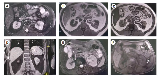
Abstract:
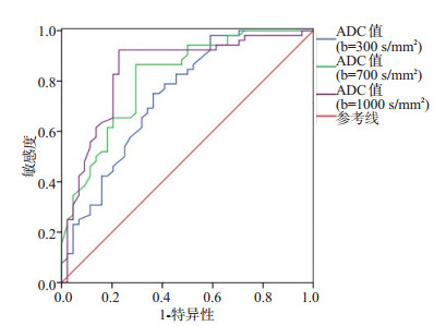
ObjectiveTo analyze the application value of magnetic resonance (MRI) diffusion weighted imaging (DWI) in the preoperative diagnosis of renal space- occupying lesions. MethodsNinty-six patients with renal space-occupying lesions admitted to our hospital from January 2019 to December 2019 were selected. Fifty-two patients with renal malignant tumors (renal malignant lesion group) and 44 patients with benign renal tumors (benign renal lesion group) were pathologically confirmed, and 96 healthy volunteers were selected as controls (normal group). They were given DWI examination. The apparent diffusion coefficient (ADC) values of each group under different diffusion sensitivity coefficient b values were compared, and the diagnostic value of ADC values under different diffusion sensitivity coefficient b values on renal spaceoccupying lesions was analyzed. ResultsThe longest diameter of lesions in renal malignant lesion group was larger than that in benign renal lesion group. In renal malignant lesion group, T1WI and T2WI showed slightly high signals, including 9 cases of uniform signals and 43 cases of high-low mixed signals of different degrees, and DWI showed high signal in tumor parenchyma and showed low signal in necrotic tumor. In renal benign lesion group, T1WI showed low signals and T2WI showed high signals, and DWI showed uniform slightly low signals. The longest diameter of the lesion in renal malignant lesion group was significantly larger than that in benign renal lesion group (P < 0.05). When the diffuse sensitivity coefficient b value was 300, 700 or 1000 s/mm2 respectively, the ADC values in malignant lesion group, benign renal lesion group and normal group were significantly different (P < 0.05), showing benign renal lesion group > normal group > renal malignant lesion group. Taking pathological biopsy results as the gold standards, the AUC values were 0.742 (95%CI: 0.642-0.842), 0.811 (95%CI: 0.725-0.897) and 0.842 (95%CI: 0.758-0.927), and sensitivities were 98.10%, 86.50% and 92.30% and specificities were 40. 90%, 70.50% and 77.30% when diffuse sensitivity coefficient b values were 300, 700 and 1000 s/mm2. And the the best cutoff values of ADC were 3.86×10³, 2.50×10³ and 1.71×103 mm2/s respectively. ConclusionDWI has good application value in the preoperative diagnosis of renal space-occupying lesions. It can be used to distinguish benign and malignant renal space-occupying lesions. ADC value has good diagnostic efficacy on renal space- occupying lesions under different diffusion sensitivity coefficient b values.
2021, 44(1): 92-95.
doi: 10.12122/j.issn.1674-4500.2021.01.18
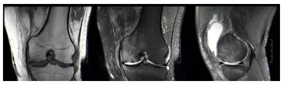
Abstract:
ObjectiveTo explore the effect and its clinical value of shear wave elastography (SWE) preliminarily on articular cartilage in patients with early knee osteoarthritis (KOA). Methods20 healthy adults and 20 early knee osteoarthritis patients who met the enrollment criteria were selected as the research objects. Routine and shear wave elastography examinations were performed. The thickness and Young's modulus of the intercondylar and medial condyle cartilage were included. The thickness and Young's modulus of cartilage within and between groups were analyzed for statistical differences. ResultsThe cartilage thickness of the intercondylar area, medial condyle in the test group were 0.225±0.025, 0.217±0.026, and those of the control group were 0.235±0.023 and 0.209±0.019 mm. There was statistically significant difference between different parts in the group (P < 0.05). There was no statistically significant difference in the same part between groups (P > 0.05). The cartilage Young's modulus of the intercondylar area, medial condyle in the test group and in the control group were 24.17±3.85, 25.94±3.55, 19.93± 2.69 and 21.59±2.57 kPa respectively. There was statistically significant difference in different parts within the group (P < 0.05). There was statistically significant difference in the same part between groups (P < 0.05). ConclusionSWE can quantitatively evaluate the mechanical properties of knee articular cartilage in vivo. The Young's modulus of intercondylar and medial condyle cartilage in early KOA patients is greater than that of the normal population.
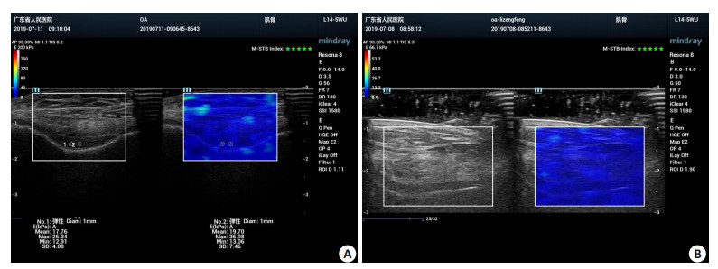
2021, 44(1): 96-102.
doi: 10.12122/j.issn.1674-4500.2021.01.19
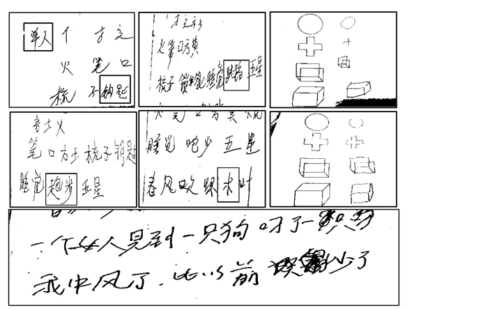
Abstract:
ObjectiveTo investigate the influence of different degrees of cholinergic pathway damage on the language function of patients with cerebral infarction in the basal ganglia. MethodsWe included 135 cases of acute basal ganglia cerebral infarction with initial onset and speech disorder in the Department of Neurology from October 2018 to October 2020. All patients were operated with the Cholinergic Pathway High Signal Scale (CHIPS) Check, according to the score. They were divided into group A (mild abnormality, CHIPS score 1-3 points, 42 cases), group B (moderate abnormality, CHIPS score 4-7 points, 53 cases) and group C (severe abnormality, CHIPS score ≥8, 40 cases). Three groups of patients were tested with the standard Chinese Aphasia Scale (ABC). Logistic regression was used to find the related factors of language damage in patients with basal ganglia cerebral infarction with cholinergic pathway damage. One-way analysis of variance was performed on each calculation item of the standard Chinese Aphasia Scale of the three groups of patients to evaluate the differences in the scores of the ABC scales of the three groups. ResultsABC scale score: (talk; comprehension; retelling; naming; reading; writing; structure and visual space; application; calculation): Group A (25.05±1.72; 204.67±5.14; 91.36±5.19; 78.31±2.34; 50.48±4.56; 87.07± 3.22; 14.48±1.31; 27.31±1.89; 22.24±1.67); Group B (18.51±1.35; 198.85±8.68; 86.94±3.77; 76.57±2.43; 46.3.25±3.65; 85.21±3.65; 1.24; 28.08±1.82; 22.19±1.42); Group C (16.35±2.09; 195.42±7.03; 85.95±2.88; 72.93±4.23; 44.35±3.41; 83.58±2.91; 12.73±2.17; 27.80±1.78; 21.85 ± 1.71). After one-way analysis of variance comparison, there are differences in the scores of 6 items in conversation, comprehension, naming, reading, writing, structure and visual space Statistical significance among three groups. In the retelling item, the difference between group C and group A was significant, and the difference between group C and group B was not significant (P=0.244). There was no significant difference in the scores of the three groups in use and calculation. Regression analysis found that age, high homocysteine, and infarct volume were risk factors for language impairment. ConclusionThe degree of language impairment in patients with cerebral infarction in the basal ganglia may be related to the degree of damage in the cholinergic pathway. Age, high homocysteine and infarct volume are risk factors for language impairment in patients with cerebral infarction in the basal ganglia.
2021, 44(1): 103-106.
doi: 10.12122/j.issn.1674-4500.2021.01.20
Abstract:
ObjectiveTo explore the early MRI findings of seromuscular-layer injury after ultrasound ablation for uterine fibroids, and analyze their clinical significance. MethodsWe retrospectively analyzed clinical data of 150 patients with uterine fibroids received high-intensity focused ultrasound (HIFU) ablation between September 2019 and September 2020. All patients received MR examination before and after operation and had complete imaging data. Changes in MRI signals before and after operation were analyzed, and the condition of seromuscular-layer injury after operation was determined according to the MRI images. The subjects were divided into the injury group (n=43) and the non-injury group (n=107). MRI results of the injury group were observed. The situation of ultrasound ablation and postoperative adverse reactions were compared between 2 groups. ResultsBefore operation, uterine fibroids showed low, equal, or high signals on T1WI, low, mixed, equal or high signals on T2WI, and enhanced scan showed enhancement. After operation, fibroids showed increased signals on T1WI, but no apparent rule of signal changes on T2WI. The enhanced scan showed no enhancement. After ultrasound ablation, the incidence of seromuscular-layer injury was 28.67% (43/150). T2WI sequence showed continuous signals of peripheral muscle layer of the fibroids, with clear boundaries. The dynamic enhanced scan showed ring enhancement of the seromuscular-layer, and local perfusion defect. The rate of seromuscular-layer injury in patients with anterior wall fibroids reached 31.40%. The irradiation time, total ablation dose, and ablation rate of fibroids were longer and higher in the injury group than in the non-injury group (P < 0.05). Adverse reactions were found in both groups after operation, and the incidence of vaginal discharge was significantly higher in the injury group than in the non-injury group (P < 0.05). ConclusionAfter HIFU ablation, patients with uterine fibroids will have seromuscular-layer injury in the early stage. Long irradiation time, high total ablation dose and high ablation rate of fibroids will increase the risk of seromuscular-layer injury, causing adverse reactions after operation. MR examination can effectively assess the condition of seromuscular-layer injury.
2021, 44(1): 107-111.
doi: 10.12122/j.issn.1674-4500.2021.01.21
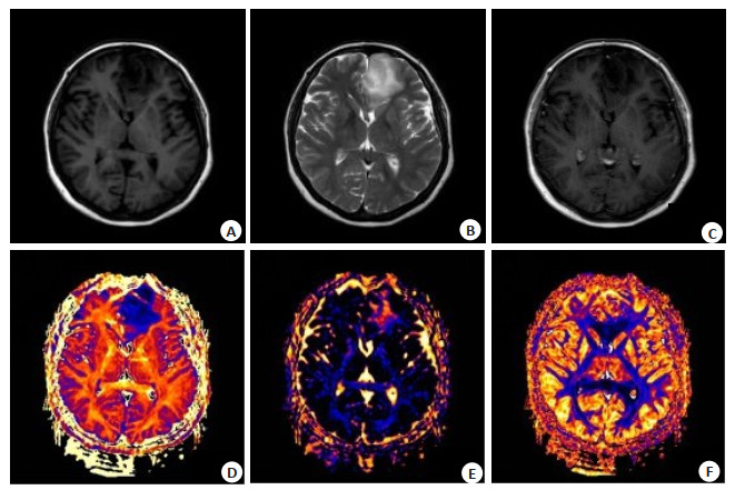
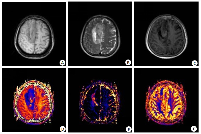
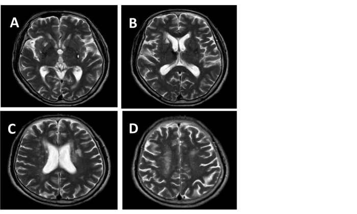
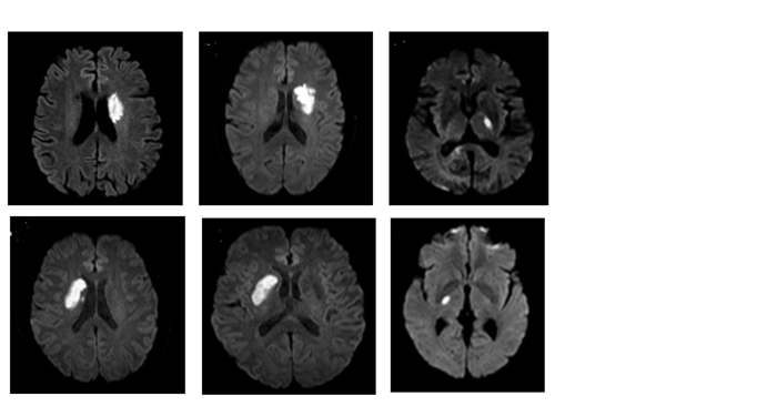
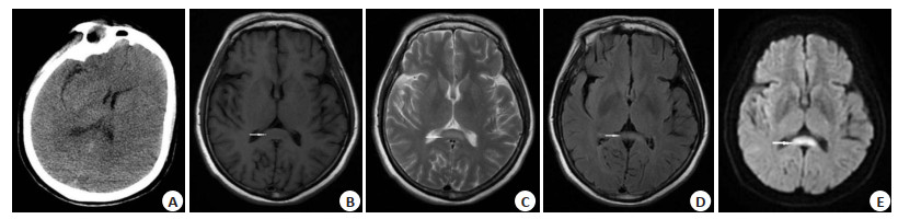
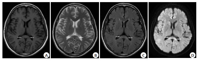
Abstract:
ObjectiveTo analyze the clinical and imaging manifestations of reversible corpus callosum pressure lesion syndrome. MethodsThe clinical and imaging of three cases with reversible splenial lesion syndrome in adults were retrospectively collected and organized. The clinical presentation and imaging manifestations were analyzed, and related literature was reviewed. ResultsTwo cases had viral encephalitis (case 1 and case 2), one case had hypoglycemic encephalopathy (case 3). The clinical manifestations of the three cases were complex and varied without any specificity. Cranial MRI together showed round-like or "boomerang" slightly, slight hypointensity on T1WI, slight hyperintensity on T2WI and FLAIR, significant hyperintensity on DWI and significant hypointensity on ADC. No obvious enhancement could be detected. The lesions in the re-examined cases reduced or disappeared. ConclusionReversible splenial lesion syndrome is one of the complex causes with no characteristic clinical symptoms, imaging manifestations of corpus callosum reversible lesions imaging syndrome. MRI can achieve the goal of early detection, early diagnosis, and assessment of therapeutic efficacy, and the prognosis is usually good after treatment of the etiology.
2021, 44(1): 112-116.
doi: 10.12122/j.issn.1674-4500.2021.01.22
Abstract:
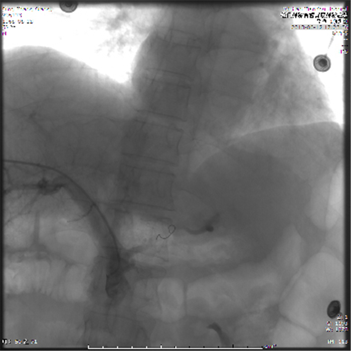
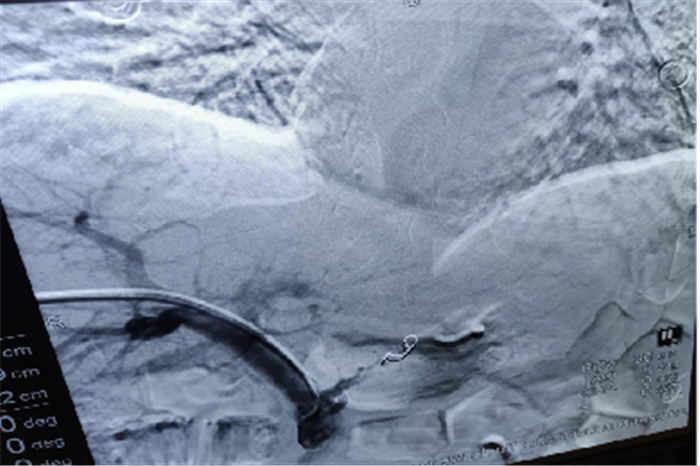
As one of the most common and dangerous complications of portal hypertension in patients with cirrhosis, esophageal and gastric varices (EV) is an independent risk factors for upper gastrointestinal bleeding when they are severe, with high fatality rate, rapid progression and easy repeated bleeding. Currently, the prevention and treatment of cirrhosis esophageal gastric varices treatment mainly includes endoscopic treatment, TIPS, surgery, Pharmacological, etc. In addition, Ultrasound guided percutaneous transhepatic portal vein embolization is a new technique for the prevention and treatment of gastric coronary vein thrombosis. The hemostasis effect is lasting and more thorough, and the patients are better tolerated. The multidisciplinary cooperation between ultrasound and DSA is expected to play an important role in the prevention and treatment of massive hemorrhage of upper digestive tract caused by portal hypertension esophagogastric varicose vein rupture.
As one of the most common and dangerous complications of portal hypertension in patients with cirrhosis, esophageal and gastric varices (EV) is an independent risk factors for upper gastrointestinal bleeding when they are severe, with high fatality rate, rapid progression and easy repeated bleeding. Currently, the prevention and treatment of cirrhosis esophageal gastric varices treatment mainly includes endoscopic treatment, TIPS, surgery, Pharmacological, etc. In addition, Ultrasound guided percutaneous transhepatic portal vein embolization is a new technique for the prevention and treatment of gastric coronary vein thrombosis. The hemostasis effect is lasting and more thorough, and the patients are better tolerated. The multidisciplinary cooperation between ultrasound and DSA is expected to play an important role in the prevention and treatment of massive hemorrhage of upper digestive tract caused by portal hypertension esophagogastric varicose vein rupture.
2021, 44(1): 117-121.
doi: 10.12122/j.issn.1674-4500.2021.01.23
Abstract:
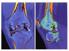
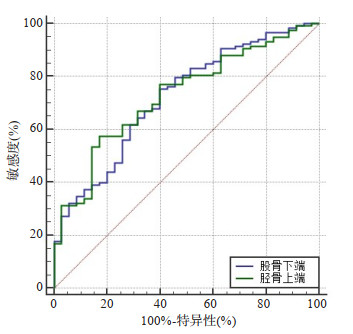
ObjectiveTo explore the value of dual-energy CT (DECT) in diagnosis of bone marrow lesions in patients with Knee Osteoarthritis (KOA). MethodsThe clinical data of 147 patients with KOA admitted between December 2016 and December 2018 were retrospectively analyzed. All patients underwent knee MRI and DECT plain scan after admission, and their knee joints were divided into 12 regions according to the anatomical structure, including each 6 regions of the lower femur and upper tibia respectively. MRI images, conventional CT and virtual non-calcium images were obtained. Using MRI results as the standard, the diagnostic value of DECT examination on patients with bone marrow lesions under different contrast material relative ratio (Rel.CM) values, and the sensitivity, specificity, accuracy rate, negative predictive value and positive predictive value were calculated. The area of interest quantitative measurement method was used to detect the CT value of virtual noncalcium images under the best Rel.CM value, and the bone marrow CT values of the positive region and the negative region on the DECT images were compared, and the receiver operating characteristic (ROC) curve was used to evaluate the diagnostic efficacy of bone marrow CT value on the DECT images on bone marrow lesions in patients with KOA. ResultsThere were no significant differences in the sensitivity, specificity, accuracy rate and positive and negative predictive values of DECT under different Rel.CM values in diagnosing bone marrow lesions in patients with KOA (P>0.05), but when Rel.CM value=1.45, the sensitivity, specificity, accuracy rate and positive and negative predictive values were the best. There were statistically significant differences in the bone marrow CT values of positive and negative bone marrow lesions in different regions under the best Rel.CM value (P < 0.05). ROC curves showed the CT values of the lower end of the femur and the upper end of the tibia can be used as predictive indicators for the diagnosis of bone marrow lesions in patients with KOA (all P < 0.05). Conclusion DECT technology has a high accuracy rate in diagnosing bone marrow lesions in patients with KOA, and it can be used as an auxiliary diagnosis by CT value.
2021, 44(1): 122-126.
doi: 10.12122/j.issn.1674-4500.2021.01.24
Abstract:
ObjectiveTo explore the value of real-time shear wave elastography (SWE) in the diagnosis of hepatitis B virus (HBV) infection and its influencing factors. MethodsA total of 137 patients with liver fibrosis diagnosed with HBV infection in our hospital were selected as the fibrosis group. Sixty patients with HBV infection without liver fibrosis during the same period were selected as the control group. The liver elastic modulus values measured by SWE of the two groups were compared. According to different pathological fibrosis stages, stratified analysis, receiver operating curve (ROC) was used to analyze the clinical value of SWE detection in differential diagnosis of liver fibrosis; logistic regression method was used to analyze the influence of SWE detection in diagnosis of liver fibrosis Influencing factors. ResultsThe measured value of hepatic elastic modulus of patients in the fibrosis group was higher than that of the control group (P < 0.05). The difference in liver elastic modulus of patients with different pathological stages of liver fibrosis was significant (P < 0.05). The measured values of hepatic elastic modulus from stage S1 to S4 gradually increased. The ROC curve was drawn and the sensitivity of SWE to determine the hepatic fibrosis value in the diagnosis of liver fibrosis was 86.13%, the specificity was 85.00%, and the missed diagnosis rate was 13.87%, the misdiagnosis rate was 15.00%. The area under the ROC curve AUC value was 0.889. Logistic regression model analysis showed the higher the pathological stage of liver fibrosis. The higher the inflammation grade was positively correlated with the correct diagnosis of hepatic elastic modulus (P < 0.05). ConclusionSWE diagnosis of HBV infection liver fibrosis as a non-invasive method has high sensitivity and specificity. Its diagnostic performance is affected by the degree of fibrosis and inflammation grade.
2021, 44(1): 127-131.
doi: 10.12122/j.issn.1674-4500.2021.01.25
Abstract:
ObjectiveTo explore and analyze the relationship between the 1.5T MR imaging features and the diagnosis of parotid tumor patients and the comparison of pathological results. Methods70 patients with early parotid tumor from May 2016 to May 2019 were selected. All patients were examined with 1.5T MR in our hospital and confirmed by pathological diagnosis. Among them, 51 cases were benign tumors (benign tumor group) and 19 cases were malignant tumors (malignant tumor group). The MR and pathological results of benign and malignant tumors, the characteristics of MR imaging results of benign and malignant tumors (including location, shape, density, contour), the types of MR time signal curve and peak time of benign and malignant groups were analyzed and compared. ResultsThere was no significant difference between MR diagnosis of parotid tumors and pathological results (P>0.05); there was significant difference between MR imaging results of parotid benign and malignant tumors (P < 0.05), and there was significant difference between the location, shape, density, contour and other manifestations of benign and malignant tumors (P < 0.05). There was significant difference between the two groups in the dynamic characteristics of tumor MR (P < 0.05), and between the benign group in the persistent type and the clearance type(clearance rate≥0.3%), flat type rate was significantly higher than malignant group (P < 0.05), malignant group clearance rate (clearance rate < 0.3%) was significantly higher than benign group (P < 0.05), benign group peak time was significantly higher than the control group (P < 0.05), with statistical significance. Conclusion1.5T MR has a high diagnostic efficiency in the diagnosis of parotid tumor patients, and it has a great value in the differentiation of benign and malignant tumors, which is worthy of clinical promotion.
2021, 44(1): 132-135.
doi: 10.12122/j.issn.1674-4500.2021.01.26
Abstract:
ObjectiveTo explore the clinical value of Multi slice spiral CT and transesophageal echocardiography in diagnosis of left atrial thrombus. MethodsA total of 150 patients with atrial fibrillation treated in our hospital from January 2019 to may 2020 were examined by MSCT and transesophageal echocardiography (TEE). The value of MSCT in the diagnosis of left atrial thrombosis was analyzed. ResultsThere were 22 cases with left atrial thrombosis, the incidence rate was 14.67%. The proportion of hypertension in patients with left atrial thrombosis was higher than that in patients without left atrial thrombosis (P < 0.05). There were no significant differences of gender, age, type and diabetes between patients with and without left atrial thrombosis (P>0.05). The kappa value of MSCT diagnosis and TEE results was 0.617 (P < 0.05). The sensitivity, specificity, accuracy, positive predictive value and negative predictive value of MSCT in diagnosing left atrial thrombosis were 77.27%, 91.41%, 89.33%, 60.71% and 95.90%. The proportion of CHA2DS2-VASc score≥2 in patients with left atrial thrombosis was significantly higher than that of patients without left atrial thrombosis (P < 0.05). The sensitivity, specificity, accuracy, positive predictive value and negative predictive value of CHA2DS2-VASc score≥2 in the diagnosis of left atrial thrombosis were 72.73%, 82.81%, 81.33%, 40.00% and 94.64%. The sensitivity, specificity, accuracy, positive predictive value and negative predictive value of MSCT combined with CHA2DS2-VASc score in the diagnosis of left atrial thrombosis were 90.91%, 93.75%, 93.33%, 71.43% and 98.36%, which were higher than those of MSCT alone, but the difference was not significant (P>0.05). The anteroposterior diameter and left-right diameter of left atrium in patients with left atrial thrombosis were significantly higher than those in patients without left atrial thrombosis (P < 0.05). The left atrial ejection fraction was significantly lower than that in patients without left atrial thrombosis (P < 0.05). ConclusionMSCT combined CHA2DS2-VASc score has good value in the diagnosis of left atrial thrombosis. Compared with MSCT, it has the advantages of no radiation, can be examined many times in a short time and is economical.
2021, 44(1): 136-140.
doi: 10.12122/j.issn.1674-4500.2021.01.27
Abstract:
ObjectiveTo review the situation of 59 cases of pneumonia in 112 patients admitted to neurosurgery and strict COVID-19 prevention and control measures within two months of the COVID-19 outbreak. MethodsA total of 112 hospitalized patients aged 3-83 years (49.5±18.9 years) in the Department of neurosurgery from January 25 to March 30, 2020 were retrospectively analyzed. In the case of lack of emergency nucleic acid detection resources, the types and chest CT manifestations of all pneumonia patients in our department were analyzed. The CT impact characteristics of different pneumonia were compared, which were distinguished from COVID- 19. We analyzed the treatment of COVID-19 highly suspected patients in our department, and summarized the experience and measures of epidemic prevention and control in our department, such as patient admission process, medical staff management, ward management and so on. Result Eighty-six patients completed chest CT examinations in the emergency department at the time of admission. Fifty-nine patients (68.6%) with pneumonia were identified. Among them, 54 patients (62.8%) had aspiration pneumonia. CT images showed 8 patients with ground glass shadow, accounting for 11.76%. Only 2 suspected patients were admitted to the neurosurgery ward, then transferred to the infection department for diagnosis and treatment. The patients were diagnosed with common pneumonia. The first throat swab nucleic acid test of all admitted patients was negative, and some suspicious patients were still negative after admission. Under the strict prevention and control of our department, there was no confirmed case of new coronary pneumonia. ConclusionThe incidence of pneumonia in neurosurgery patients is high. Specialist physicians should strengthen their understanding of pneumonia and their ability to read films, identify early chest CT changes in pneumonia. The strict protective measures, especially the suffocation operation implemented and the good ventilation conditions, are harmful to the nerves.
2021, 44(1): 141-145.
doi: 10.12122/j.issn.1674-4500.2021.01.28
Abstract:
ObjectiveTo explore the application value of CT and magnetic resonance diffusion weighted imaging (MRI-DWI) in preoperative evaluation of esophageal cancer. MethodsRelevant data of 107 patients with esophageal cancer who were treated at the hospital from August 2017 to July 2019 were retrospectively analyzed. All patients underwent surgical treatment. CT and MRI-DWI were performed before surgery, and pathological examinations were performed after surgery. Value of CT and MRIDWI in T staging and N staging were analyzed. Lengths of esophageal cancer measured by CT and MRI-DWI were compared with the actual length. ResultsUsing pathological diagnosis as the golden standard, Kappa values of CT in diagnosis of T1, T2, T3 and T4 stages were 0.719, 0.695, 0.843 and 0.851, which of MRI-DWI were 0.958, 0.948, 0.925 and 0.921. Kappa values of CT and MRI-DWI in diagnosis of N stage were 0.806 and 0.977, respectively. Length of esophageal cancer measured by CT was significantly larger than that by MRI- DWI. There was no significant difference between the length of esophageal cancer measured by MRI-DWI and the actual length (P>0.05). Pearson correlation analysis showed that lengths of esophageal cancer measured by CT and MRI-DWI were highly correlated with the actual length (r=0.816, 0.991, P < 0.05). ConclusionBoth CT and MRI-DWI can be used to evaluate preoperative staging and lesion length of patients with esophageal cancer, but MRI-DWI is better for preoperative staging and lesion length evaluation.
2021, 44(1): 146-150.
doi: 10.12122/j.issn.1674-4500.2021.01.29
Abstract:
ObjectiveTo explore the value difference between multi- slice spiral CT (MSCT) scan and upper gastrointestinal angiography in the diagnosis and treatment of acquired esophageal fistula. MethodsEighty-one patients with high suspicion of esophageal fistula after radiotherapy and chemotherapy were selected as the research objects. After gastroesophageal examination, 56 patients were diagnosed as secondary esophageal fistula after radiotherapy, and 25 patients did not develop secondary esophageal fistula. All patients before receiving gastroesophageal examination received upper gastrointestinal angiography and MSCT scan. The gastroesophageal examination results were used as the gold standard to determine the value of upper gastrointestinal angiography and MSCT scan in diagnosing esophageal fistula after radiotherapy. ResultsAmong 56 patients diagnosed with secondary esophageal fistula, the results of gastroesophageal endoscopy were used as the gold standard. MSCT's coincidence rate of fistula location in trachea, left main bronchus, right main bronchus, left middle lung bronchus, and right upper lung bronchus were all reaching 100%. The upper gastrointestinal angiography only diagnosed the fistula location in the trachea with a diagnostic coincidence rate of 100%. Overall, the MSCT diagnosis of the fistula location was higher than the upper gastrointestinal angiography (P < 0.05). The detection rates of MSCT for window fistula, tract fistula, linear esophageal fistula, and irregular- shaped esophageal fistula were 50.00%, 23.21%, 12.50%, 10.71%, respectively. The detection rates of window fistulas, tube fistulas, linear esophageal fistulas, and irregular-shaped esophageal fistulas by gastrointestinal angiography were 44.64%, 19.64%, 8.93%, and 7.14%, respectively. The results of gastroesophageal endoscopy were used as the gold standard Establishing a four-grid table. MSCT had a sensitivity of 96.43% and a specificity of 96.00% in diagnosing esophageal fistula. The upper gastrointestinal esophagography had a sensitivity of 80.36% and a specificity of 88.00%. ConclusionCompared with MSCT scanning and upper gastrointestinal angiography in acquired esophageal fistula, the former has higher detection rate and diagnostic value, and has the characteristics of non-invasive, quick and convenient examination.
2021, 44(1): 151-154.
doi: 10.12122/j.issn.1674-4500.2021.01.30
Abstract:
ObjectiveTo explore the value of spiral CT in the diagnosis of preoperative staging of senile gastric cancer. MethodsA total 110 elderly patients with gastric cancer treated in our hospital from January 2017 to January 2020 were selected. Spiral CT and gastroscopy were performed in our hospital, the difference of spiral CT, upper gastric cancer detection rates were analyzed. And the consistency of spiral CT preoperative T and N staging and pathologic results was examined. ResultsThe detection rates of gastric cancer by spiral CT and gastroscopy were 92.73% and 95.45% respectively and the difference was not statistically significant (P>0.05). The Kappa value of consistent with the preoperative T staging of spiral CT and postoperative pathological diagnosis was 0.893 (P < 0.05), the accuracy rate was 92.73%. The Kappa value of consistent with the preoperative N staging of spiral CT and postoperative pathological diagnosis was 0.927 (P < 0.05), the accuracy rate was 95.45%. ConclusionSpiral CT and gastroscopy have good results in the diagnosis of gastric cancer in the elderly, among which spiral CT has a good value in preoperative T and N stage.
2021, 44(1): 155-158.
doi: 10.12122/j.issn.1674-4500.2021.01.31
Abstract:
ObjectiveTo explore the application value of MRI in the preoperative guidance and evaluation of surgical effect sofanterior cruciate ligament (ACL) reconstruction combined with partial synovectomy. MethodsEighty patients with ACL injury admitted to our hospital from January 2017 to January 2019 were selected. The patients received ACL reconstruction and partial synovectomy. We selected 40 uninjured people with MRI examination as control group. MRI examination was performed in observation group before and after surgery and in control group. The ACL angle, PCL included angle, PCL index, Blumensaat angle, and tibial anterior movement index were recorded in the two groups. Observation group received MRI examination, anterior drawer test, Lachman test, and axis shift test in the hospital during one year after surgery. Lysholm score and Tegner score were used to evaluate the knee joint function, and the MRI score of ACL graft was calculated. ResultsThe ACL angle in observation group was higher than that before surgery while the PCL index, Blumensaat angle. The tibial anterior movement were lower than those before surgery (P < 0.05). There were no significant differences compared with those in control group (P>0.05). The PCL included angle after surgery was higher than that before surgery, and higher than that in control group (P < 0.05). Among the 80 patients, 70 cases (87.50%) were negative for the anterior drawer test, Lachman test and axis shift test, and 10 cases (12.50%) were positive for 1 testor or more. The integrity and signal of the graft were graded according to Rak's method, showing 73 cases (92.25%) of grade 1 and 7 cases (8.75%)of grade 2. Lysholm knee joint score with 88.63±6.51 points and Tegner knee joint motor score with 8.02±1.56 point, which were higher than those before surgery (P < 0.05). The MRI score of ACL graft was 72.13±10.02 points. Pearson correlation analysis results showed that MRI score of ACL graft was significantly positively correlated with Lysholm knee joint score and Tegner knee joint motor score (r=0.675, 0.742, P < 0.05). ConclusionACL reconstruction combined with partial synovectomy has good therapeutic effects on patients with ACL injury. It can restore knee joint function and stability. MRI is a powerful tool for preoperative guidance and evaluation of postoperative surgical effects.
2021, 44(1): 159-162.
doi: 10.12122/j.issn.1674-4500.2021.01.32
Abstract:
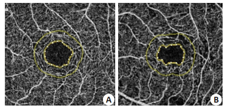
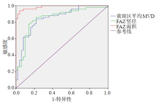
ObjectiveTo investigate the value of macular morphological changes in the early diagnosis of diabetic retinopathy (DR). MethodsA total of 108 patients with type 2 DR treated in our hospital from January 2018 to June 2019 were selected. There were 68 cases of non proliferative DR (NPDR group), 40 cases of proliferative DR (PDR group) and 50 cases of simple type 2 diabetes mellitus (DM group). The optical coherence tomography blood flow imaging (OCTA) was performed in each group, the blood flow density (MVD), macular fovea avascular area (FAZ) and macular retinal thickness were compared among the groups. ResultsThe MVD of macular area in NPDR group and PDR group were (0.48±0.02)% and (0.47±0.03)%, which were significantly lower than those in DM group (P < 0.05). The vertical diameter of FAZ in NPDR group and PDR group were 0.37±0.03 mm and 0.38±0.02 mm respectively, which were significantly higher than those in DM group (P < 0.05). The area of FAZ in PDR group was 0.49±0.04 mm2, which was significantly higher than that in DM group and NPDR group (P < 0.05). There was no significant difference of mean macular retinal thickness between DM group, NPDR group and PDR group (P>0.05). The area under ROC curve of MVD, FAZ vertical diameter and FAZ area in the diagnosis of NPDR and PDR were 0.845, 0.840 and 0.922, respectively (P < 0.05). The cut-off values were 0.50%, 0.37 mm and 0.43 mm2, respectively. The sensitivity were 80.50%, 79.80% and 88.50%, and the specificity were 78.00%, 75.00% and 84.00%, respectively. ConclusionIn DR patients, the MVD of macular area decreases, while the vertical diameter and area of FAZ are enlarged, which has certain application value in the diagnosis.
2021, 44(1): 163-166.
doi: 10.12122/j.issn.1674-4500.2021.01.33
Abstract:
ObjectiveTo explore the differential diagnostic value of abdominal MRI dynamic contrast-enhanced imaging (DCEI) combined with diffusion- weighted imaging (DWI) on benign and malignant liver tumors. MethodsThe clinical data of 64 patients (91 lesions) with liver tumors from March 2019 to March 2020 in our hospital were retrospectively analyzed. Sixty-one malignant lesions and 30 benign lesions were confirmed by surgical pathology. All patients were given MRI DCEI and DWI, and the features of findings of liver lesions in the above sequences were observed, and the differential diagnostic value of MRI DCEI combined with DWI on benign and malignant liver tumors was evaluated. ResultsThe proportion of patients with fast in and fast out in DCEI and high signal in DWI of malignant lesions was higher than that of benign lesions (P < 0.05). The sensitivity and accuracy of DCEI combined with DWI were higher than those of DCEI or DWI (P < 0.05). The specificity was not significantly different from that of DCEI (P>0.05), but it was higher than that of DWI (P < 0.05). ConclusionThe features of DCEI and DWI findings are significantly different in benign and malignant liver tumors. DCEI combined with DWI can significantly improve the diagnostic accuracy and sensitivity in the diagnosis of liver tumors.
2021, 44(1): 167-170.
doi: 10.12122/j.issn.1674-4500.2021.01.34
Abstract:
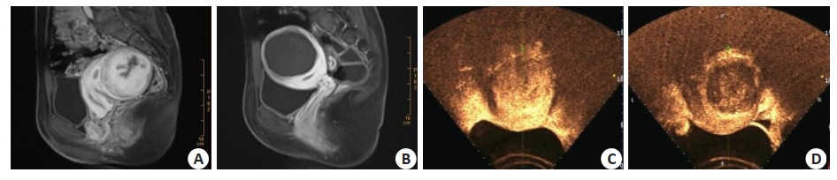
ObjectiveTo compare the application value of enhanced MRI and contrast-enhanced ultrasound in efficacy observation of high-intensity focused ultrasound (HIFU) for uterine fibroids. MethodsBetween February 2019 and February 2020, 102 patients with uterine fibroids underwent HIFU treatment in our hospital were selected for contrast- enhanced ultrasound and enhanced MRI pre- and postoperatively. The effects of the two detection methods in the evaluation of efficacy were compared. ResultsThere was no significant difference in the average diameter and volume of fibroids before and after treatment by the two detection methods (P>0.05). The enhanced MRI could clearly show the size and boundary of fibroids, while contrast- enhanced ultrasound showed relatively insufficient automatic fibroids. The image quality score and the ablation rate of Enhanced MRI were higher than those of contrast-enhanced ultrasound (P < 0.05). Enhanced MRI and contrastenhanced ultrasound were effective in presenting ablation areas and residual lesions. There was no significant difference in the total effective rate of HIFU for uterine fibroids between the two (P>0.05). ConclusionEnhanced MRI is superior to contrastenhanced ultrasound in the qualitative diagnosis of uterine fibroids, while contrast-enhanced ultrasound examination is more repeatable and flexible. Both are necessary examinations before and after HIFU treatment.c
2021, 44(1): 171-173.
doi: 10.12122/j.issn.1674-4500.2021.01.35
Abstract:
ObjectiveTo analyze the value of MSCT and B ultrasound in the diagnosis of acute renal colic caused by ureteral calculi. MethodsSixty cases of ureteral calculi treated in our hospital from May 2018 to December 2019 were retrospectively selected for B-ultrasound and MSCT imaging data. The diagnostic rate of B-ultrasound and MSCT for different ureteral calculi was analyzed, the relationship between the diagnostic sensitivity of B-ultrasound to ureteral calculi and the size of calculi was analyzed, and the sensitivity of B-ultrasound to the diagnosis of other MSCT signs caused by ureteral calculi was analyzed in further step. ResultsThe detection rate of MSCT in upper, middle and lower ureteral calculi was higher than that of Bultrasound(98.3% vs 70%, P < 0.05). The difference between the sensitivity of B-ultrasound to calculi with diameter ≥6 mm and < 6 mm was significant (χ2=3.951, P < 0.05). The effect of MSCT on hydronephrosis caused by ureteral calculi and the detection rate of ureter distortion and perirenal exudation was significantly higher than that of ultrasonography (P < 0.05). ConclusionMSCT is more reliable in the diagnosis of acute renal colic caused by ureteral calculi in outpatient or emergency department.
2021, 44(1): 174-178.
doi: 10.12122/j.issn.1674-4500.2021.01.36
Abstract:
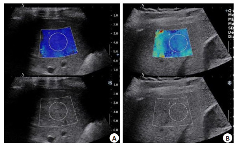
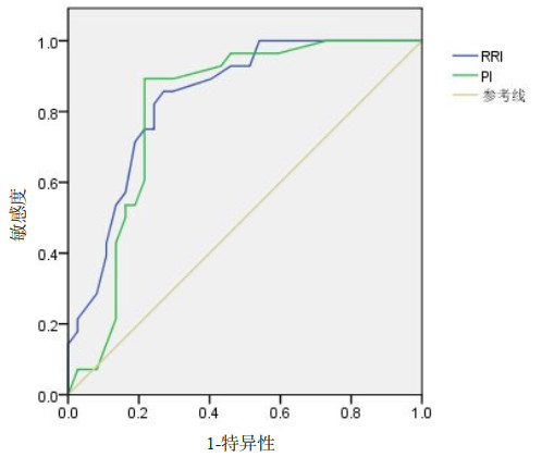
ObjectiveTo explore the value of bedside ultrasound in evaluating acute kidney injury (AKI) after severe trauma. MethodsNinety patients with severe trauma treated in ICU of the hospital between December 2018 and July 2020 were enrolled. The patients were divided into AKI group (n=39) and non-AKI group (n=51), according to the occurrence of AKI after entering the ICU. All patients received bedside ultrasound examination. The general clinical data, renal function indexes, renal resistive index (RRI) and pulsatility index (PI) at different time points were compared between two groups. Meanwhile, the correlations among RRI, PI and renal function indexes were analyzed. The receiver operating characteristic (ROC) curve was used to evaluate the diagnostic value of RRI and PI in AKI. ResultsThe AKI group had higher APACHE Ⅱ score, SOFA score and 28-day mortality rate than the non-AKI group (P < 0.05). AKI group had significantly higher serum CysC and β2-MG levels than the non-AKI group on the 1st, 3rd, 5th and 7th day (P < 0.05). Besides, there were significant differences in serum β2-MG levels in AKI group at different time points (P < 0.05). On the first day after entering the ICU, no significant differences were found in RRI and PI between the 2 groups (P>0.05). However, on the 3rd, 5th, and 7th day after entering the ICU, AKI group had significantly higher RRI and PI than the non-AKI group (P < 0.05). The significant differences were found in RRI and PI in AKI group at different time points (P < 0.05). RRI and PI were significantly positively correlated with serum CysC and β2-MG levels (P < 0.05). Both RRI and PI had good effect in the diagnosis of AKI in patients with severe trauma, and their AUC values were 0.838 and 0.809, respectively. ConclusionThe ultrasound has good value in evaluating AKI after severe trauma. RRI and PI levels can reflect the severity of AKI to a certain extent, which can be used to assist clinical diagnosis of AKI.
2021, 44(1): 179-183.
doi: 10.12122/j.issn.1674-4500.2021.01.37
Abstract:
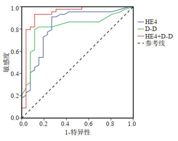
ObjectiveTo analyze the diagnostic value of ultrasound combined with serum HE4 and D-dimer in ovarian cancer based on ROC curve. MethodsSeventy patients with ovarian lesions in the hospital from January 2016 to June 2020 were enrolled, including 44 cases of ovarian cancer (malignant group) and 26 cases of benign ovarian lesions (benign group), and 20 healthy individuals (control group). Serum HE4 and D-D levels and ultrasound imaging characteristics of three groups were compared, and the value of ultrasound combined with serum HE4 and D-D in the diagnosis of ovarian cancer was analyzed. ResultsSerum HE4 and D-D levels had significant difference among three groups (P<0.05). Serum HE4 level among three groups was the highest in malignant group, followed by benign group and control group (P<0.05). Serum D-D level among three groups was the highest in malignant group (P<0.05), while the D-D level showed no significant difference between benign group and control group (P>0.05). With the increase of tumor stage, there was an increasing trend in the levels of serum HE4 and D-D in malignant group, with statistic difference (P<0.05). When the critical values of HE4 and D-D were 97.12 pmol/L and 0.45 mg/L respectively, the two substances had the highest efficiency in the diagnosis of ovarian cancer, while the diagnostic efficacy of combined detection of HE4 and D-D was higher than that of single detection. Taking pathological examination results as "gold standard", the ultrasound score of benign group was significantly lower than that of malignant group (P<0.05). And there were significant differences in ultrasound signs such as the composition, separation, and thickness RI and PI between benign group and malignant group (P<0.05). The sensitivity, specificity and accuracy of ultrasound combined with serum HE4 and D-D in diagnosing ovarian cancer were the highest, being 92.31%, 93.18% and 92.86%, respectively. ConclusionUltrasound, serum HE4 and D-D are of certain value in diagnosis of malignant ovarian cancer, and the combined detection of the three can effectively improve the diagnosis efficacy and provide a more reliable reference for clinical diagnosis.
2021, 44(1): 184-188.
doi: 10.12122/j.issn.1674-4500.2021.01.38
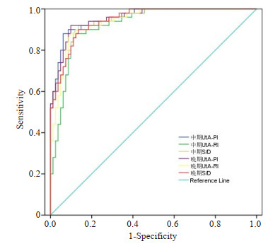
Abstract:
ObjectiveTo analyze the correlation between the UtA-PI, UtA-RI and S/D values measured by ultrasound in pregnant women of different pregnancy periods and the hypertensive disorder complicating pregnant (HDCP) women's disease prediction and maternal and infant outcomes. MethodsA total of 162 HDCP patients who reach the inclusion criteria from January 2018 to December 2019 in our hospital were selected as the observation group, and 100 normal pregnant women were used as the control group. Ultrasound examination was performed in the first trimester (10-14 weeks), the second trimester (20-23 weeks), and the third trimester (30-31 weeks) to record UtA-PI, UtA-RI and S/D. The relationship between UTA PI, UTA RI and S/D was analyzed by logistic regression model. The ROC curve was used to analyze the value of UTA PI, UTA RI and S/D in predicting HDCP. ResultsThe UtA-PI, UtA-RI and S/D values of the observation group during the second and third trimester were significantly higher than those of the control group, and the difference was significant (P<0.05). The sensitivity and specificity of UtA-RI and S/D values for differential diagnosis of HDCP are higher than 85%, and the AUC was greater than 0.9. The values of UtA-PI, UtA-RI and S/D of HDCP pregnant women in the second and third trimester pregnancy abnormal pregnancy groups were higher than those of normal pregnancy group (P<0.05). UtA-PI, UtA-RI and S/D values of ultrasonography of HDCP pregnant women during the second and third trimesters were independent of the outcome of pregnancy Threats (P<0.05). ConclusionThe value of UTA-PI, UTA-RI and S/D measured by ultrasound can effectively predict HDCP. The value of UTA-PI, UTA-RI and S/D are independent threat factors affecting pregnancy outcome.
2021, 44(1): 189-192.
doi: 10.12122/j.issn.1674-4500.2021.01.39
Abstract:
ObjectiveTo explore and analyze the effect of neuroacid combined with Flunarizine Hydrochloride Capsule on cognitive impairment caused by leukoaraiosis. MethodsEighty-six patients with cognitive impairment caused by leukoaraiosis were selected and randomly divided into routine group and combined group, 43 cases in each group.The routine group was treated with flunarizine hydrochloride capsule alone.The combined group was treated with nerve acid and Flunarizine Hydrochloride capsules.The baseline data of the two groups were compared. The changes of cognitive function, clinical efficacy before and after treatment and incidence of adverse reactions were compared. ResultsThe baseline data of the combined group were similar to those of the routine group (P>0.05). Before treatment, the scores of visual space and execution, attention, naming, language, abstraction, delayed memory and orientation dimension Montreal Cognitive Assessment Scale (MoCA) were similar to those of the routine group (P>0.05), but after treatment, the scores of MoCA in the combined group were higher than those before treatment (P < 0.05), and after treatment, the above dimensions of MoCA in the combined group were higher than those before treatment (P < 0.05). The scores and total scores were higher than those of the routine group (P < 0.05). The clinical efficacy distribution of the combined group was significantly different from that of the routine group (P < 0.05). The total effective rate of the former group was 86.05%, which was significantly higher than that of the latter group (58.14%) (P < 0.05). The incidence of adverse reactions in the combined group was 11.63%, which was similar to 6.98% in the routine group (P>0.05), and the patients were tolerable. ConclusionNeuric acid combined with flunarizine hydrochloride capsules can improve the cognitive function and enhance the curative effect of patients with cognitive impairment caused by leukoaraiosis, which is safe and reliable.
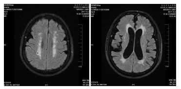
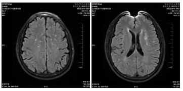
2021, 44(1): 193-197.
doi: 10.12122/j.issn.1674-4500.2021.01.40
Abstract:
Immuno-positron emission tomography (Immuno-PET) is high efficient, noninvasive technique to in vivoimaging. This technique can not only visualize the tumor location, but also identify and quantify the changes of tumor immunological receptor (such as PD-L1). It has a promising prospect in programmed death ligand 1 (PD-L1) targeted imaging and evaluation on prognosis. In recent years, immuno-PET in PD-L1-targeted in vivo imaging becomes a hotspot in research. This review would summarize the principle of various new PD-L1 probes, application, common nuclides, properties of novel molecular tracers, imaging effects of immuno-PET in vivo(animal and human body).
Immuno-positron emission tomography (Immuno-PET) is high efficient, noninvasive technique to in vivoimaging. This technique can not only visualize the tumor location, but also identify and quantify the changes of tumor immunological receptor (such as PD-L1). It has a promising prospect in programmed death ligand 1 (PD-L1) targeted imaging and evaluation on prognosis. In recent years, immuno-PET in PD-L1-targeted in vivo imaging becomes a hotspot in research. This review would summarize the principle of various new PD-L1 probes, application, common nuclides, properties of novel molecular tracers, imaging effects of immuno-PET in vivo(animal and human body).
2021, 44(1): 198-201.
doi: 10.12122/j.issn.1674-4500.2021.01.41
Abstract:
Temporal lobe epilepsy (TLE) is the most common focal epilepsy in adults. There are still inherent difficulties in identifying epileptic regions in these patients only by relying on ordinary MRI. However, with improved localization techniques, TLE can often be correctly identified. In recent years, the study of TLE has shifted to "functional" MRI. Working with the brain in a "static state". There are many ways to analyze fMRI data from the quiescent state of patients with TLE, and we will provide an extensive overview of them in this review.
Temporal lobe epilepsy (TLE) is the most common focal epilepsy in adults. There are still inherent difficulties in identifying epileptic regions in these patients only by relying on ordinary MRI. However, with improved localization techniques, TLE can often be correctly identified. In recent years, the study of TLE has shifted to "functional" MRI. Working with the brain in a "static state". There are many ways to analyze fMRI data from the quiescent state of patients with TLE, and we will provide an extensive overview of them in this review.
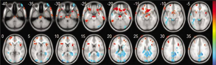
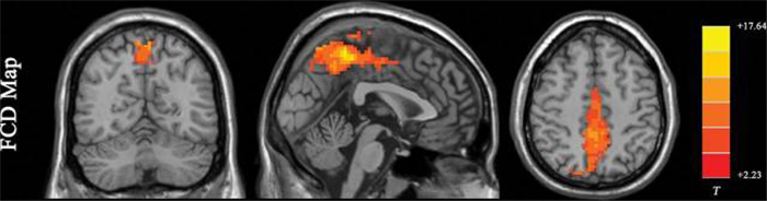
2021, 44(1): 202-205.
doi: 10.12122/j.issn.1674-4500.2021.01.42
Abstract:
Medical infrared thermal imaging is a non-contact, non-radiation, relatively cheap method. It will change the human body infrared radiation into digital and image signals, showing the temperature of the body parts accurately, and is especially suitable for pain chronic, sensitive, and wandering. It has a certain reference value for the diagnosis of pain and the evaluation of curative effect. This article reviews the working principle of infrared thermal imaging and its application in medical pain, including soft tissue pain, herpes zoster and herpetic neuralgia, arthritis, cervical and lumbar diseases, primary osteoporosis and so on. Finally, we summarize the advantages and disadvantages of infrared thermal imaging in clinical application. In the future, infrared thermal imaging will play an irreplaceable role on pain diagnosis and treatment process.
Medical infrared thermal imaging is a non-contact, non-radiation, relatively cheap method. It will change the human body infrared radiation into digital and image signals, showing the temperature of the body parts accurately, and is especially suitable for pain chronic, sensitive, and wandering. It has a certain reference value for the diagnosis of pain and the evaluation of curative effect. This article reviews the working principle of infrared thermal imaging and its application in medical pain, including soft tissue pain, herpes zoster and herpetic neuralgia, arthritis, cervical and lumbar diseases, primary osteoporosis and so on. Finally, we summarize the advantages and disadvantages of infrared thermal imaging in clinical application. In the future, infrared thermal imaging will play an irreplaceable role on pain diagnosis and treatment process.
2021, 44(1): 206-208.
doi: 10.12122/j.issn.1674-4500.2021.01.43
Abstract:
Transient ischemic attack (TIA) is defined as transient neurological dysfunction caused by focal ischemia of brain, spinal cord or retina without acute infarction. The only basis for the differential diagnosis of TIA or acute cerebral infarction is whether there is infarction or not, which has no relationship with the duration of clinical symptoms. In clinic, TIA is usually diagnosed by clinical symptoms, and no positive findings are found in routine MR examination. 3D-ASL is a relatively popular and prominent perfusion imaging technology, which can produce quantitative perfusion images. As a powerful clinical means to study brain function, 3D-arterial spin labeling (3D-ASL) has been paid more and more attention and is in constant development. Clinical studies have shown that 3d- asl can provide the basis for the diagnosis of TIA. When combined with DWI, MRA or DSC-PWI, it can provide reliable basis for the diagnosis, treatment, prognosis and risk assessment of TIA, and different diagnostic modes can be selected for patients with different physical tolerance. Therefore, this paper will review the current situation of 3D-ASL combined with multimodal MRI in TIA patients with different physical tolerance.
Transient ischemic attack (TIA) is defined as transient neurological dysfunction caused by focal ischemia of brain, spinal cord or retina without acute infarction. The only basis for the differential diagnosis of TIA or acute cerebral infarction is whether there is infarction or not, which has no relationship with the duration of clinical symptoms. In clinic, TIA is usually diagnosed by clinical symptoms, and no positive findings are found in routine MR examination. 3D-ASL is a relatively popular and prominent perfusion imaging technology, which can produce quantitative perfusion images. As a powerful clinical means to study brain function, 3D-arterial spin labeling (3D-ASL) has been paid more and more attention and is in constant development. Clinical studies have shown that 3d- asl can provide the basis for the diagnosis of TIA. When combined with DWI, MRA or DSC-PWI, it can provide reliable basis for the diagnosis, treatment, prognosis and risk assessment of TIA, and different diagnostic modes can be selected for patients with different physical tolerance. Therefore, this paper will review the current situation of 3D-ASL combined with multimodal MRI in TIA patients with different physical tolerance.
2021, 44(1): 209-213.
doi: 10.12122/j.issn.1674-4500.2021.01.44
Abstract:
In recent years, driven by the wave of visualization, point of Care ultrasound(POCUS)has become more and more widely used in anesthesia and perioperative due to its advantages of simplicity, speed, practicality, and low cost.With being widely used in guiding vascular puncture and nerve block, modern anesthesiology pays more and more attention to the application of POCUS in perioperative evaluation and monitoring. This can not only improve the ability of anesthesiologists to accurately evaluate critically ill patients before surgery but also better optimize perioperative management and ensure patient safety. This article summarizes the application of POCUS in the evaluation and monitoring of five parts of the heart, lungs, airway, stomach, and eyes during the perioperative period in recent years, and provides a reference for the use of POCUS in the perioperative period. It also provides a theoretical basis for further promoting POCUS into the normative training of anesthesiolog
In recent years, driven by the wave of visualization, point of Care ultrasound(POCUS)has become more and more widely used in anesthesia and perioperative due to its advantages of simplicity, speed, practicality, and low cost.With being widely used in guiding vascular puncture and nerve block, modern anesthesiology pays more and more attention to the application of POCUS in perioperative evaluation and monitoring. This can not only improve the ability of anesthesiologists to accurately evaluate critically ill patients before surgery but also better optimize perioperative management and ensure patient safety. This article summarizes the application of POCUS in the evaluation and monitoring of five parts of the heart, lungs, airway, stomach, and eyes during the perioperative period in recent years, and provides a reference for the use of POCUS in the perioperative period. It also provides a theoretical basis for further promoting POCUS into the normative training of anesthesiolog