


Imaging manifestations and diagnostic value of MRI for corpus callosum injury caused by traumatic brain injury
-
摘要:
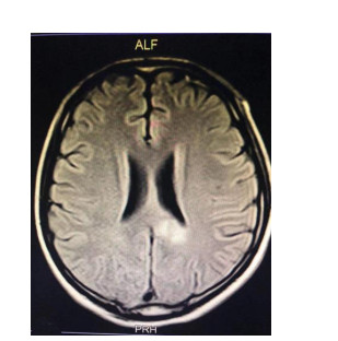
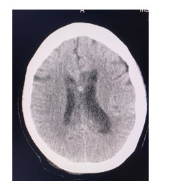
目的分析颅脑创伤致胼胝体损伤的MRI影像学表现及诊断价值。 方法回顾性分析本院2016年2月~2020年11月诊治的103例胼胝体损伤患者一般资料,均为颅脑创伤所致,且均在本院接受MRI和CT检查。观察患者MRI和CT影像学表现,并比较MRI和CT的诊断效能,并分析不同类型胼胝体损伤的MRI参数值。 结果MRI检查显示99例患者增强扫描无明显强化、边缘清楚,4例患者存在强化或边缘不清晰,其中45例非出血性损伤患者,T1WI呈现等信号或者低信号,T2WI和DWI均呈现稍高信号,同时各扫描序列中均未见出血信号,58例出血性损伤患者,各扫描序列上均呈混杂信号,且可见出血信号;MRI和CT对膝部损伤、压部损伤以及体膝部损伤检出率比较无显著差异(P>0.05),但MRI对体部损伤检出率明显高于CT(P < 0.05);MRI和CT对出血性损伤诊断准确率比较无显著差异(P>0.05),但MRI对非出血性损伤、胼胝体萎缩、软化灶以及胶质增生诊断准确率显著高于CT(P < 0.05);两组患者DWI序列显示以及脑弥漫性轴突损伤评分均无显著差异(P>0.05),但出血性损伤T2WIFLAIR、T2WI/T1WI显示明显高于非出血性损伤(P < 0.05)。 结论MRI检查应用于颅脑创伤所致胼胝体损伤较CT可显示更多的病灶,可鉴别诊断出血性损伤与非出血性损伤病灶,同时可准确定位胼胝体损伤具体部位,具有较好的临床应用价值。 Abstract:ObjectiveTo analyze the imaging manifestations and diagnostic value of MRI for corpus callosum injury caused by traumatic brain injury. MethodsThe general data of 103 patients with corpus callosum injury caused by traumatic brain injury who were diagnosed and treated in the hospital from February 2016 to November 2020 were retrospectively analyzed. All of the patients received MRI and CT examinations, and the imaging were analyzed. The diagnostic efficiencies of MRI and CT were compared, and MRI parameters of different types of corpus callosum injuries were analyzed. ResultsMRI examination showed no obvious enhancement but clear edges in 99 patients, and enhancement or unclear edges in 4 patients after enhanced scan. There were 45 patients with non-hemorrhagic injury, showing equal signal or low signal on T1WI, slightly higher signals on both T2WI and DWI. Besides, no bleeding signal was observed in each scan sequence. For 58 patients with hemorrhagic injury, there were mixed signals on each scan sequence, with bleeding signals. There were no significant differences between MRI and CT in the detection rates of genu injury, splenium injury, and body genu injury (P>0.05), but the detection rate of body injury by MRI was significantly higher than that by CT (P < 0.05). There was no significant difference between MRI and CT in the diagnostic accuracy for hemorrhagic injury (P>0.05), but the diagnostic accuracy rate of MRI was significantly higher than that of CT for non-hemorrhagic injury, corpus callosum atrophy, softening lesions and gliosis (P < 0.05). There were no significant differences in DWI sequence display and DAI scores between the two groups (P>0.05). The T2WIFLAIR and T2WI/T1WI display of hemorrhagic injury was significantly better than that of non-hemorrhagic injury (P < 0.05). ConclusionCompared with CT, MRI can show more lesions in the corpus callosum caused by traumatic brain injury, and it can be used to differentiate and diagnose hemorrhagic and non-hemorrhagic lesions, and accurately locate the specific location of corpus callosum injury. -
Key words:
- traumatic brain injury /
- corpus callosum injury /
- MRI /
- diagnostic value
-
表 1 MRI和CT对胼胝体损伤部位检出率比较
Table 1. Comparison of the detection rates of corpus callosum injuries at different sites between MRI and CT [n(%)]
检查方法 膝部损伤 体部损伤 压部损伤 体膝部 CT 23(22.33) 22(21.36) 12(11.65) 22(21.36) MRI 30(29.13) 35(33.98) 13(12.62) 25(24.27) χ2 1.245 4.099 0.046 0.248 P 0.265 0.043 0.831 0.618  下载: 导出CSV
表 2 MRI和CT对胼胝体损伤类型的诊断准确率比较
Table 2. Comparison of the diagnostic accuracy of MRI and CT for different types of corpus callosum injuries [n(%)]
检查方法 出血性损伤 非出血性损伤 胼胝体萎缩 软化灶 胶质增生 CT 50(48.54) 29(28.16) 10(9.71) 20(19.42) 12(11.65) MRI 58(56.31) 45(43.69) 25(24.27) 38(36.89) 32(31.07) χ2 1.246 5.399 7.744 7.775 11.560 P 0.264 0.020 0.005 0.005 0.001
下载: 导出CSV
表 3 出血性损伤和非出血性损伤患者MRI序列显示以及ADI评分情况
Table 3. MRI findings and ADI scores of patients with hemorrhagic injury and non-hemorrhagic injury
分组 DWI (n) T2WI-FLAIR (n) T2WI/T1WI (n) 脑弥漫性轴突损伤评分(分) 出血性损伤 58 58 58 1.64±0.43 非出血性损伤 45 42 40 1.51±0.38 t/χ2 - 3.983 6.773 1.600 P - 0.046 0.009 0.113
下载: 导出CSV
-
[1] Currie S, Saleem N, Straiton JA, et al. Imaging assessment of traumatic brain injury[J]. Postgrad Med J, 2016, 92(1083): 41-50. doi: 10.1136/postgradmedj-2014-133211 [2] McDonald S, Rushby JA, Dalton KI, et al. The role of abnormalities in the corpus callosum in social cognition deficits after Traumatic Brain Injury[J]. Soc Neurosci, 2018, 13(4): 471-9. doi: 10.1080/17470919.2017.1356370 [3] Cicuendez M, Castaño-León A, Ramos A, et al. Prognostic value of corpus callosum injuries in severe head trauma[J]. Acta Neurochir, 2017, 159(1): 25-32. doi: 10.1007/s00701-016-3000-4 [4] Kontzialis M, Soares BP, Huisman TAGM. Lesions in the splenium of the corpus callosum on MRI in children: a review[J]. J Neuroimaging, 2017, 27(6): 549-61. doi: 10.1111/jon.12455 [5] 于航, 刘雪雁. 31例儿童可逆性胼胝体压部损伤综合征临床分析[J]. 国际儿科学杂志, 2019, 46(1): 73-6. doi: 10.3760/cma.j.issn.1673-4408.2019.01.018 [6] Skandsen T, Kvistad KA, Solheim O, et al. Prevalence and impact of diffuse axonal injury in patients with moderate and severe head injury: a cohort study of early magnetic resonance imaging findings and 1-year outcome[J]. J Neurosurg, 2010, 113(3): 556-63. doi: 10.3171/2009.9.JNS09626 [7] 杨擎. 伴可逆性胼胝体压部损伤的临床轻度脑炎/脑病的MRI研究进展[J]. 国际医学放射学杂志, 2019, 42(3): 299-302. https://www.cnki.com.cn/Article/CJFDTOTAL-GWLC201903011.htm [8] Lieb JM, Ahlhelm FJ. Agenesis of the corpus callosum[J]. Radiologe, 2018, 58(7): 636-45. doi: 10.1007/s00117-018-0388-2 [9] 王建武, 冯学彬, 彭如臣. 脑弥漫性轴索损伤的临床特征和MRI、CT诊断(附47例分析[) J]. 中国CT和MRI杂志, 2015, 13(6): 4-7. doi: 10.3969/j.issn.1672-5131.2015.06.002 [10] Moe HK, Limandvik Myhr J, Moen KG, et al. Association of cause of injury and traumatic axonal injury: a clinical MRI study of moderate and severe traumatic brain injury[J]. J Neurosurg, 2019: 1-9. http://www.ncbi.nlm.nih.gov/pubmed/31604329 [11] Figueira Rodrigues Vieira G, Guedes Correa JF. Early computed tomography for acute post-traumatic diffuse axonal injury: a systematic review[J]. Neuroradiology, 2020, 62(6): 653-60. doi: 10.1007/s00234-020-02383-2 [12] 田种红. 螺旋CT在胼胝体发育不全诊断中的应用价值[J]. 中国急救医学, 2018, 38(z2): 298. doi: 10.3969/j.issn.1002-1949.2018.z2.298 [13] 金新安, 陈伟, 高明生, 等. CT和MRI诊断胼胝体损伤的对比研究[J]. 影像诊断与介入放射学, 2011, 20(3): 174-7. doi: 10.3969/issn.1005-8001.2011.03.004 [14] Humble SS, Wilson LD, Wang L, et al. Prognosis of diffuse axonal injury with traumatic brain injury[J]. J Trauma Acute Care Surg, 2018, 85(1): 155-9. doi: 10.1097/TA.0000000000001852 [15] 吴晓芬, 王璐, 吴晨颖. CT与MRI在弥漫性颅脑轴索损伤诊断中的应用价值评价[J]. 临床和实验医学杂志, 2020, 19(2): 209-11. doi: 10.3969/j.issn.1671-4695.2020.02.028 [16] Yiannakkaras C, Konstantinou N, Constantinidou F, et al. Whole brain and corpus callosum diffusion tensor metrics: How do they correlate with visual and verbal memory performance in chronic traumatic brain injury[J]. J Integr Neurosci, 2019, 18(2): 95. doi: 10.31083/j.jin.2019.02.144 [17] 邵雪非, 刘清祥, 方新运, 等. MRI检查中SWI和DTI序列对弥漫性轴索损伤诊断和预后评价的作用[J]. 中华创伤杂志, 2018, 34(8): 711-6. doi: 10.3760/cma.j.issn.1001-8050.2018.08.007 [18] 明小春, 龙世亮, 梁永升, 等. 常规MRI联合DWI及SWI在脑弥漫性轴索损伤中的应用价值[J]. 广东医学, 2018, 39(S1): 88-90. https://www.cnki.com.cn/Article/CJFDTOTAL-GAYX2018S1030.htm [19] 蔡桂兰, 王瑞金, 乔杉杉, 等. 弥散加权成像高信号的胼胝体病变原因及临床和影像学特征[J]. 临床与病理杂志, 2018, 38(10): 2116-23. doi: 10.3978/j.issn.2095-6959.2018.10.010 [20] Grover H, Qian Y, Boada FE, et al. MRI evidence of altered callosal sodium in mild traumatic brain injury[J]. AJNR Am J Neuroradiol, 2018, 39(12): 2200-4. doi: 10.3174/ajnr.A5903 -

 点击查看大图
计量
- 文章访问数: 695
- HTML全文浏览量: 297
- PDF下载量: 5
- 被引次数: 0