

 Find Duplicates
 Check Document
 Submission(new)
 Experts Office
 Editorial Office
2023 Vol. 46, No. 5
Display Method:
2023, 46(5): 779-786.
doi: 10.12122/j.issn.1674-4500.2023.05.01
Abstract:
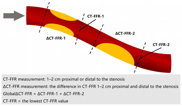
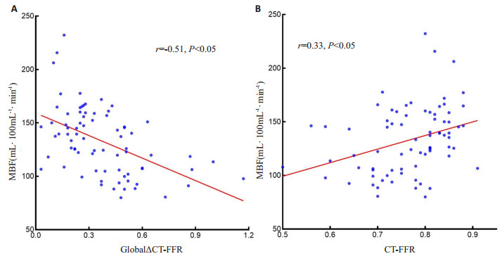
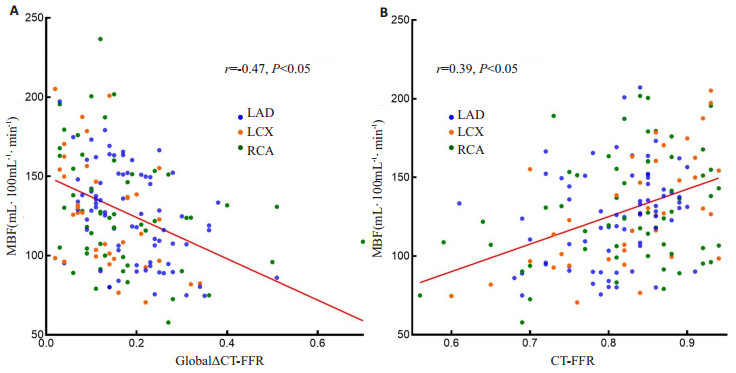
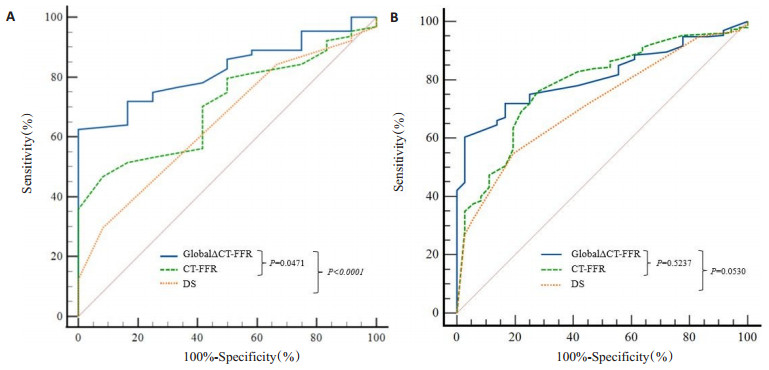
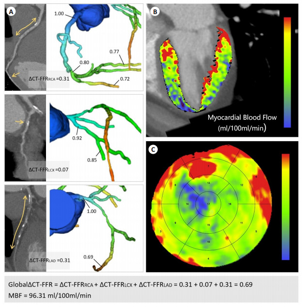
Objective To assess the predictive capability of GlobalΔCT-FFR in determining abnormal myocardial blood flow through an examination of the correlation between GlobalΔCT-FFR and myocardial blood flow. Methods A retrospective inclusion was conducted on a cohort of 76 patients who underwent dynamic computed tomographic myocardial perfusion + coronary computed tomographic angiography between 2019 and 2021, as a result of suspected coronary artery disease. The relationship between GlobalΔCT-FFR and myocardial blood flow (MBF) was assessed using Univariate Spearman correlation. We were assessed the sensitivity, specificity, diagnostic accuracy, positive predictive value, and negative predictive value of GlobalΔCT-FFR, CT-FFR and coronary computed tomographic angiography diameter stenosis (DS) for myocardial blood flow abnormalities at both per-patient and per-vessel level, utilizing MBF as a reference standard. The ROC curve was constructed, and the area under the curve (AUC) was calculated. Results At the per-patient level, there was a moderate negative correlation between GlobalΔCT-FFR and mean MBF (r=-0.51, P < 0.05), CT-FFR exhibited a weak positive correlation with mean MBF (r=0.33, P < 0.05). At the per-vessel level, GlobalΔCT-FFR showed a moderate negative correlation with mean MBF (r=-0.47, P < 0.05), CT-FFR demonstrated a weak positive correlation with mean MBF (r=0.39, P < 0.05). At the per-patient level, the AUC for GlobalΔCT-FFR, CT-FFR, and DS was found to be 0.82(95%CI: 0.72-0.90, P < 0.05), 0.71(95% CI: 0.59-0.81, P < 0.05) and 0.65(95% CI: 0.53-0.76, P < 0.05), respectively. At the per-vessel level, the AUC for GlobalΔCT-FFR, CT-FFR, and DS was determined to be 0.81(95% CI: 0.75-0.86, P < 0.05), 0.78(95% CI: 0.72-0.83, P < 0.05) and 0.71(95% CI: 0.65-0.77: P < 0.05), respectively. In the evaluation of ROC curves, GlobalΔCT-FFR exhibited superior performance compared to CT-FFR and DS in per-patient analysis (P=0.0471, P < 0.0001). In per-vessel analysis, GlobalΔCT-FFR demonstrated similar performance to CT-FFR and DS (P=0.5237, P=0.0530). Conclusion The diagnostic performance of GlobalΔCT-FFR in predicting abnormal myocardial blood flow has been confirmed, suggesting its potential as an alternative indicator for quantitatively measuring myocardial blood flow.
2023, 46(5): 787-791.
doi: 10.12122/j.issn.1674-4500.2023.05.02
Abstract:
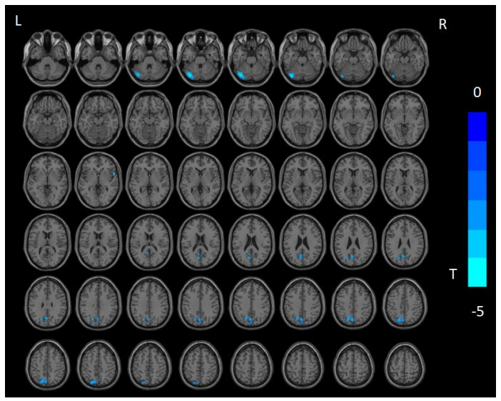
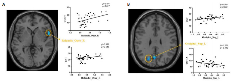
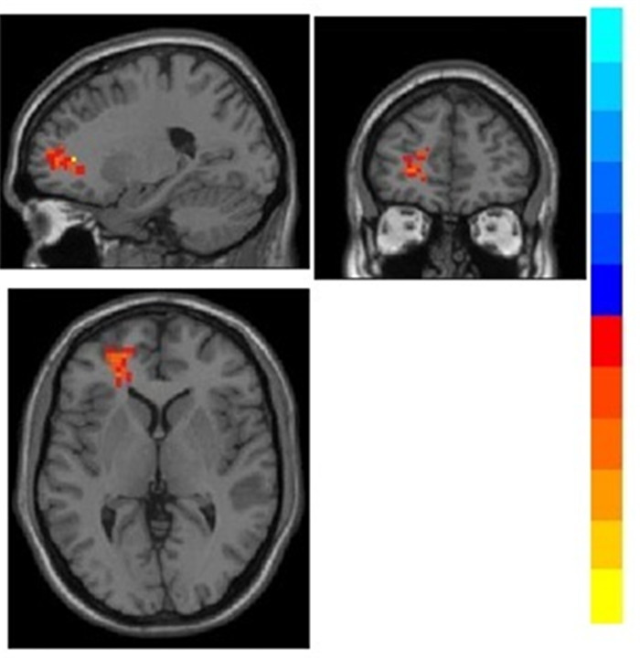
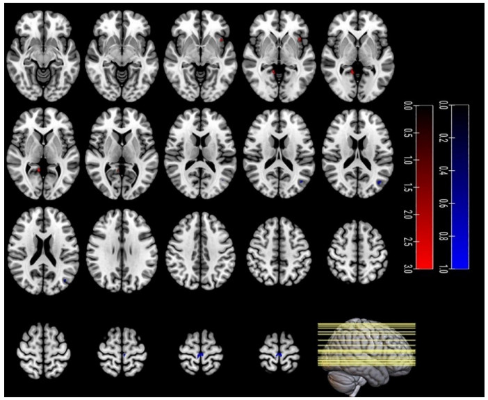
Objective To investigate the changes in amplitude of low-frequency (ALFF) and the correlations between ALFF and clinical cognitive evaluations in subjective cognitive decline (SCD) patients, in order to gain a valuable time window for delaying and preventing the progress of Alzheimer's disease. Methods Forty-five patients with SCD (SCD group) and forty well-matched healthy controls (healthy control group) were recruited in this study, thirty-five and thirth-three case were included in ALFF value analysis, respectively. All subjects were examined by functional MRI and assessed with multiple clinical scales. The changes of brain area between SCD and healthy control group were compared by ALFF analysis method. In addition, linear regression analysis was used to evaluate the correlation between ALFF values and clinical scores of all subjects. Results The ALFF values of the left superior occipital gyrus, left superior parietal gyrus, left cuneus and precuneus, left cerebellum and right Rolandic operculum were lower in the SCD group compared with healthy control group, the difference was statistically significant (P < 0.05). Among the SCD patients, the ALFF values of right Rolandic operculum were positively correlated with the Wechsler logical memory test and Boston naming test scores. The Boston naming test scores were also positively correlated with the ALFF values of left superior occipital gyrus. Additionally, the ALFF values of left superior occipital gyrus showed a significant negative correlation with trail making test A scores (P < 0.05). Conclusion SCD patients have abnormal brain activity signals, and the ALFF values of abnormal brain regions are correlated with some clinical scale evaluations. ALFF may be considered as a characteristic biomarker for early detection of these patients.
2023, 46(5): 792-797.
doi: 10.12122/j.issn.1674-4500.2023.05.03
Abstract:
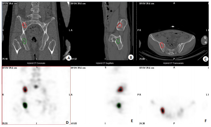
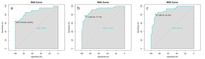
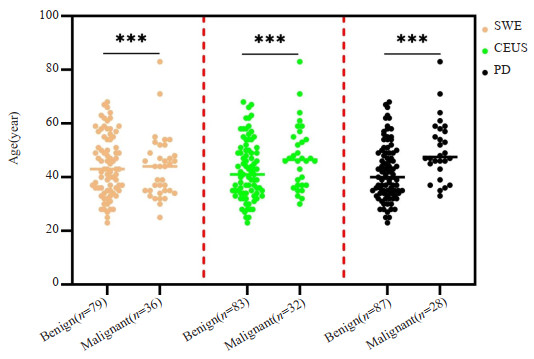
Objective To investigate the diagnostic value of standardized uptake value (SUV) of quantitative SPECT/CT based on the maximum expectation reconstruction algorithm in metastatic bone tumors. Methods A total of 75 patients who underwent quantitative SPECT/CT bone scanning in the Department of Nuclear Medicine of our hospital from March to May 2022 were retrospectively analyzed. The SUV of 87 bone hypermetabolism lesions was delineated, and the SUV of the spine of patients with normal bone metabolism was delineated as the control group (43 spinal vertebrae). The maximum standardized uptake value (SUVmax), mean standardized uptake value (SUVmean) and minimum standardized uptake value (SUVmin) of benign lesions, malignant lesions and control group were measured. A non-parametric test was used for comparison between groups, and ROC curve was used to obtain the best threshold value. Results According to pathology and/or imaging follow-up of 6 to 12 months or more, 48 lesions were diagnosed as bone metastasis and 39 lesions as benign lesions. SUVmax 30.15(20.75, 45.43) g/mL, SUVmean 16.65(11.75, 23.10) g/mL, SUVmin 11.6(8.40, 15.27) g/mL in the bone metastasis group, and SUVmax 13.5(10.4, 15.27) g/mL, SUVmean 8.22(5.89, 9.56) g/mL, SUVmin 5.83(2.63, 6.89) g/mL in the benign group, while the SUVmax 7.47±1.99 g/mL, SUVmean 3.74 ± 0.68 g/mL, SUVmin 1.06 ± 0.46 g/mL in the control group, respectively. The SUV of the bone metastasis group was significantly higher than that of the benign lesion group and the control group (P < 0.001). ROC curve analysis showed that the area under the curve of SUVmax, SUVmean and SUVmin were 0.876, 0.912 and 0.901, respectively. When the cut-off value of SUVmax was 26.6 g/mL, the specificity was 100% and the sensitivity was 64.6%. When the cut-off value of SUVmin was 8.1 g/mL, the sensitivity and specificity were 81.2% and 89.7%, respectively. The cutoff value of SUVmean was 11.5 g/mL, sensitivity and specificity were 77.1% and 92.3%, respectively. The detection rate of combined quantitative SPECT/CT bone fusion imaging and SUVmean value was higher than that of conventional SPECT/CT bone fusion imaging (χ2=11.576, P=0.001), and the diagnostic coincidence rate was 75.86% and 94.25%, respectively. Conclusion The SUV of quantitative SPECT/CT has a certain clinical application value in the differential diagnosis of benign and malignant bone lesions, which complements the qualitative analysis of bone metastases and benign bone lesions. Quantitative SPECT/CT bone fusion imaging +SUVmean is more accurate than conventional SPECT/CT bone fusion imaging in the diagnosis of tumor bone metastasis.
2023, 46(5): 798-804.
doi: 10.12122/j.issn.1674-4500.2023.05.04
Abstract:
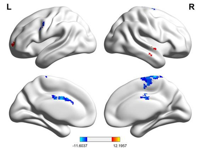
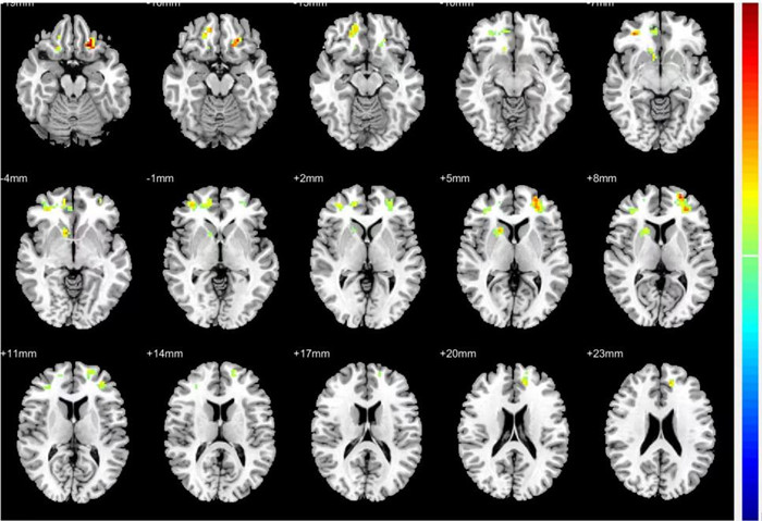
Objective To investigate the characteristics of spontaneous brain function activity in the resting state of primary insomnia (PI) patients using a combined dynamic and static low-frequency amplitude functional linkage method. Methods The scores and fMRI data of clinical scales (Pittsburgh sleep quality index scale, depression self-rating scale, anxiety self-rating scale, insomnia patients also completed the insomnia severity index scale) were collected from 20 PI patients (PI group) and 20 healthy volunteers (control group). The differences in spontaneous brain functional activity between the two groups were analyzed using dynamic and static low frequency amplitude (ALFF) methods, further analysing the correlation between the signal values of the differential brain regions and insomnia. A seed-based functional connectivity analysis was performed between the differential brain regions based on dynamic ALFF and the remaining voxels of the whole brain. Results Compared to normal controls, the PI group had increased dynamic ALFF values in the left superior frontal gyrus, suggesting increased temporal variability in intrinsic brain activity in this region. A significant negative correlation was found with insomnia severity index (r=-0.463, P=0.04). Compared to the control group, the PI group showed increased static ALFF values in the right middle temporal gyrus and left superior frontal gyrus and decreased static ALFF values in the left cingulate gyrus, left precentral gyrus and right frontal supplementary motor area. Correlation analysis with whole brain voxels was performed using the left superior frontal gyrus as the seed point, and the main brain regions with increased functional connectivity in the PI group relative to the HC group were: left orbital middle frontal gyrus, right caudate nucleus, right orbital middle frontal gyrus, and left dorsolateral superior frontal gyrus. Conclusion The presence of localised changes in brain activity in PI patients in terms of dynamic low-frequency amplitude correlates with the insomnia index, and combined functional connectivity can provide a more comprehensive picture of abnormal brain function activity in PI patients. It is suggested that fMRI can evaluate to some extent the altered spontaneous neurological activity in PI patients.
2023, 46(5): 805-810.
doi: 10.12122/j.issn.1674-4500.2023.05.05
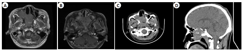
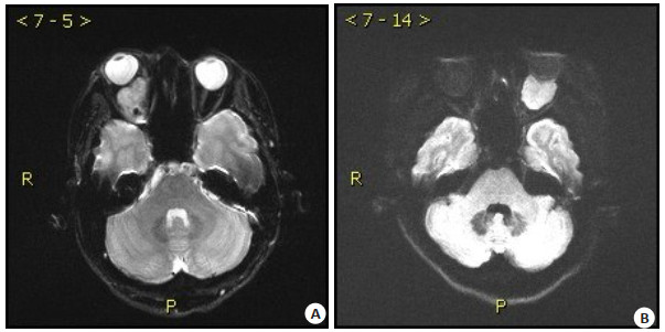
Abstract:
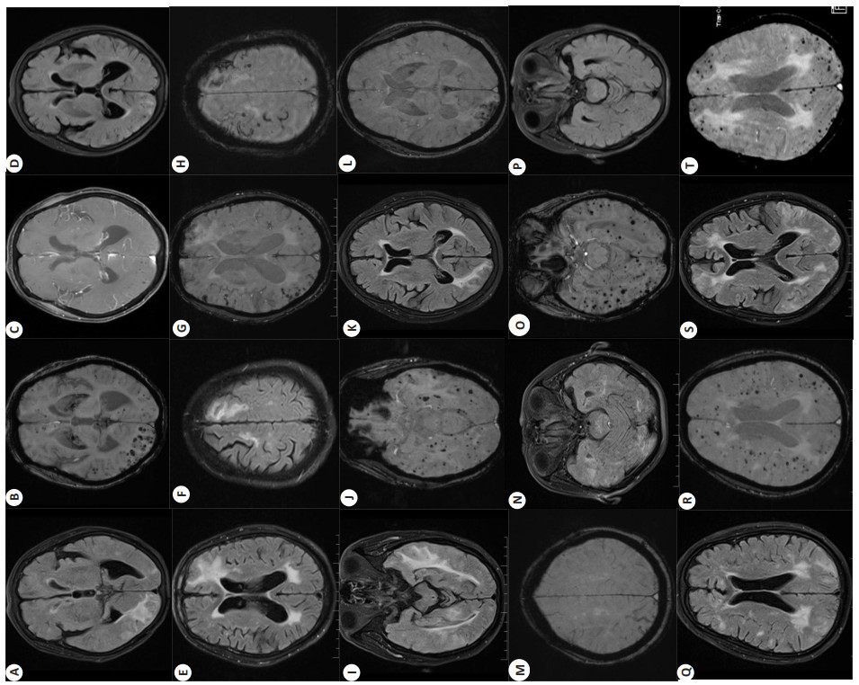
Objective To investigate the clinical manifestations, imaging findings, and prognosis of patients with cerebral amyloid angiopathy-related inflammation. Methods The clinical data of 7 patients with cerebral amyloid angiopathy-related inflammation admitted to our hospital were retrospectively analyzed, including general data, clinical manifestations, head MRI examination, blood and cerebrospinal fluid(CSF) examination, drug treatment and prognosis. Results Clinical manifestations: 5 cases with acute onset, 2 cases with subacute onset. 4 patients had focal neurological deficits, 5 patients had acute cognitive impairment, 3 patients had headache, 1 patient was diagnosed with epilepsy based on clinical manifestations and EEG, and all patients had no disorder of consciousness. MRI examination of the head: the SWI of all patients showed a large number of scattered and diffuse cerebral microhemorrhages in the cortex and subcortex. The SWI of 5 patients showed iron deposition on the surface of the cortex, and 2 patients had convex subarachnoid hemorrhage. All patients had T2WI and flair showed asymmetric high white matter signals in the cortex and subcortex. Enhanced scanning of the pia mater showed enhancement of the pia mater in 3 patients. After treatment, Flair showed that the abnormal white subcortex substance signal area was reduced in 5 patients. Blood and CSF tests: 6 cases of apolipoprotein E gene screening, including 3 cases of ε4/ε4 genotype, 2 cases of ε2/ε4 genotype and 1 case of ε3/ε4 genotype; 4 patients completed the lumbar puncture test of CSF, 3 patients had slightly elevated CSF protein, 2 patients had slightly elevated red blood cells in CSF and 2 patients had decreased Aβ40 and Aβ42 in CSF. Drug treatment: 4 patients received glucocorticoid therapy, and 3 patients received only symptomatic and supportive treatment. Prognosis: The clinical symptoms and imaging of 4 patients treated with glucocorticoid improved significantly, and the clinical symptoms and imaging of 1 patient were improved by symptomatic and supportive treatment only. Conclusion The main clinical features of cerebral amyloid angiopathy-related inflammation patients are focal neurologic deficits, cognitive dysfunction and headache. The main magnetic resonance imaging features were cortical and subcortical asymmetric white matter hyperintensity and cerebral microhemorrhages. Some patients had varying degrees of leptomeningeal enhancement on the enhanced scan. Patients may benefit from glucocorticoid therapy, but in some mild patients, clinical symptoms and imaging may also improve spontaneously.
2023, 46(5): 811-816.
doi: 10.12122/j.issn.1674-4500.2023.05.06
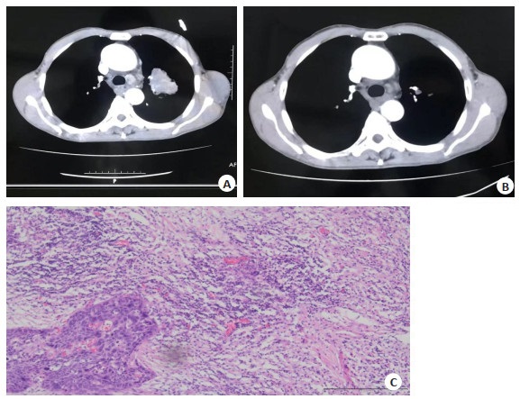
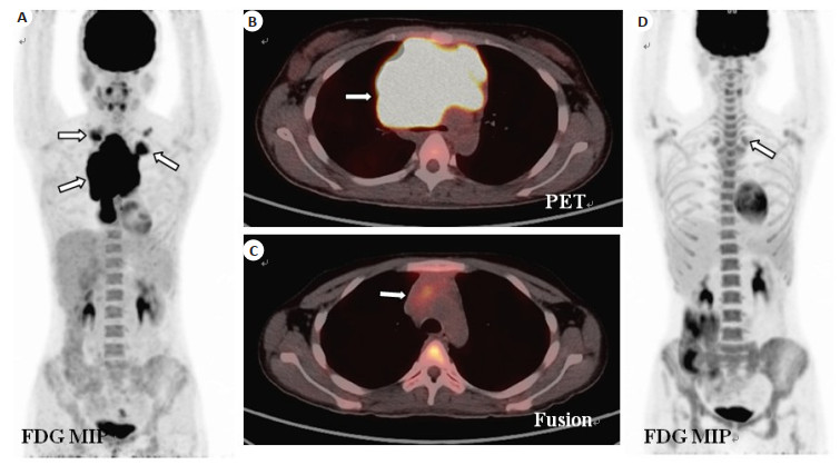
Abstract:
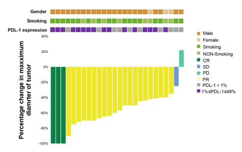
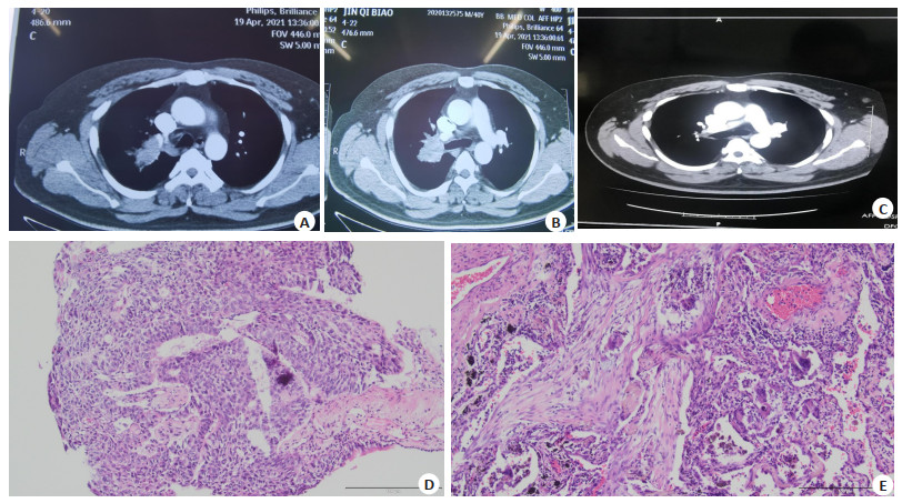
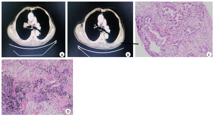
Objective To evaluate the short-term efficacy and safety of sintilimab combined with chemotherapy neoadjuvant in the treatment of stage Ⅲ non-small cell lung cancer. Methods The clinical data of 26 patients with stage Ⅲ non-small cell lung cancer treated with Sintilimab combined with chemotherapy neoadjuvant therapy in our hospital from January 2021 to July 2023 were analyzed. The imaging and pathological effects of the patients were evaluated. The safety of immunotherapy combined with chemotherapy neoadjuvant therapy was observed. Results Twenty-three patients completed radical surgery with a surgical resection rate of 88.5%, and 22 patients underwent R0 resection with a R0 resection rate of 95.7%. Objective response rate of imaging results was 92.3% (complete response rate was 11.5%, partial response rate was 80.8%). The disease control rate was 96.1%, the pathological complete response rate was 26.1%, and the main pathological response rate was 60.9%. Common adverse reactions: The incidence of leukopenia was 42.3%, peripheral neurotoxicity was 46.2%, gastrointestinal adverse reactions were 23.1%, thyroid dysfunction was 11.5%, and the incidence of grade 3 or above adverse reactions was about 7.7%. No serious immune related adverse reactions had occurred. Conclusion The combination of sintilimab and neoadjuvant chemotherapy has a significant therapeutic effect on stage Ⅲ non-small cell lung cancer, with a high R0 resection rate, high pathological response rate, and tolerable safety.
2023, 46(5): 817-822.
doi: 10.12122/j.issn.1674-4500.2023.05.07
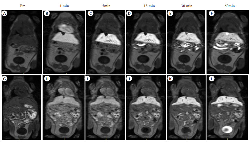
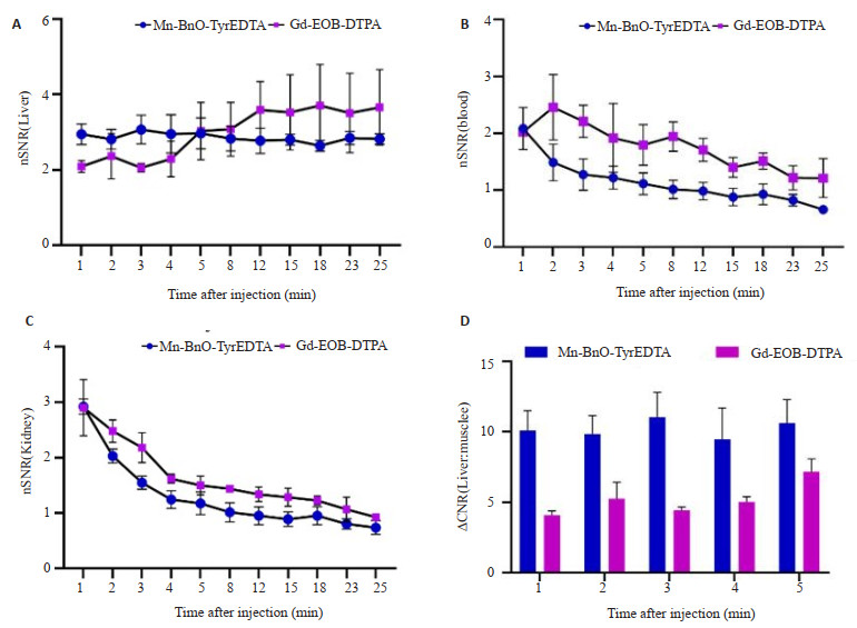
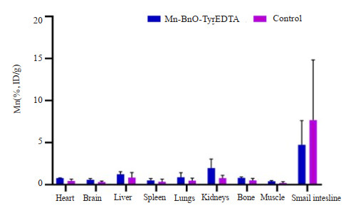
Abstract:
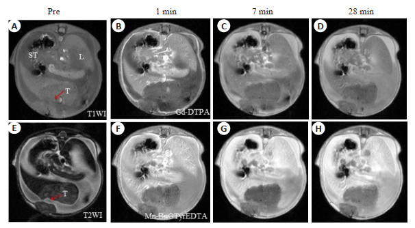
Objective To evaluate the liver targeting of manganese benzyltyrosine ethylenediamine tetraacetic acid (Mn-BnO-TyrEDTA) MRI contrast agent and its preliminary application in mouse liver cancer model. Methods Mice were given by the same dose of Mn-BnO-TyrEDTA and disodium gadolinium serate (Gd-EOB-DTPA) via a caudal vein, and dynamic enhanced MRI scans were performed for 1 h to monitor the distribution and clearance of contrast agents. Simultaneously injecting liver cancer cell H22 (1×10⁵/30 μL) into mouse liver and matrix adhesive (25 μL) using a mixed solution to establish a liver cancer model and perform MRI imaging. Gd-DTPA was used as the control group. Results After injecting Mn-BnO-TyrEDTA for 1 min, the liver began to strengthen, and the signal intensity reached its peak at 3 min and continued until 30 min; After 1 min, the blood and kidney signal intensity began to significantly decrease, and after 3 min, the contrast agent entered the bladder. By comparing the normalized signal‑to‑noise ratio and contrast‑to‑noise ratio for 3 min, it was determined that the liver targeting values of the two contrast agents were 3.07±0.38 and 11±4.0 for Mn-BnO-TyrEDTA and 2.06±0.11 and 4.4±0.4 for Gd-EOB-DTPA, respectively, with statistical differences (P<0.05). Compared with normal liver parenchyma, the intrahepatic lesions in the mouse liver cancer model showed significantly low signal intensity. Conclusion Mn-BnO-TyrEDTA has liver targeting, and it is cleared through both liver and kidney pathways. Its liver targeting is significantly stronger than Gd-EOB-DTPA at 1-5 min. There is a good signal difference between the intrahepatic lesions of the mouse liver cancer model and the surrounding normal liver parenchyma.
2023, 46(5): 823-828.
doi: 10.12122/j.issn.1674-4500.2023.05.08
Abstract:
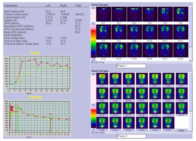
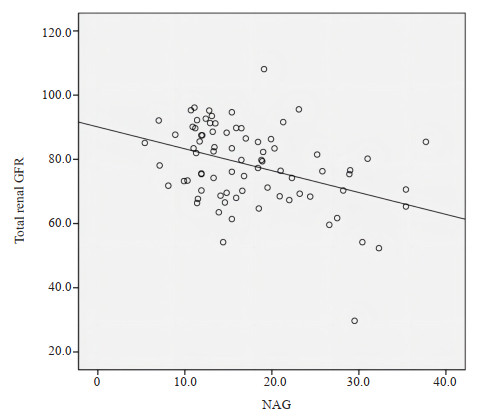
Objective To explore the clinical significance of 99mTc-DTPA renal dynamic imaging and urinary N-acetyl-β-D-glucosamine (NAG) in the early stage of renal damage in middle-aged and elderly patients with essential hypertension. Methods Eighty middle-aged and elderly patients with essential hypertension were selected as the observation group, with an average age of 63.0±8.6 years old. They were divided into grade 1 hypertension group (H1 group), grade 2 hypertension group (H2 group) and grade 3 hypertension group (H3 group) according to blood pressure level. According to urinary albumin / creatinine ratio, patients were divided into normal albuminuria group (NA group, n=53) and microalbuminuria group (MA group, n=27). At the same time, 20 middle-aged and elderly healthy subjects were selected as the control group (NC group), with an average age of 61±9.6 years old. Renal dynamic imaging was performed in all patients. Glomerular filtration rate (GFR), peak time (tp) and half excretion time (t1/2) were calculated. Urinary NAG was detected by random clean midstream urine. Fasting blood samples were taken to measure serum cystatin C (Cys-C) and serum creatinine. The above indexes were compared between groups according to the grade of hypertension, and NA, MA and control groups were compared at the same time. Results Compared with the control group, GFR was decreased, tp and t1/2 were prolonged, and urinary NAG was increased in hypertension groups.Compared with the hypertension group, with the increase of blood pressure, GFR decreased, tp, t1/2prolonged, urinary NAG and Cys-C increased (P<0.05). Cys-C in H2 and H3 groups was higher than that in control group (P<0.05). There was no significant difference in Cys-C between H1 group and control group (P > 0.05). Comparison of NA and MA groups with control group in patients with hypertension, GFR decreased, peak time and half excretion time prolonged, urinary NAG and Cys-C increased in NA and MA group (P<0.05). There was no significant difference in serum creatinine between the NA and MA groups and the control group (P > 0.05). Compared with NA group, GFR in MA group was lower, tp and t1/2 were longer, urinary NAG and Cys-C were higher in MA group than in NA group, but there was no significant difference in serum creatinine between the two groups (t=0.885, P > 0.05). There was a negative correlation between GFR and urinary NAG in hypertension group (r=-0.39, P<0.01). Conclusion Renal dynamic imaging combined with urinary NAG has good value in the diagnosis of early renal damage in patients with essential hypertension.
2023, 46(5): 829-835.
doi: 10.12122/j.issn.1674-4500.2023.05.09
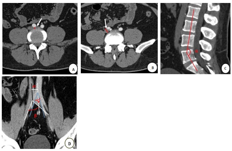
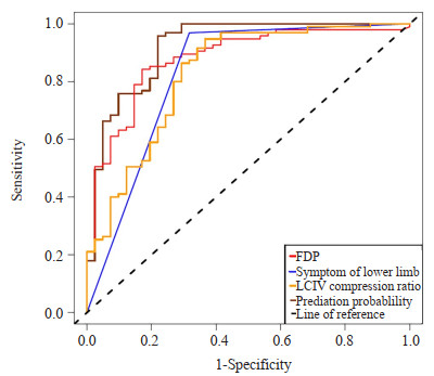
Abstract:
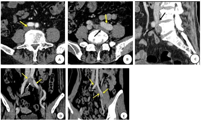
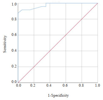
Objective To explore the incidence and risk prediction model of lower extremity deep venous thrombosis in patients with atypical left common iliac vein compression based on baseline enhanced CT combined with clinical data. Methods A total of 137 patients with atypical left common iliac vein (LCIV) compression diagnosed for the first time in Shunde Hospital of Southern Medical University from June 2017 to October 2022 were retrospectively analyzed. These patients did not receive any medication or physical measures to prevent the formation of DVT during the diagnosis and treatment period. Based on the occurrence of lower limb DVT during follow-up, the patients were divided into the non-DVT group (n=41) and the DVT group (n=96). The statistical differences in baseline CT findings and clinical data between the two groups were compared, and independent risk factors were selected through univariate and multivariate logistic regression analysis. The predictive efficacy of each risk factor was analyzed using ROC curve analysis. Results The incidence of DVT secondary to atypical LCIV was 70% (96/137). Univariate binary logistic regression analysis showed that LCIV minimum short diameter, right common iliac vein minimum short diameter, LCIV compression ratio, lower lumbar anterior flexion angle, symptoms of lower limb, D-dimer and fibrin degradation product (FDP) were risk factors for DVT secondary to atypical LCIV (P<0.05). Multivariate Logistic regression analysis showed that FDP (OR=1.05, 95% CI=1.01- 1.1, P=0.002), symptom of lower limb (OR=12.59, 95% CI: 2.78- 57.12, P<0.001) and LCIV compression ratio (OR=1.05, 95% CI: 1.01-1.09, P=0.02) were independent risk factors. ROC curve analysis showed that the AUC of FDP, symptom of lower limb and LCIV compression ratio were 0.879, 0.826 and 0.827, respectively. The AUC of the combined prediction model was 0.921, and the sensitivity and specificity were 95.79% and 78.05%, respectively. Conclusion The incidence of secondary DVT with atypical LCIV is high. FDP, symptom of lower limb, LCIV compression ratio are independent risk predicators for their occurrence. The prediction model combined with the three independent risk factors has high predictive efficacy, providing personalized risk assessment for patients with secondary DVT with atypical LCIV.
2023, 46(5): 836-840.
doi: 10.12122/j.issn.1674-4500.2023.05.10
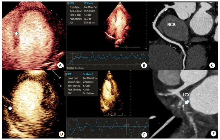
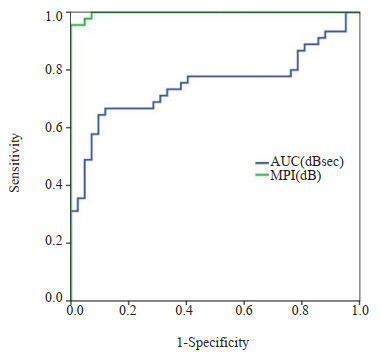
Abstract:
Objective To explore the correlation between quantitative myocardial contrast echocardiography (MCE) and the degree of coronary artery stenosis, and evaluate its predictive value on the the degree of coronary artery stenosis. Methods A total of 35 patients with suspected coronary heart disease were continuously enrolled in our hospital. All patients underwent MCE, coronary angiography and/or coronary CT angiography. According to coronary angiography and/or coronary CT angiography results, the patients were divided into two groups: coronary artery disease group (coronary artery stenosis ≥75%, n=20) and control group (coronary artery stenosis <75%, n=15). Clinical indicators and two-dimensional ultrasound were compared between two groups. In addition, quantitative parameters of MCE were measured according to the number of coronary arteries, and ROC curve evaluate the sensitivity and specificity of MCE parameters in predicting coronary artery stenosis ≥75%. Results The proportion of hypertension and smoking history were higher in the coronary group than that in the control group (50.00% vs 13.33%, 65.00% vs 26.67%, P<0.05), and two-dimensional ultrasound showed a high incidence of segmental wall motion abnormalities (90.00% vs 46.67%, P<0.05). Contrast-enhanced ultrasound showed that there were significant differences in peak myocardial intensity and area under the curve between the two groups (P<0.001). ROC curve analysis showed that peak myocardial intensity and area under the curve had a predictive value of 5.91 dB (sensitivity and specificity were 97.8% and 95.2%) and 28.94 dB sec (sensitivity and specificity were 66.7% and 74.8%) in predicting the degree of coronary artery stenosis ≥75%. Conclusion Contrast-enhanced ultrasound quantitative analysis is non-invasive, simple and suitable for long-term follow-up. It can accurately evaluate myocardial ischemia and is helpful for clinical judgement coronary artery stenosis degree and guidance for subsequent treatment.
2023, 46(5): 841-846.
doi: 10.12122/j.issn.1674-4500.2023.05.11
Abstract:
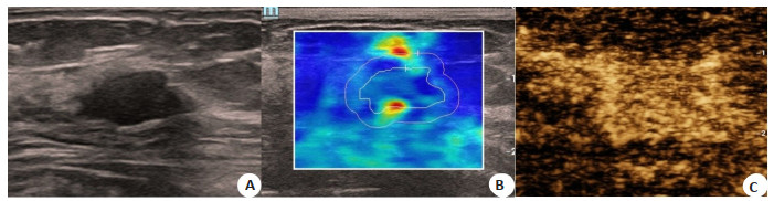
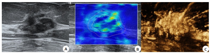
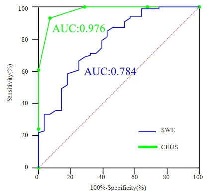
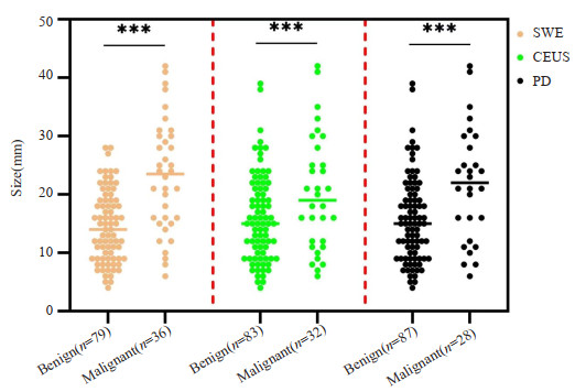
Objective To compare the diagnostic value of ultrasonic elastography and contrast-enhanced ultrasound in the diagnosis of breast BI-RADS 4a nodules. Method A total of 115 patients diagnosed as BI-RADS category 4a by routine ultrasonic detection in our hospital from January 2019 to February 2023 were included. Each lesion was analyzed, and pathological diagnosis were taken as the gold standard to construct diagnostic ROC curves of the two detection methods. The value of the two methods in the diagnosis of benign and malignant breast BI-RADS 4a nodules of BI-RADS was compared. Results Among 115 patients, there were 87 benign lesions and 28 malignant lesions. The Kappa values of ultrasonic elastography and contrast-enhanced ultrasound scores were small and inconsistent (P=0.258). Using pathological results as the gold standard, contrast score had higher diagnostic value (sensitivity, specificity and accuracy were 0.812, 0.976 and 0.956, respectively), and was more consistent with the gold standard than elastic imaging. The area under the diagnostic curve of contrast-enhanced ultrasound score was 0.976, higher than the value of elastic imaging (0.784). Three different diagnostic methods showed that the age of patients with malignant nodules was higher than that of patients with benign nodules, and the maximum diameter of nodules was larger. Conclusion Compared with ultrasonic elastography, contrast-enhanced ultrasound can significantly improve the accuracy of benign and malignant diagnosis of breast BI-RADS 4a nodules of BI-RADS, reduce the risk of preoperative biopsy, and is worthy of clinical promotion.
2023, 46(5): 847-852.
doi: 10.12122/j.issn.1674-4500.2023.05.12
Abstract:
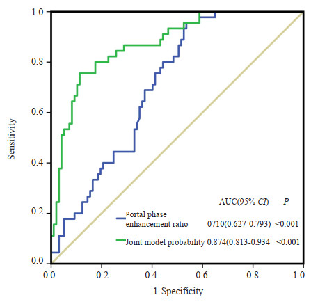
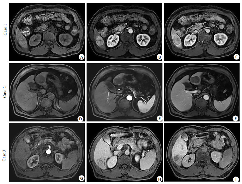
Objective To analyze the MRI features and improve the diagnostic rate of atypical enhanced primary hepatocellular carcinoma (HCC). Methods A total of 142 patients with pathologically confirmed HCC at Shenzhen People's Hospital from 2019 to 2021, who concurrently received preoperative contrast-enhanced MRI examination, were included in this retrospective study. Patients were divided into two groups based on MRI features: Typical enhancement group (n=97, those who met the "fast in and fast out" criteria) and atypical enhancement group (n=45, those who did not meet the "fast in and fast out" criteria). A series of imaging parameters, including enhancement pattern, tumor diameter, non- rim arterial phase enhancement, enhancement capsule, non-rim washout, halo enhancement, intratumoral fat or bleeding, intratumoral nodules, mosaic sign, tumor infiltration into blood vessels, lesion signal intensity on arterial phase, lesion signal intensity on portal phase, apparent diffusion coefficient, and portal phase enhancement rate, were independently assessed by two expert radiologists. Variables with statistical significance in the univariate analysis were further incorporated in the multivariate Logistic regression model to establish a prediction model for identifying atypical enhanced HCC. The ROC curve was used to evaluate the sensitivity and specificity of the prediction model. Results There was no significant difference between the two groups in non- rim arterial phase enhancement, enhanced capsule, intratumoral fat or bleeding, intratumoral nodules, mosaic sign, vascular infiltration, tumor diameter and apparent diffusion coefficient value (P>0.05), while there were statistical differences in non-rim washout (P=0.005), halo enhanced (P=0.005) and portal phase enhanced rate (P=0.001). Multivariate logistic regression analysis showed that the absence of non- rim washout (OR=27.995, 95% CI: 3.910- 200.462, P=0.001) and portal phase enhancement rate (OR=1.034, 95% CI: 1.018-1.051, P < 0.001) were independent influencing factors for atypical enhancement in HCC. A combined prediction model was established based on the non-rim washout and portal phase enhancement rate. ROC analysis showed that the prediction ability of the combined observation (non- rim washout plus portal phase enhancement rate) for atypical enhanced HCC was overall satisfying with the AUC of 0.874. Conclusion Non-rim washout enhancement and portal venous phase enhancement rate are independent influencing factors for atypical enhancement HCC, and the combination of these two parameters can improve the diagnostic efficacy of HCC.
2023, 46(5): 853-857.
doi: 10.12122/j.issn.1674-4500.2023.05.13
Abstract:
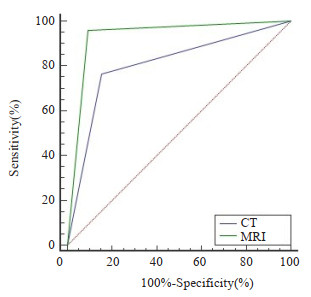
Objective To explore the CT and MRI signs and value of diagnostic in skull base bone invasion in nasopharyngeal carcinoma. Methods A total of 130 patients with nasopharyngeal carcinoma treated in our hospital from April 2018 to July 2022 were selected as the research subjects. All patients were performed with CT and MRI. The pathological results were counted, qualitative diagnosis was taken as the gold standard, the diagnostic results of CT and MRI for skull base bone invasion were recorded. ROC curve was used to analyze the diagnostic value of CT and MRI for skull base bone invasion of nasopharyngeal carcinoma. Results All 130 patients had squamous cell carcinoma by biopsy, including 88 cases of differentiated non keratinized squamous cell carcinoma, 13 cases of differentiated keratinized squamous cell carcinoma, and 29 cases of undifferentiated squamous cell carcinoma. A total of 98 cases of skull base bone invasion were detected by qualitative diagnosis, with a detection rate of 75.38%. 80 cases of skull base bone invasion were detected by CT, with a detection rate of 61.54%. 97 cases of skull base bone invasion were detected by MRI, with a detection rate of 74.62%. The detection rate of MRI examination was higher than that of CT examination (P < 0.05). The diagnostic sensitivity, specificity and accuracy of MRI examination for skull base bone invasion of nasopharyngeal carcinoma were 95.92%, 90.62%, 94.62%, respectively, which were higher than 76.53%, 84.37%, 78.46% of CT examination (P < 0.05). Conclusion Compared with the CT examination, MRI examination has a higher detection rate of skull base bone invasion in nasopharyngeal carcinoma, which has higher diagnostic value.
2023, 46(5): 858-862.
doi: 10.12122/j.issn.1674-4500.2023.05.14
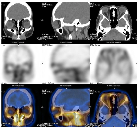
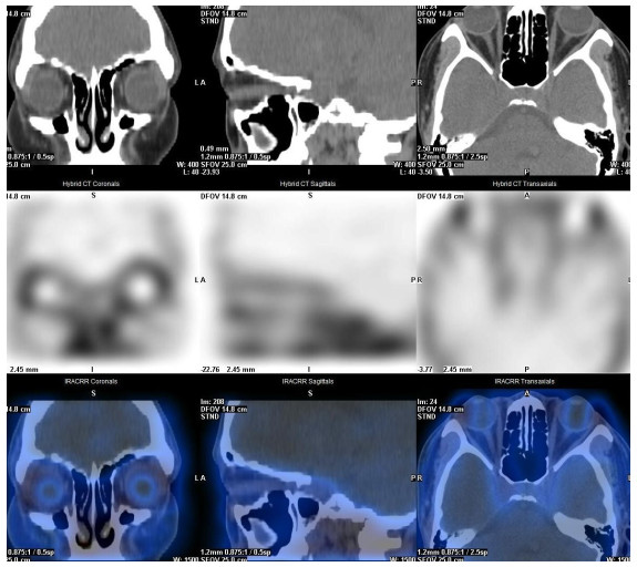
Abstract:
Objective To assess the clinical value of the 99mTc-DTPA orbital SPECT/CT imaging technique for quantitative analysis of extraocular muscles in the inflammatory activity of thyroid associated ophthalmopathy (TAO). Methods Forty-four patients with TAO who underwent 99mTc-DTPA orbital SPECT/CT imaging in our hospital were retrospectively analyzed, and the patients with TAO were classified into an active group [clinical activity score (CAS)≥3] and an inactive group (CAS < 3) according to CAS scores, with 23 patients (46 eyes) in the active group and 21 patients (42 eyes) in the inactive group. In the consensus on SPECT images corrected for CT attenuation, the uptake values were determined by placing a region of interest within the area of the highest uptake in each extraocular muscle. To determine the background uptake values, a circular region of interest was placed on the occipital lobe at the level of the optic nerve. The UR was calculated as the ratio of the maximum uptake value in the region of interest to the maximum uptake value in the background. And the highest UR value of extraocular muscles was considered as the most severely involved extraocular muscle. The URmax value of extraocular muscles was selected for statistical analysis of intergroup variability, which was used to evaluate the diagnostic efficacy for the diagnosis of TAO activity. Results The URmax of the extraocular muscles was significantly higher in patients with active TAO than inactive TAO, and the difference was statistically significant. (P < 0.05). The results showed a significant correlation between the URmax and CAS (r=0.799, P < 0.05). The URmax of the extraocular muscles and CAS were in agreement in assessing the clinical activity of TAO (Kappa value=0.915, P < 0.05); The area under the ROC curve was 0.975, and the URmax threshold for distinguishing between active and inactive periods was 5.2, with a sensitivity of 87.5% and a specificity of 100%. Conclusion The measurements of quantitative extra-ocular muscles analysis technique used in this study is able to directly show the inflammatory response of extra-ocular muscles, which can accurately localize the involved extra-ocular muscles and enable more precise outlining region of interest, as well as measure the uptake ratio of each extra-ocular muscle, and more accurately assess the degree of inflammatory.
2023, 46(5): 863-867.
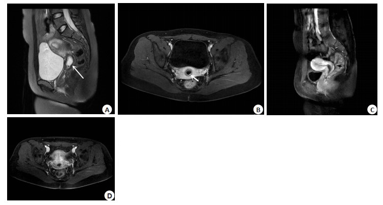
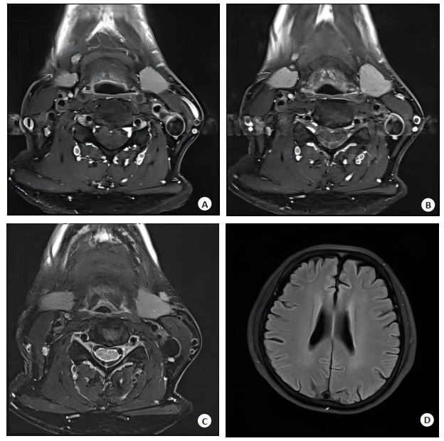
doi: 10.12122/j.issn.1674-4500.2023.05.15
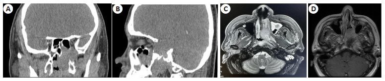
Abstract:
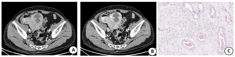
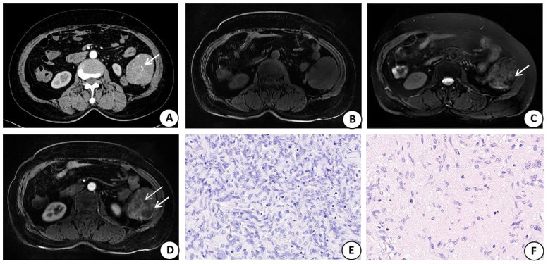
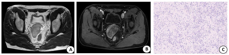
Objective To analyze the imaging features of primary solitary fibrous tumors (SFT) in the abdomen and pelvis, according to the pathology. Methods We retrospectively analyzed the imaging data of 12 cases with pathologically confirmed abdominal and pelvic SFT, 4 cases underwent CT examination, and 2 cases underwent MRI examination. 6 cases underwent CT and MRI examination at the same time, and the pathological results were compared with the analysis of the pathological results. Results On plain CT scan, the mass showed equal / low density. There were 3 cases of calcification and 1 case of hemorrhage. On MRI plain scan, 8 cases showed isohypotension on T1WI, 1 case showed uniform hypotension on T2WI / T2 lipid pressure, the other 7 cases showed high and low mixed signals, 4 cases showed "Yin-Yang" pattern, 4 cases showed flow empty vessels in tumor. Enhanced scanning: 12 cases were inhomogeneous enhancement, 10 cases were "map like" enhancement, 2 cases were piebald like enhancement, of which 1 case was continuous enhancement, 1 case was gradual enhancement, and the remaining 10 cases were "fast in and slow out" enhancement. Tortuous blood vessels were seen in the tumors in 6 cases, namely "earthworm digging into the earth" sign. Except for necrotic cystic areas, the solid components of the tumor examined by MRI were divided into two categories: Type A nodules, T1WI isointense, T2WI / T2 hypopolipid signal, no enhancement on enhanced examination; Type B nodules, low signal intensity on T1WI and high signal intensity on T2WI / T2 lipid pressure, showed mild to moderate / significant enhancement on enhanced examination. 5 cases of mass were 2 types of nodules coexisting, and 3 cases were type B nodules. Pathology showed that 5 cases of tumor cells showed atypical or malignant SFT. The positive rate of CD34 was 100%, STAT6 and Bcl-2 were 91.7%, Vimentin was 83.3%, and CD99 was 75%. Conclusion There are characteristic imaging features in abdominal and pelvic SFT, and different imaging features in the mass correspond to the pathological tissue components. When the diameter of the mass > 10 cm or the irregular calcium focus in the tumor, we should be vigilant against the possibility of malignant transformation of the tumor. Combined with immunohistochemical markers is of great significance in the diagnosis and differential diagnosis of the disease.
2023, 46(5): 868-872.
doi: 10.12122/j.issn.1674-4500.2023.05.16
Abstract:
Objective To evaluate the value of diffusion-weighted MRI using readout-segmented echo-planar imaging (RS-EPI) sequence in the diagnosis of orbit tumors and tumorlike lesions. Methods A retrospective study was performed on 60 patients with orbital tumors or tumorlike lesions in our hospital from December 2016 to December 2018. The patients were imaged on a 3T MRI scanner (MAGNETOM Trio Tim). The RS-EPI and single-shot echo-planar imaging (SS-EPI) sequences were compared in terms of image quality. The diagnostic efficacy between RS-EPI sequence and SS-EPI sequence for orbital tumors and tumorlike lesions were compared. Results Fat suppression in the RS-EPI sequence showed no statistical difference to that in the SS-EPI sequence (P > 0.05), while RS-EPI sequence was was superior to SS-EPI sequence in terms of normal anatomical structure display, ghosting artifact, overall image quality (P < 0.001). The apparent diffusion coefficient (ADC) values of vitreous body and brainstem yielded no statistical difference between two methods (P > 0.05). The geometric distortion ratio in both anterior-posterior and right-left direction, as well as signal-to-noise ratio of RS-EPI sequence were all lower than those of SS-EPI sequence (P < 0.001). The diagnostic sensitivity of RS-EPI sequence for orbital tumors and tumorlike lesions was significantly higher than that of SS-EPI sequence (P < 0.05). ROC curve denoted that the area under the curve of RS-EPI sequence in the diagnosis of orbital tumors and tumorlike lesions was significantly larger than that of SS-EPI sequence (P < 0.001). Conclusion MRI using RS-EPI sequence can effectively improve the image quality and diagnostic sensitivity of orbital tumors and tumorlike lesions.
2023, 46(5): 873-878.
doi: 10.12122/j.issn.1674-4500.2023.05.17
Abstract:
Objective To investigate the efficacy of Deauville visual 5-point scale (Deauville 5-PS), the ratio of the maximum standard uptake (SUVmax) of a lesion to the liver (LLR), and the change rate of SUVmax (ΔSUVmax) before and during chemotherapy for interim 18F-FDG PET/CT in the prognosis prediction of large B- cell lymphoma (DLBCL). Methods We conducted a retrospective analysis of medical data from 117 treatment-naive DLBCL patients, and applied Deauville 5-PS, LLR and ΔSUVmax to analyze the status of mid-term chemotherapy PET/CT. ROC curve was used to calculate the optimal critical values of LLR and ΔSUVmax for predicting the progression-free survival (PFS) and overall survival (OS). Kaplan-Meier survival analysis and univariate and multivariate Cox proportional hazards regression model were applied to analyze the prognosis of 2-year PFS rate and OS rate. Results Among the 117 patients with DLBCL, 46 (39.3%) progressed and 34 (29.1%) died. At Deauville 5-PS, LLR and ΔSUVmax set to 4, 1.81, and 75%, respectively, the specificity, positive predictive value and accuracy of ΔSUVmax in predicting the PFS and OS of DLBCL patients were found to be higher than those with LLR alone, which was higher than that for Deauville 5-PS alone. However, Deauville 5-PS alone had the highest sensitivity for predicting PFS and OS in DLBCL patients. The 2-year PFS and OS rates of patients in the Deauville 5-PS < 4 group and ≥4 group were 80.8%, 93.4% and 49.3%, 65.6%, respectively (P < 0.001). The 2-year PFS and OS rates of patients with LLR < 1.81 and ≥1.81 were 82.4%, 89.4% and 27.2%, 56.3%, respectively (P < 0.001). The 2-year PFS and OS rates of patients with ΔSUVmax>75% and ΔSUVmax≤75% were 86.2%, 93.1% and 20.5%, 48.9%, respectively(P < 0.001). Univariate analysis revealed that Deauville 5-PS, LLR, and ΔSUVmax were all prognostic factors of PFS and OS in DLBCL patients (P < 0.01). Multivariate analysis showed that ΔSUVmax and international prognostic index scores served as independent risk factors for PFS and OS in DLBCL patients. Conclusion All three evaluation methods of Deauville 5-PS, LLR and ΔSUVmax of interim PET/CT can predict the prognosis of DLBCL patients, among which ΔSUVmax show better predictive efficacy than Deauville 5-PS and LLR and ΔSUVmax had an independent predictive value for the prognosis of DLBCL.
2023, 46(5): 879-883.
doi: 10.12122/j.issn.1674-4500.2023.05.18
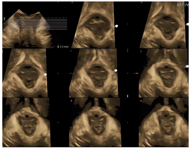
Abstract:
Objective To analyze the value of multimodal pelvic floor ultrasound parameters in evaluating maternal birth trauma and postpartum stress urinary incontinence (SUI). Methods A total of 200 parturients admitted to Hainan Traditional Chinese Medicine Hospital from January to December 2022 were selected.The parturients were divided into the normal urine control group (n=144) and the SUI group (n=56). All women were examined with transperineal pelvic floor ultrasound on day 42 after delivery. Women in the SUI group were examined with pelvic floor ultrasound at 3 months and 6 months after delivery. The two groups were compared in terms of bladder neck descent (BND), urethral rotation angle (URA), retrovesical angle (RVA), pelvic diaphragmatic hiatal area, the maximum descent of uterus and rectal ampulla, urethral funnel formation, and the presence or absence of levator ani muscle injury. The correlation between pelvic floor ultrasound parameters and SUI was analyzed. Results There was no statistically significant difference in the descent of uterus or rectal ampulla between the groups (P > 0.05). BND, URA, RVA and pelvic diaphragmatic hiatal area in the SUI group were larger than those in the normal urine control group (P < 0.05). The urethral funnel formation rate and the incidence of levator ani muscle injury were higher than those in the normal urine control group (P < 0.05). There was no statistically significant difference in the descent of uterus or rectal ampulla among patients with SUI at different time points (P > 0.05). BND, URA, RVA, pelvic diaphragmatic hiatal area, urethral funnel formation rate, and the incidence of levator ani muscle injury showed a downward trend at 3 months and 6 months after delivery (P < 0.05). BND, URA, RVA and pelvic diaphragmatic hiatal area were positively correlated with SUI (P < 0.05). Conclusion Multimodal pelvic floor ultrasound parameters such as BND, URA, RVA and pelvic diaphragmatic hiatal area are closely related to SUI. They provide important guidance for the prevention and treatment of SUI.
2023, 46(5): 884-888.
doi: 10.12122/j.issn.1674-4500.2023.05.19
Abstract:
Objective To investigate the changes in gray matter volume of the brain before and after acupuncture treatment in patients with knee osteoarthritis (KOA) based on voxel-based morphometry, and then to analyze the efficacy mechanism of acupuncture. Methods We prospectively included 25 patients with KOA, and acupuncture treatment was performed on patients with KOA, who were evaluated using visual analogue scale, self-rating anxiety scale, self-rating depression scale and Montreal cognitive assessment rating scales before and after acupuncture treatment, and scanned high-resolution T1WI. The voxel-based morphometry method was used to analyze the differences in the gray matter structure of the brain in KOA patients before and after acupuncture treatment. The correlation with the results of the rating scales was analyzed. Results KOA patients showed relief of pain, anxiety, and depression after acupuncture treatment (P < 0.001), and cognitive function was improved (P < 0.001). After treatment brain regions with increased gray matter volume included the right lingual gyrus and left insula; brain regions with decreased gray matter volume included the left middle occipital gyrus and paracentral lobule. (P < 0.001 at the level of the somatosensory image, P < 0.05 at the level of the clump). Conclusion The clinical symptoms of KOA patients could be significantly relieved by acupuncture. The structural changes of multiple brain regions in KOA patients are also found after acupuncture. It is speculated that the efficacy of acupuncture may be achieved through the interaction of multiple brain regions.
2023, 46(5): 889-894.
doi: 10.12122/j.issn.1674-4500.2023.05.20
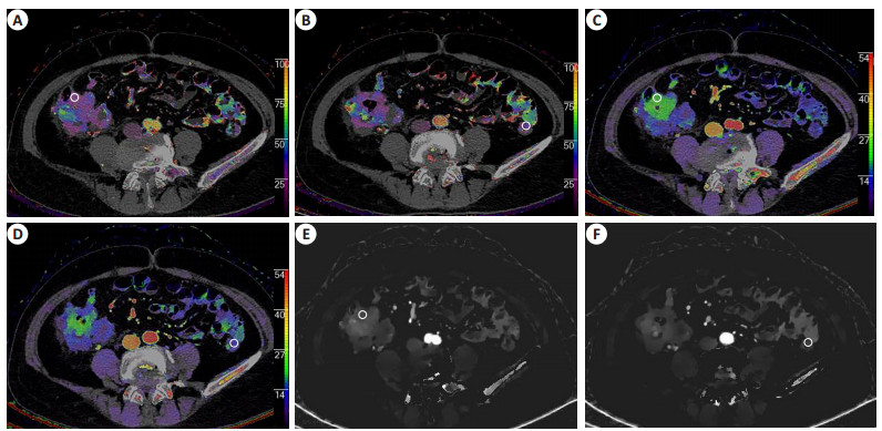
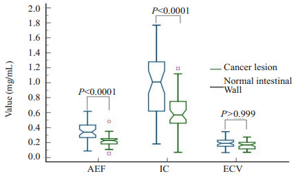
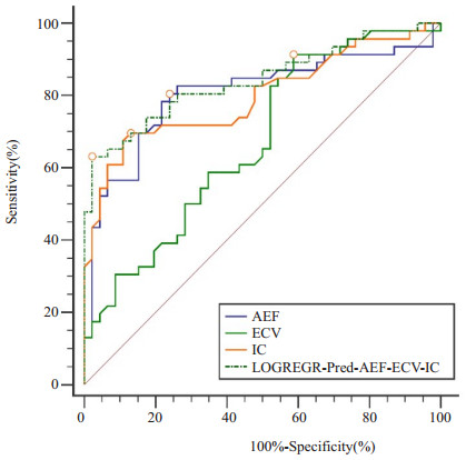
Abstract:
Objective To explore the diagnostic value of quantitative contrast-enhanced parameters of spectral CT in colorectal cancer. Methods Spectrum base images of 46 patients with colorectal cancer who underwent three- phase enhancement of abdominal and pelvic spectral CT were processed to generate the water-free iodine images, arterial enhancement fraction (AEF) and extracellular volume (ECV) images, and the iodine concentration (IC) value, AEF value and ECV value were measured on the colorectal lesions and the normal colorectal walls. The independent sample t test and Mann-Whitney U test were applied for inter-group comparisons, and ROC curve was used to evaluate the diagnostic efficacy. Results The AEF value and IC value presented significantly highly in the patients than that in normal intestinal wall (0.338±0.122 vs 0.225±0.072, 1.007±0.402 mg/mL vs 0.599±0.229 mg/mL, P < 0.0001). ECV showed no significant difference between colorectal lesion and the normal intestinal wall(P > 0.999). The areas under the ROC curve of AEF, IC, ECV and three indicators combination in the diagnosis of colorectal cancer were 0.806, 0.797, 0.671 and 0.842, respectively, and the Jordan index were 0.565, 0.565, 0.326 and 0.609, respectively. The cutoff value of three indicator combination in the diagnosis of colorectal cancer was 0.660 with the sensitivity of 63.04% and the specificity of 97.83%, and the logistic regression equation was Y=13.75×AEF+16.30×ECV-6.70. Conclusion The combined diagnostic model of spectral CT quantitative contrast-enhanced parameter of AEF, ECV and IC has certain value in the diagnosis of colorectal cancer.
2023, 46(5): 895-899.
doi: 10.12122/j.issn.1674-4500.2023.05.21
Abstract:
Objective To explore the application effects of PET/CT in the diagnosis of lesion distal metastasis and evaluation of prognosis in patients with chemoradiotherapy after rectal cancer surgery. Methods Retrospective analysis was conducted on the data of 70 patients with rectal cancer surgery in the hospital from March 2019 to March 2020. All patients received adjuvant therapy with chemoradiotherapy after surgery, and PET/CT technology was used to detect the therapeutic effect. The prognosis of patients was evaluated according to whether recurrence or metastasis occurred after treatment. With postoperative pathological detection as the gold standard, the diagnostic value of CT and PET/CT on lesion distal metastasis in patients with rectal cancer was compared. Patients with recurrence and metastasis were included in the recurrence metastasis group (n=26), and those without recurrence and metastasis were included in the good prognosis group (n=44). The clinical data of the two groups were compared. Multivariate Logistic regression analysis was used to analyze the related factors affecting prognosis of patients with chemoradiotherapy after rectal cancer surgery. Results Among the 70 patients at 3 year after surgery, distal metastasis was positive in 21 cases and negative in 49 cases by pathological diagnosis, distal metastasis was positive in 22 cases and negative in 48 cases by CT diagnosis, including 4 cases of missed diagnosis and 5 cases of misdiagnosis. The diagnostic sensitivity, specificity, accuracy rate, positive predictive value, negative predictive value and Kappa value were 80.95%, 88.37%, 87.14%, 77.27%. 91.67% and 0.698. PET/CT diagnosis showed 23 cases of positive distal metastasis and 47 negative cases, including 2 cases of missed diagnosis and 4 cases of misdiagnosis, and the diagnostic sensitivity, specificity, accuracy rate, positive predictive value, negative predictive value and Kappa value were 90.47%, 91.84%, 91.43%, 82.61%, 95.74% and 0.801. The age, maximum lesion diameter, TNM stage, maximum standardized uptake value (SUVmax), minimum standardized uptake value (SUVmin) and retention index (RI) in recurrence metastasis group were significantly higher than those in good prognosis group (P < 0.05). There were no statistical differences in gender, BMI and pathological type between the two groups (P > 0.05). Multivariate Logistic regression analysis showed that age, maximum lesion diameter, TNM stage, SUVmax, SUVmin and RI were risk factors affecting the prognosis of postoperative chemoradiotherapy in patients with rectal cancer (P < 0.05). Conclusion PET/CT has a high diagnostic value on lesion distal metastasis in patients with chemoradiotherapy after rectal cancer surgery. SUVmax, SUVmin and RI are risk factors affecting the prognosis of patients with postoperative chemoradiotherapy. It can be used in clinical prediction of postoperative recurrence and metastasis.
2023, 46(5): 900-904.
doi: 10.12122/j.issn.1674-4500.2023.05.22
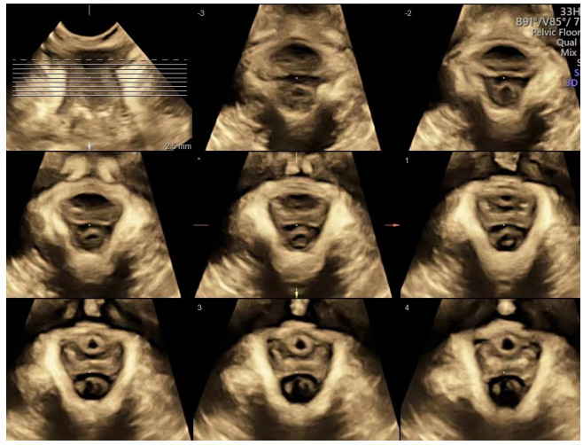
Abstract:
Objective To evaluate the curative effect of electrical stimulation biofeedback in female patients with pelvic floor dysfunction (PFD) based on four-dimensional pelvic floor ultrasound. Methods A total of 120 female patients with postpartum PFD who underwent electrical stimulation biofeedback for 4 courses in the hospital were enrolled as the research objects from January 2021 to January 2023.All underwent four-dimensional pelvic floor ultrasound examination before and after treatment.The clinical curative effect was evaluated.The grading of pelvic floor muscle strength, 1 h urine pad test, systolic blood pressure of pelvic floor muscle and four-dimensional ultrasound parameters (bladder neck movement and urethral rotation angle under rest and Valsalva maneuver state) were compared before and after treatment.The relationship between bladder neck movement, urethral rotation angle and grading of pelvic floor muscle strength, systolic blood pressure of pelvic floor muscle was analyzed by Pearson correlation analysis. Results In the 120 patients, total response rate of electrical stimulation biofeedback was 84.17%.After treatment, grading of pelvic floor muscle strength and systolic blood pressure of pelvic floor muscle were significantly increased, score of 1 h urine pad test was significantly decreased, the difference was statistically significant (P < 0.001).After treatment, levator ani thickness (under rest and Valsalva maneuver state) was significantly increased, left-right diameter and area of levator hiatus, posterior bladder angle, bladder neck movement and urethral rotation angle were significantly decreased, the difference was statistically significant (P < 0.001).After treatment, bladder neck movement was negatively correlated with grading of pelvic floor muscle strength and systolic blood pressure of pelvic floor muscle (r=-0.746, -0.781, P < 0.001), and urethral rotation angle was also negatively correlated with them (r=-0.779, -0.792, P < 0.001). Conclusion Four-dimensional pelvic floor ultrasound can better evaluate pelvic floor muscle function in PFD patients before and after electrical stimulation biofeedback.
2023, 46(5): 905-909.
doi: 10.12122/j.issn.1674-4500.2023.05.23
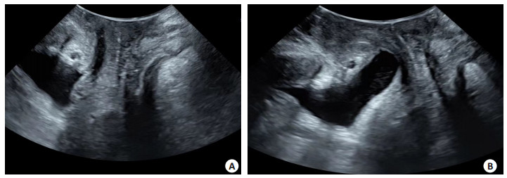
Abstract:
Objective To explore the application value of MRI in high-intensity focused ultrasound ablation (HIFU) ablation of intrauterine ectopic pregnancy. Methods The data of five patients with intrauterine ectopic pregnancy treated by HIFU ablation in Foshan Women and Children Hospital from December 2016 to December 2021 were analyzed retrospectively.All the patients had median age of 35 years old and underwent MRI before and after operation.We observed the preoperative and postoperative MRI images with location, size, shape, growth mode and signal intensity characteristics of gestational sacs, the changes and enhancement of myometrium at the attachment of gestational sacs, and MRI findings after HIFU. Results MRI before HIFU showed that 4 cases were cesareanscar pregnancy (CSP) and 1 case was cervical pregnancy.2 cases gestational sac signal were homogeneous and 3 cases were inhomogeneous.In CSP patients, the anterior uterine wall muscle layer was significantly thinner, with a thickness of 1-2 mm.In 1 CSP patient, there was no enhancement of pregnancy sac, but multiple thickening and tortuous enhanced vascular shadow were seen near the uterus.In the other 3 CSP patients, there was obvious enhancement of strip-like and patchy lesions in the pregnancy sac.In cervical pregnancy, the wall of the pregnancy sac was uniformly strengthened.MRI after HIFU showed that 4 cases of pregnancy sac were smaller than before, and 1 case had no significant change.The signal intensity of 5 cases ware higher/inhomogeneous than before.No enhancement lesion was found in 1 case of CSP pregnancy sac before operation.1 case of CSP patient had no enhancement before operation, but the periuterine thickening and tortuous enhancement vessels decreased after operation, and the enhancement degree was weakened.In the other 2 cases of CSP gestational sac, there were still some cord-like and patchy enhancement, but the enhancement range was smaller than before, and some necrosis changes were found.There was no enhancement of gestational sac wall in patients with cervical pregnancy. Conclusion MRI preoperative examination can clearly determine the position and signal of gestational sac, which is helpful to accurately locate HIFU before operation, and can show the relationship between gestational sac and surrounding tissues.Postoperative dynamic contrast-enhanced scanning can judge the therapeutic effect of HIFU.
2023, 46(5): 910-914.
doi: 10.12122/j.issn.1674-4500.2023.05.24
Abstract:
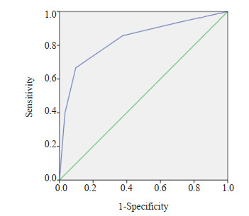
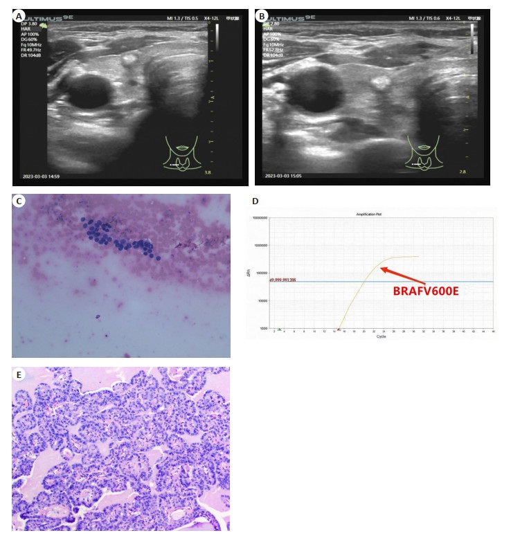
Objective To investigate the value of 2020 Chinese Thyroid Imaging Report and Data System (C-TIRADS) combined with BRAFV600E testing in the differential diagnosis of benign and malignant category Ⅲ nodules in Bethesda system for reporting thyroid cytopathology (BSRTC). Methods A total of 156 thyroid nodules in 124 patients with BSRTC category Ⅲ who underwent ultrasound-guided fine needle aspiration biopsy at the Second Affiliated Hospital of Fujian Medical University from October 2020 to October 2022 were selected.Each nodule underwent C-TIRADS classification and BRAFV600E testing and was confirmed by histopathology.The diagnostic efficacy of C-TIRADS, BRAFV600E testing, and their combination in BSRTC category Ⅲ nodules were assessed according to histopathology findings. Results According to histopathology findings, 93 benign nodules and 63 malignant nodules in 156 nodules, 48 positive and 108 negative for BRAFV600E testing.The area under the ROC curve of C-TIRADS classification was 0.834(95%CI: 0.765-0.902).The sensitivities of C-TIRADS classification and BRAFV600E testing were 73.0% and 76.2%, the specificity were 88.2% and 98.9%, the positive predictive value were 80.7% and 97.9%, the negative predictive value were 82.8% and 85.2%, the accuracy were 82.1% and 89.1%, respectively.The sensitivity, specificity, positive predictive value, negative predictive value and accuracy of the two combined diagnosis were 90.4%, 96.8%, 95.0%, 93.8% and 94.2%, respectively.The combination of two methods significantly increased the sensitivity (χ2=6.435, P=0.011; χ2=4.629, P=0.032) and negative predictive value (χ2=5.588, P=0.018; χ2=3.875, P=0.049) when compared with C-TIRADS and BRAFV600E testing in BSRTC category Ⅲ nodules. Conclusion C-TIRADS classification and BRAFV600E testing have great diagnostic efficacy in BSRTC category Ⅲ nodules, and the combination of two methods can improve the diagnostic sensitivity and negative predictive value, suggesting the great value in differentiating BSRTC category Ⅲ nodules.
2023, 46(5): 915-918.
doi: 10.12122/j.issn.1674-4500.2023.05.25
Abstract:
Objective To evaluate the value of high-resolution magnetic resonance vessel wall imaging for recurrence of cerebral infarction triggered by carotid plaques. Methods Patients with recurrent cerebral infarction and patients with initial cerebral infraction in our hospital from June 2021 to June 2022 were enrolled as study group (n=60) and control group (n=60).All patients received high-resolution magnetic resonance vessel wall imaging.Then comparison was conducted on the clinical characteristics, carotid plaque burden and compositional features. Results Study group had notably increased luminal area and larger total vessel area and vessel wall normalization index than control group (P < 0.05), while no statistical difference was found in vessel wall area and luminal stenosis rate between two groups (P > 0.05).The proportions of carotid plaques of calcification, lipoid-rich necrotic core, intraplaque hemorrhage and fibrous cap rupture were higher in study group than in control group (P < 0.05), while the proportion of intraplaque calcification and luminal stenosis > 50% demonstrated no statistical difference between two groups (P > 0.05).The area of lipoid-rich necrotic core, intraplaque calcification and intraplaque hemorrhage were significantly larger in study group than in control group (P < 0.001). Conclusion Application of high-resolution magnetic resonance vessel wall imaging can effectively display the stenosis degree of the vessel wall, carotid plaque burden and compositional features.It provide a basis for the prevention of cerebral infarction triggered by carotid plaques, thus effectively reducing the recurrence rate of cerebral infarction.
2023, 46(5): 919-923.
doi: 10.12122/j.issn.1674-4500.2023.05.26
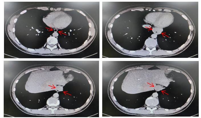
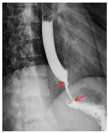
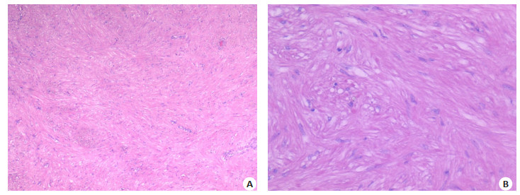
Abstract:
Objective To explore the efficacy and safety of ultramicro invasive surgery in the treatment of huge esophageal tumors, and to provide new ideas and new methods for the treatment of esophageal ultrainvasive surgery. Methods A retrospective analysis was made on two patients with large esophageal masses who were treated by single-port thoracoscope combined with esophagoscope and double-mirror combined with ultramicroscopic surgery in our hospital.We counted the operative time, intraoperative blood loss, postoperative chest tube indwelling time, drainage volume, length of hospital stay, pathological results and the incidence of serious postoperative complications in two patients. Results The huge esophageal masses of two patients were successfully resected by double mirror combined with ultramicroscopic surgery.The average operation time was about 230 min, and the average intraoperative bleeding was about 20 mL.Postoperative retention time of chest tubes was 3 d, the average drainage volume was about 395 mL, and the average postoperative hospital stay was 7 d.There were no serious postoperative complications such as thoracotomy, esophageal fistula and pulmonary infection.Postoperative pathological examination showed esophageal leiomyoma.Both patients were cured and discharged from hospital. Conclusion Single-aperture thoracoscope combined with esophagoscope ultramicroscopic surgery provides a new direction and technical support for the surgical treatment of giant esophageal leiomyoma.Compared with single-aperture/multi-aperture thoracoscope or combined esophagoscope, its efficacy and safety have more obvious advantages.
2023, 46(5): 924-929.
doi: 10.12122/j.issn.1674-4500.2023.05.27
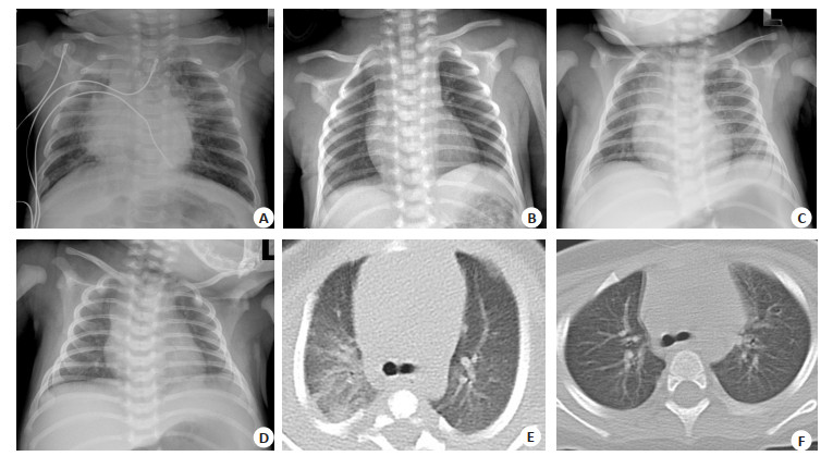
Abstract:
Objective To investigate the clinical characteristics and chest CT or X-ray features of family-clustered children infected with the Omicron variant of severe acute respiratory syndrome coronavirus 2(SARS-CoV-2). Methods A retrospective analysis was performed on the medical data of 241 children with SARS-CoV-2 Omicron variant infection who were admitted to Guangdong Second Provincial General Hospital, the designated hospital for COVID-19 from October 2022 to December 2022.The children were divided into 76 cases of 0-1 year, 80 cases of 1-3 years old and 85 cases of 3-6 years old according to the age of infection.The clinical characteristics, results of laboratory examination, chest CT or X-ray findings and outcome of the three groups were compared. Results Among 241 children, 203 cases (84.2%) were mild, 38 cases (15.8%) were severe, no moderate or critical.Fever was the most common symptom (97.5%), followed by cough (54.4%), diarrhea (29.9%), nasal congestion and/or runny nose (25.7%), and convulsion (15.4%).Children aged 3-6 years old were prone to upper respiratory symptoms, with the lowest fever duration and the shortest nucleic acid negative conversion time.Children aged 0-1 year mainly showed symptoms of cough, diarrhea and rash, with the lowest peak of fever.Infants aged 0-1 year were more likely to present radiological phenotype of pneumonia than older children.X-rays showed patchy and/or cloudy shadows in both lung fields, while chest CT showed thickened lung texture, decreased transparency of lung fields, and linear or patchy high-density shadows with blurred boundaries.Children aged 1-3 years old had the highest incidence of severe COVID-19 infection (22.5%), and they were more likely to have convulsions than those aged 0-1 year.Compared with mild cases, severe cases had lower cycle threshold value of SARS-CoV-2 N gene, lower lymphocyte count, higher neutrophil-to-lymphocyte ratio and higher serum amyloid A level.Among children aged 3-6 years, compared with those who received 0 or 1 dose of the vaccine, children who received 2 doses of the vaccine had a lower risk of fever lasting more than 2 d (OR=0.373, 95%CI: 0.152-0.912, P=0.029), a lower risk of diarrhea (OR=0.298, 95% CI: 0.096-0.922, P=0.030), a shorter time for nucleic acid negative conversion (OR=0.359, 95% CI: 0.149-0.867, P=0.021), and a lower incidence of severe cases (OR=0.237, 95%CI: 0.061-0.922, P=0.040). Conclusion Fever and cough are the main symptoms of children with family clustering SARS-CoV-2 Omicron variant infection, and chest CT can show pulmonary inflammatory changes.Children aged 1-3 years old are more likely to have convulsions than those aged 0-1 year.The neutrophil-to-lymphocyte ratio and serum amyloid A levels are helpful in predicting the severity of infection.Vaccination can reduce the risk of severe infection in children aged 3-6 years, decrease the viral load and shorten nucleic acid negative conversion time.
2023, 46(5): 934-937.
doi: 10.12122/j.issn.1674-4500.2023.05.29
Abstract:
Alzheimer's disease is a neurodegenerative disease occurring in middle-aged and elderly people, characterized by memory impairment and cognitive decline. At present, most of the multimodal imaging studies on Alzheimer's disease mainly focus on local specific brain regions, and there is a lack of in-depth exploration of whole brain network patterns. This paper summarizes the research on multimodal imaging of Alzheimer's disease based on machine learning and brain network. First, it introduces the definition of Alzheimer's disease and the limitations of machine learning technology in brain disease imaging research. Secondly, the general process of machine learning in brain network prediction is described, including: feature extraction, feature selection and feature dimensionality reduction, model construction and model evaluation; Finally, the research results of machine learning in Alzheimer's disease brain network of gray matter structure, white matter structure, resting-state functional brain network and multimodal fusion brain network are introduced in turn. By combing the research results in recent years, this paper has made the following three prospects for the future development direction of this field: large sample multi- center research, interpretable deep learning technology, and the establishment of longitudinal prediction models.
Alzheimer's disease is a neurodegenerative disease occurring in middle-aged and elderly people, characterized by memory impairment and cognitive decline. At present, most of the multimodal imaging studies on Alzheimer's disease mainly focus on local specific brain regions, and there is a lack of in-depth exploration of whole brain network patterns. This paper summarizes the research on multimodal imaging of Alzheimer's disease based on machine learning and brain network. First, it introduces the definition of Alzheimer's disease and the limitations of machine learning technology in brain disease imaging research. Secondly, the general process of machine learning in brain network prediction is described, including: feature extraction, feature selection and feature dimensionality reduction, model construction and model evaluation; Finally, the research results of machine learning in Alzheimer's disease brain network of gray matter structure, white matter structure, resting-state functional brain network and multimodal fusion brain network are introduced in turn. By combing the research results in recent years, this paper has made the following three prospects for the future development direction of this field: large sample multi- center research, interpretable deep learning technology, and the establishment of longitudinal prediction models.
2023, 46(5): 938-941.
doi: 10.12122/j.issn.1674-4500.2023.05.30
Abstract:
Hepatocellular carcinoma (HCC) is a serious disease threatening the life and health of Chinese. Immunotherapy has been proved to be a promising new method for the treatment of HCC, but only 15%-20% of patients respond to it clinically. Through activating immune cells, inducing apoptosis and inhibiting inflammation, photodynamic therapy can improve the tumor microenvironment to improve the efficacy of HCC immunotherapy. This article reviews the current research status of photodynamic therapy in the treatment of HCC, the mechanism of action on tumor microenvironment and the combined immunotherapy for HCC, aiming to provide new ideas and directions for the clinical comprehensive treatment of HCC.
Hepatocellular carcinoma (HCC) is a serious disease threatening the life and health of Chinese. Immunotherapy has been proved to be a promising new method for the treatment of HCC, but only 15%-20% of patients respond to it clinically. Through activating immune cells, inducing apoptosis and inhibiting inflammation, photodynamic therapy can improve the tumor microenvironment to improve the efficacy of HCC immunotherapy. This article reviews the current research status of photodynamic therapy in the treatment of HCC, the mechanism of action on tumor microenvironment and the combined immunotherapy for HCC, aiming to provide new ideas and directions for the clinical comprehensive treatment of HCC.
2023, 46(5): 942-947.
doi: 10.12122/j.issn.1674-4500.2023.05.31
Abstract:
Contrast-enhanced ultrasound is comparable to CT and MRI in its ability to diagnose focal liver lesions. Currently, SonoVue and Sonazoid were the commonly used contrast agents applied to focal liver lesions, the former being a pure blood pool agent and the latter adding a Kupffer phase to liver's triple-vessel phase, but whether the ultrasonographic feature and application value of the two agents are equivalent has not been reported systemically. This article reviews the progress of the SonoVue and Sonazoid application in terms of their physicochemical properties, the diagnostic and differential diagnostic values in the diagnosis of benign and malignant lesions in the liver, guiding interventional treatment and safety performance.
Contrast-enhanced ultrasound is comparable to CT and MRI in its ability to diagnose focal liver lesions. Currently, SonoVue and Sonazoid were the commonly used contrast agents applied to focal liver lesions, the former being a pure blood pool agent and the latter adding a Kupffer phase to liver's triple-vessel phase, but whether the ultrasonographic feature and application value of the two agents are equivalent has not been reported systemically. This article reviews the progress of the SonoVue and Sonazoid application in terms of their physicochemical properties, the diagnostic and differential diagnostic values in the diagnosis of benign and malignant lesions in the liver, guiding interventional treatment and safety performance.
Research progress of nomogram based on MR-radiomics in extramural vascular invasion of rectal cancer
2023, 46(5): 948-952.
doi: 10.12122/j.issn.1674-4500.2023.05.32
Abstract:
In recent years, nomograms have been widely used in the diagnosis, treatment, and prognosis of rectal cancer. Some related radiomics studies have also introduced nomogram models, and the number is increasing year by year, especially the nomogram models based on magnetic resonance radiomics. Through a literature review, the author found that most of the nomogram models based on MR Radiomics were better than single radiomics in predicting extramural vascular invasion of rectal cancer, but there were differences in research methods and results. This article summarized several main research methods and comprehensively discussed the research status and prospects of the nomogram based on MR radiomics in the diagnosis of extramural vascular invasion of rectal cancer.
In recent years, nomograms have been widely used in the diagnosis, treatment, and prognosis of rectal cancer. Some related radiomics studies have also introduced nomogram models, and the number is increasing year by year, especially the nomogram models based on magnetic resonance radiomics. Through a literature review, the author found that most of the nomogram models based on MR Radiomics were better than single radiomics in predicting extramural vascular invasion of rectal cancer, but there were differences in research methods and results. This article summarized several main research methods and comprehensively discussed the research status and prospects of the nomogram based on MR radiomics in the diagnosis of extramural vascular invasion of rectal cancer.
2023, 46(5): 953-956.
doi: 10.12122/j.issn.1674-4500.2023.05.33
Abstract:
Chest CT is the most commonly used imaging method for the differentiation of benign and malignant pulmonary nodules currently, but it exists radiation burden. MRI has no radiation risk, and it can be multi-parametric imaging and has been widely used throughout the whole body. However, the application of MRI in pulmonary nodules is somewhat limited because of the reason such as low signal-to-noise ratio due to the low proton signal of the lung, susceptibility artifacts at the interface between the lung and adjacent soft tissues, and cardiopulmonary motion artifacts. Following with the constant development of MRI anti-motion artifact techniques, ultrashort echo time sequences, functional MRI and radiomics/artificial intelligence techniques, MRI shows huge potential for the differentiation of benign and malignant pulmonary nodules. This article reviews the application value of MRI in the qualitative and quantitative differentiation of benign and malignant pulmonary nodules, MRI can be used as a good complementary examination tools on the basis of CT and PET/CT examination for the differentiation of benign and malignant pulmonary nodules.
Chest CT is the most commonly used imaging method for the differentiation of benign and malignant pulmonary nodules currently, but it exists radiation burden. MRI has no radiation risk, and it can be multi-parametric imaging and has been widely used throughout the whole body. However, the application of MRI in pulmonary nodules is somewhat limited because of the reason such as low signal-to-noise ratio due to the low proton signal of the lung, susceptibility artifacts at the interface between the lung and adjacent soft tissues, and cardiopulmonary motion artifacts. Following with the constant development of MRI anti-motion artifact techniques, ultrashort echo time sequences, functional MRI and radiomics/artificial intelligence techniques, MRI shows huge potential for the differentiation of benign and malignant pulmonary nodules. This article reviews the application value of MRI in the qualitative and quantitative differentiation of benign and malignant pulmonary nodules, MRI can be used as a good complementary examination tools on the basis of CT and PET/CT examination for the differentiation of benign and malignant pulmonary nodules.