

 Find Duplicates
 Check Document
 Submission(new)
 Experts Office
 Editorial Office
2023 Vol. 46, No. 4
Display Method:
2023, 46(4): 575-582.
doi: 10.12122/j.issn.1674-4500.2023.04.01
Abstract:
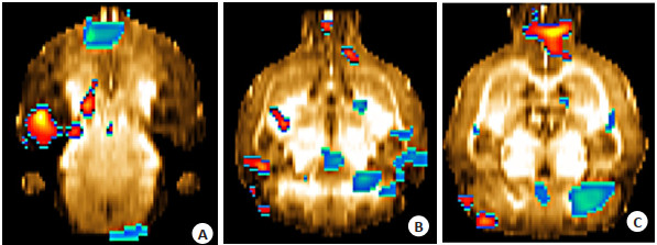
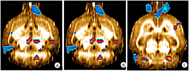
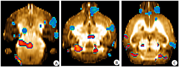
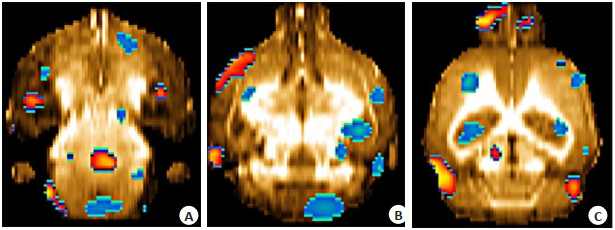
Objective To investigate the effects of Chaihu Shugan powder on the behavior of rats with liver-qi stagnation depression and to explore the potential antidepressant mechanism of Chaihu Shugan powder through the improvement of brain glucose metabolism. Methods Forty male Wistar rats were randomly divided into control group, model group, low-dose Chaihu Shugan powder group, and high-dose Chaihu Shugan powder group, with 10 rats per group. A rat model of liver-qi stagnation was established by using chronic restraint stress combined with solitary rearing. The high and low dose Chaihu Shugan powder groups were administered 1.04 g/mL and 0.52 g/mL concentrated solutions by gavage, respectively. The control and model groups were treated with normal saline by gavage once daily for 21 days from the beginning of the experiment. Results Behavioral indicators: Compared with the control group, the sucrose preference of the rats in the model group significantly decreased (P < 0.05), and the immobility time in the open field test significantly increased (P < 0.05). Brain metabolism indicators: The brain regions with increased metabolism in the model group were the bilateral medulla oblongata and left cerebellum (P < 0.05), while the brain regions with decreased metabolism included bilateral corpus callosum, bilateral M1, bilateral M2, left striatum, right thalamus, right hippocampus, right prefrontal cortex, right cingulate cortex, right S1, right S2, and right insular cortex (P < 0.05). In comparison with the model group, both high-and low-dose groups showed a significant increase in sucrose preference (P < 0.05) and a significant decrease in immobility time in the open field test (P < 0.05). In the low-dose group, the brain regions with increased metabolism encompassed bilateral striatum, bilateral hippocampus, bilateral corpus callosum, bilateral prefrontal cortex, left inferior colliculus, left M2, right auditory cortex, right thalamus, right M1, right S1, right S2, right insular cortex (P < 0.05), whereas the brain regions with decreased metabolism included left medulla, left visual cortex, and left cerebellum (P < 0.05). The high-dose group displayed increased metabolism in the right prefrontal cortex (P < 0.05). No significant differences were observed in other regions of interest (P>0.05). Conclusion The pathogenesis of liver-qi stagnation syndrome may involve chronic stress combined with isolation, resulting in decreased glucose metabolism in brain regions related to emotional cognition and behavioral regulation, leading to decompensation and concurrent glucose uptake competition in the left striatum and cerebellar brain regions. Chaihu Shugan powder can improve the behavior of rats with liver-qi stagnation depression by exerting its antidepressant effect primarily through enhancement of emotional cognition and behavioral-related brain regions, and ameliorating glucose uptake competition in the left striatum and cerebellar brain regions.
2023, 46(4): 583-590.
doi: 10.12122/j.issn.1674-4500.2023.04.02
Abstract:
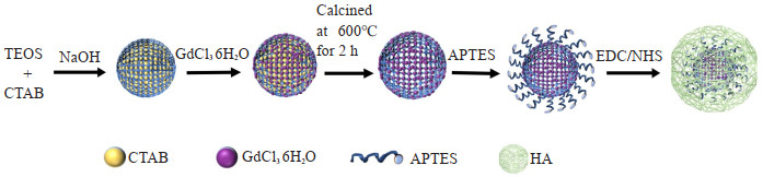
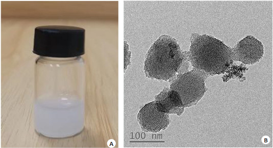
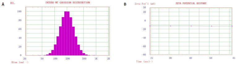
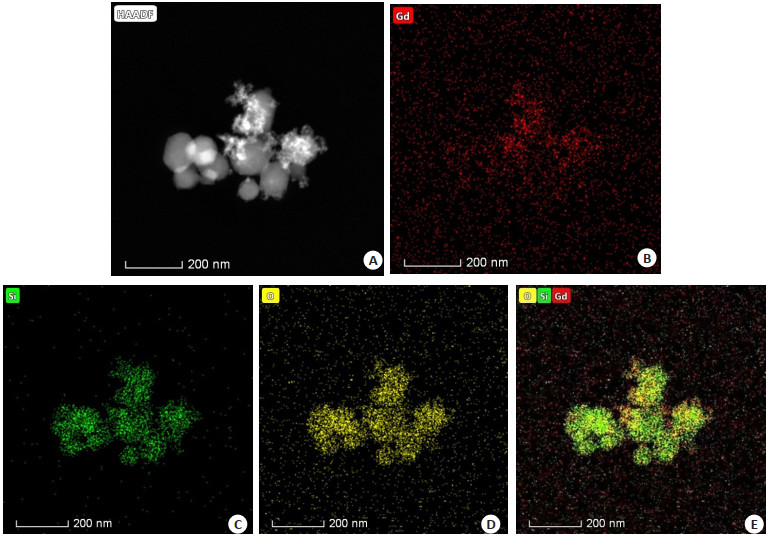
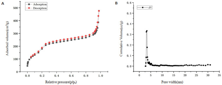
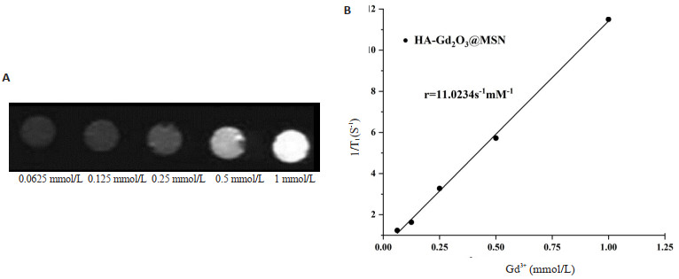
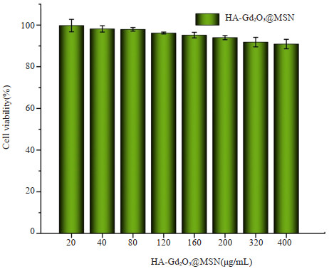
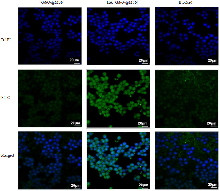
Objective To synthesize a hyaluronic acid (HA)-modified mesoporous silica nanoprobe (MSN) for MRI, which is HAGd2O3@MSN. Its performance and imaging characteristics were preliminarily studied, so as to provide a new technical tool for the diagnosis of atherosclerosis. Methods The cetyltrimethylammonium bromide (CTAB) was utilized as the template, and tetraethyl orthosilicate and CTAB were formulated into MSN under alkaline conditions. GdCl36H2O was incrementally introduced to produce the Gd2O3@MSN. Subsequently, the CTAB template was eliminated through calcination at 600 ℃ for 2 h, which led to the formation of the molecular probe Gd2O3@MSN. The HA-Gd2O3@MSN was then prepared through amide condensation. The morphology of HA-Gd2O3@MSN was examined by transmission electron microscopy, while its hydrodynamic diameter and zeta potential were measured by dynamic light scattering technique. The imaging results were observed by a 3.0T MR scanner and the relaxation rate of the molecular probe Gd2O3@MSN was analyzed using data from the inductively coupled plasma mass spectrometry testing. Results The hydrodynamic diameter of the synthesized probe HA-Gd2O3@MSN was 223.5±10.5 nm, the zeta potential of HA-Gd2O3@MSN was -13.04 mV, with its relaxation rate being 11.023 mmol· L-1· s-1. As the concentration of the molecular probe increased incrementally, the T1 signal was correspondingly enhanced. Through in vitro cellular studies, we demonstrated that HA-Gd2O3@MSN was capable to bind and target the CD44 receptors on the surface of macrophages in a HA-dependent manner. Furthermore, the HA-coated nanoprobes were found to be less toxic in cytotoxicity assays. Conclusion The nanoprobe HA-Gd2O3@MSN, with its high T1 relaxation rate, low cytotoxicity, excellent targeting capacity and good MR imaging enhancement effects, it lays the foundation for identification of atherosclerotic plaques at earlier stages.
2023, 46(4): 591-596.
doi: 10.12122/j.issn.1674-4500.2023.04.03
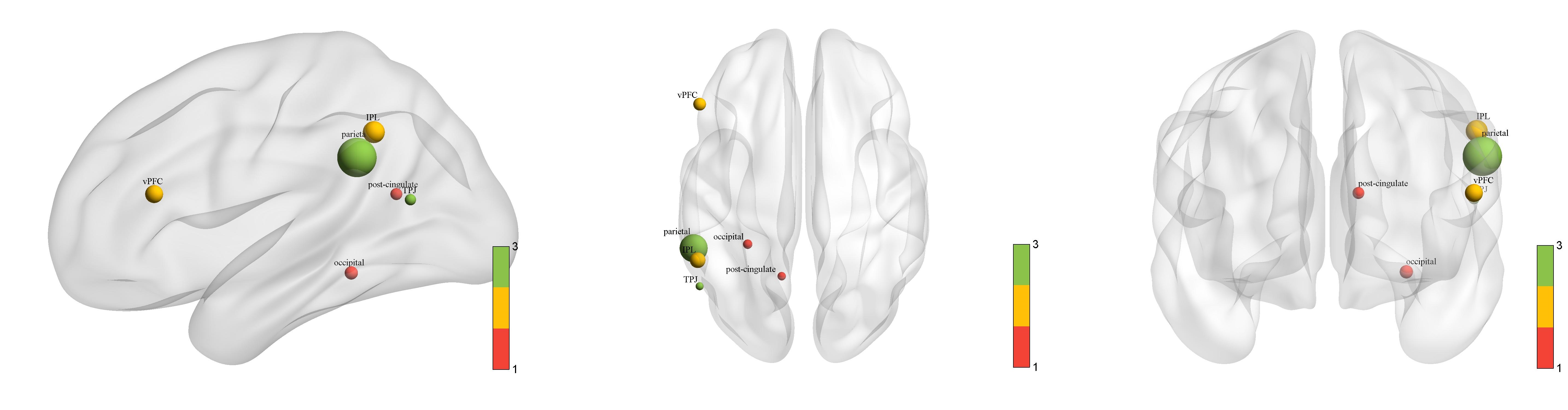
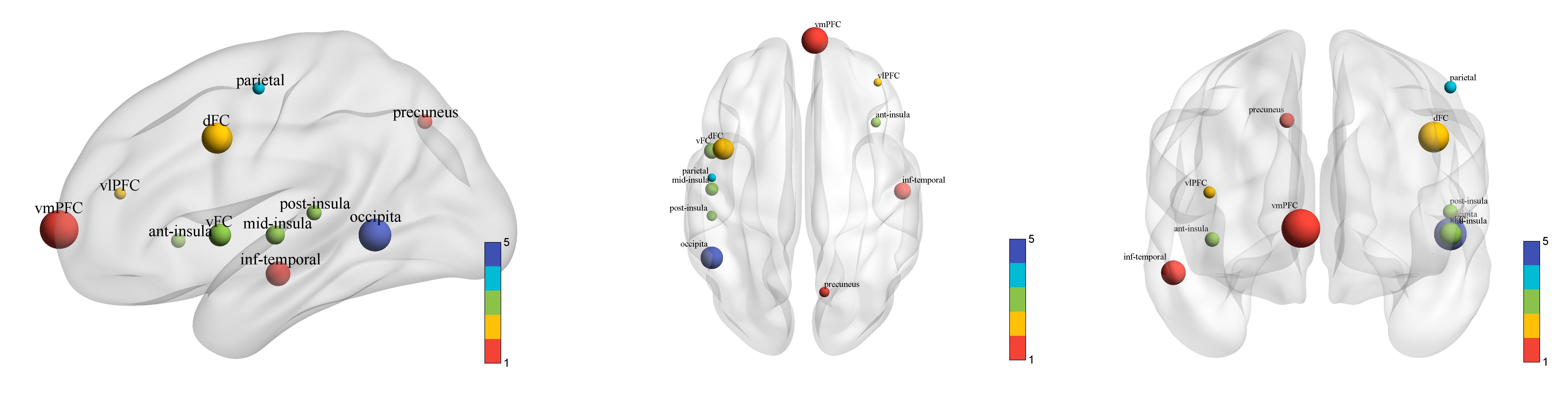
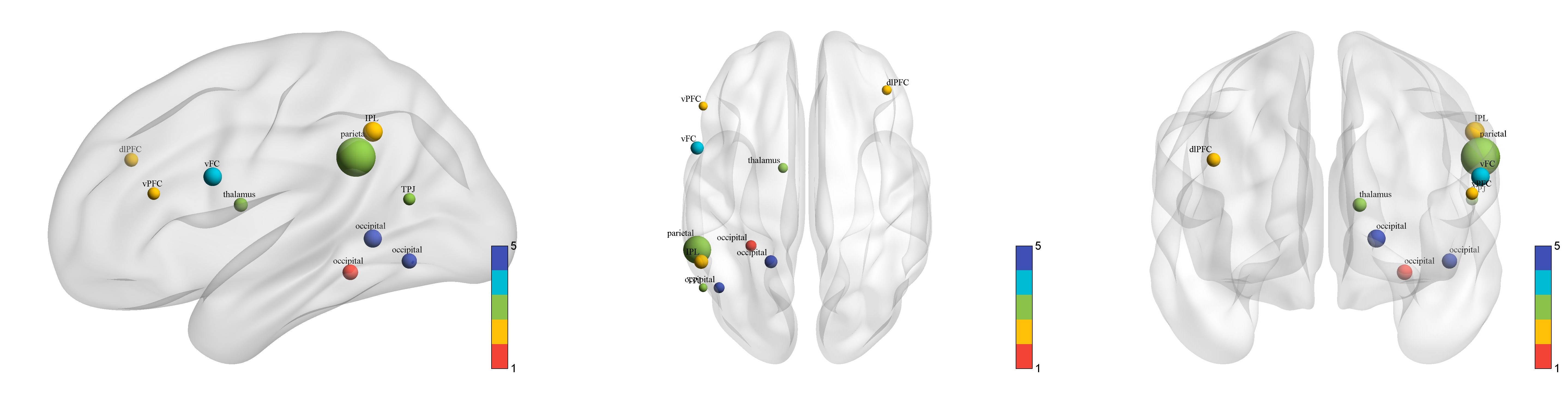
Abstract:
Objective To explore the changes in brain functional connectivity in patients with major depressive disorder of different genders by using resting- state functional magnetic resonance imaging. Methods A total of 106 patients with major depressive disorder were included, while 106 healthy control volunteers were recruited. After collecting resting fMRI images of all subjects, the data were preprocessed and functional network construction was constructed using GRETNA software. The network attributes were calculated, and the differences between groups were compared by two- sample t test (Boferroni multiple comparison correction). Results Compared with the control group, the global attributes of the experimental group increased, and there were statistical differences in the local efficiency of global attributes (P=0.0003) and small world (P=0.041) in women, and the global efficiency (P=0.0098) and local efficiency (P=0.0098) in men. The node clustering coefficients of the left posterior cingulate gyrus, left ventral prefrontal cortex, left parietal inferior gyrus, left parietal lobe, and left temporoparietal junction cortex in female patients with depression were significantly lower than those in men (P < 0.05). The nodality values of the right inferior temporal gyrus, right ventral lateral pre frontal cortex, left dorsal frontal cortex, right anterior insulin, left ventral frontal cortex, left/right middle insulin, left posterior insulin, and left parietal lobe were lower than those of men (P < 0.05). The local efficiency of the nodes of the left post cingulate, right dorsolateral prefrontal cortex, left ventral prefrontal cortex, left parietal gyrus, left parietal gyrus, left parietal region, left temporoparietal junction cortex, and left occipital lobe were lower in female depression than in men (P < 0.05). Conclusion The decline area of female depressed patients is mainly the emotional control and visual control area. The main decline area of male depression patients is the pressure control and reward control area. This has the potential to be a neural mechanism for gender differences.
2023, 46(4): 597-604.
doi: 10.12122/j.issn.1674-4500.2023.04.04
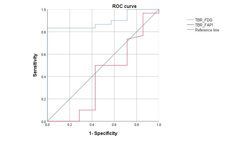
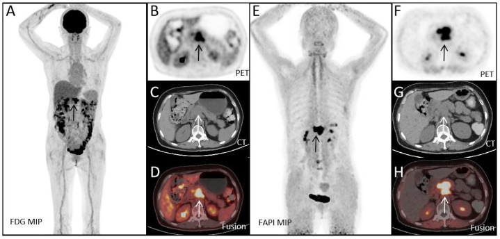
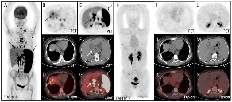
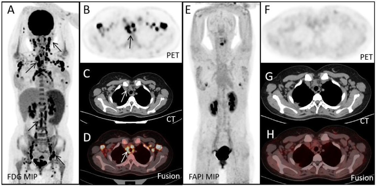
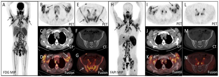
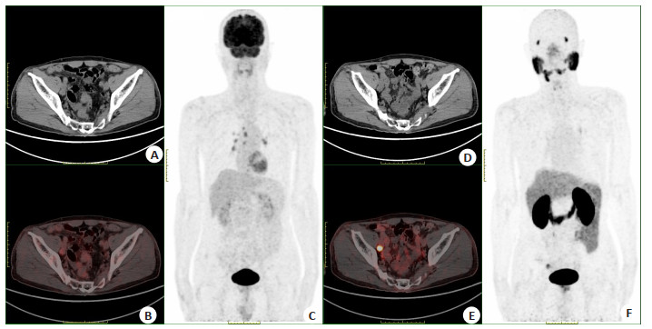
Abstract:
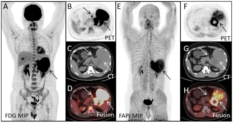
Objective To compare the diagnostic value of 18F-FDG PET/CT and 68Ga-FAPI PET/CT in lymphoma and explore the application prospect of 68Ga-FAPI PET/CT in lymphoma diagnosis. Methods A retrospective analysis was performed on 37 patients with lymphoma who were undiagnosed from January 2020 to December 2022 in our hospital. The differences in maximum standard uptake value (SUVmax) and target-to-background ratio (TBR) values between 18F-FDG PET/CT and 68Ga-FAPI PET/CT were compared. The correlation between SUVmax and TBR values of the two examination methods and Ki67 was analyzed. The differences between the two examination methods in lymphoma staging and infiltration diagnosis were compared. Results Among the 37 patients, 30 patients were diagnosed with lymphoma. The diagnostic efficiency of 18F-FDG PET/CT was higher than that of 68Ga-FAPI PET/CT. In lymphoma patients, SUVmax-FDG>SUVmax-FAPI (17.35 vs 4.80)(P < 0.05). In lymphoma patients, TBR-FDG>TBR-FAPI (29.35 vs 7.05)(P < 0.05). There was a significant positive correlation between SUVmax-FDG, TBR-FDG and Ki67 (SUVmax-FDG: R2=0.28, P < 0.05;TBR-FDG: R2=0.19, P < 0.05), while there was no significant correlation between SUVmax-FAPI, TBR-FAPI and Ki67 (P>0.05). In detecting lymphoma infiltration, 18F-FDG PET/CT was superior to 68Ga-FAPI PET/CT. Conclusion The SUVmax and TBR values of 18F-FDG PET/CT for diagnosing lymphoma are higher than those of 68Ga-FAPI PET/CT. They have better efficacy in the diagnosis and staging of lymphoma, providing improved guidance for clinical diagnosis and treatment, but 68Ga-FAPI PET/CT still plays a significant role in the diagnosis of lymphoma and has important clinical value.
2023, 46(4): 605-608.
doi: 10.12122/j.issn.1674-4500.2023.04.05
Abstract:
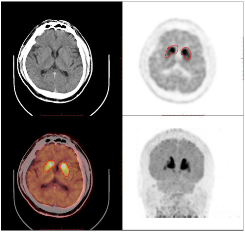
Objective To investigate the clinical value of PET/CT imaging of cerebral dopamine transporter imaging agent 11C-methyl-N-2β-carbomethoxy-3β-(4-flurophenyl) tropanel(11C-β-CFT) in the early diagnosis of Parkinson's disease. Methods The 11C-CH3I generated by the first step of the reaction was converted into Triflate-11CH3 online and the precursor 2β-carbomethoxy-3β-(4-flurophenyl) demethylatropanel was reacted at room temperature, and after the reaction was completed, the reaction solution was transferred to the C18 column with water for purification, and the product 11C-β-CFT was eluted from the C18 column with ethanol and mixed with water to obtain the final product. Five patients with mild Parkinson's disease underwent brain PET/CT imaging about 60 minutes after intravenous injection of 11C-β-CFT. Results The total synthesis time of 11C-β-CFT was about 16 minutes, the clarification purity>98%, the specific activity>2 GBq/μmol, and the uncorrected synthesis yield was (25.0±5.0)%. Brain PET/CT in patients with mild Parkinson's disease showed a significant decrease in bilateral uptake of 11C-β-CFT in the anterior and posterior shell nuclei, most notably in the diseased side of the crustal nucleus. Conclusion The synthesis speed, high yield, and purity and specific activity of the fully automated preparation of 11C-β-CFT can meet the clinical needs. The above imaging results show that the imaging agent can accurately reflect the condition and help in the early diagnosis of Parkinson's disease.
2023, 46(4): 609-613.
doi: 10.12122/j.issn.1674-4500.2023.04.06
Abstract:
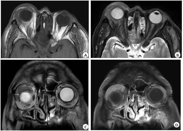
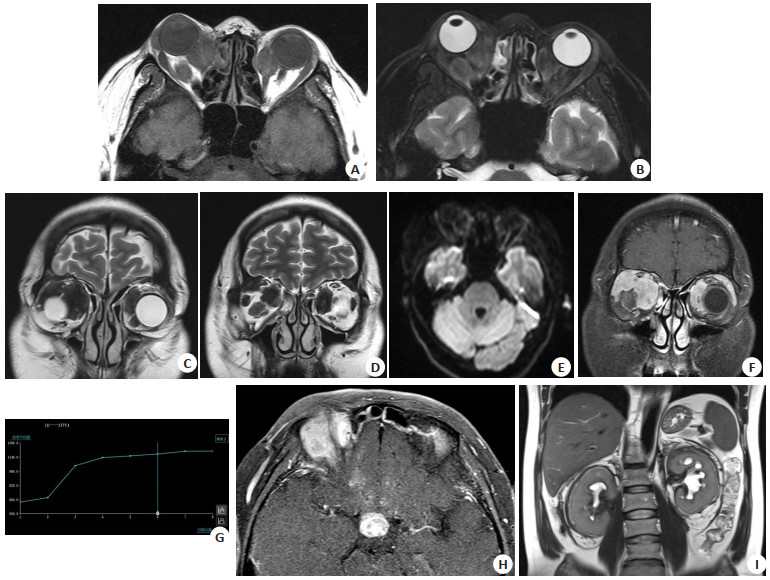
Objective To analyze the MRI features of adult orbital xanthogranuloma and improve recognition of the disease. Methods The MRI images of 11 patients were retrospectively analyzed. We analyzed the location, unilateral or bilateral involvement, shape, margins, orbital structures, T1WI and T2WI signal characteristics, diffusion-weighted imaging characteristics, apparent diffusion coefficient and enhancement patterns. Results Eleven cases of orbital xanthogranuloma were confirmed by surgical pathology, including 6 cases of adult-onset xanthogranuloma, 4 cases of necrobiotic xanthogranuloma, and 1 case of Erdheim-Chester disease. Of the 6 cases of adult-onset xanthogranuloma, 3 involved unilateral eyelids, 1 involved the right nasolacrimal duct, 1 involved bilateral eyelids and lacrimal glands and 1 involved right eyelid, pterygopalatine fossa and left temporal muscle. Of the 4 cases of necrobiotic xanthogranuloma, 2 involved the left eyelid, and 2 involved bilateral eyelids, nasal and temporal regions. The case of Erdheim-Chester disease involved multiple bilateral orbital lesions and also affected the pituitary gland, pericardium, and kidneys. The lesions of 4 cases had clear boundaries, while those of 7 cases had diffuse boundaries. Ten cases showed low signal intensity on T1WI, high signal intensity on T2WI and fatsuppressed imaging, and 1 case showed low signal intensity on T1WI, T2WI and fat-suppressed imaging. Diffusion-weighted imaging showed low signal intensity (n=7). The value of apparent diffusion coefficient was 1.18×10⁻³ mm2/s (n=7). Dynamic contrast- enhanced MRI showed continuous rising patterns (type Ⅰ) in all 6 cases. Conclusion Orbital xanthogranuloma typically presents as eyelid swelling without ulceration, involving eyelids and subcutaneous tissue, the anterior and posterior orbit, and lacrimal glands, and may involve extraocular muscle tendons but not optic nerves. No bone destruction was observed in the involved orbit. MRI can assist in the diagnosis of this disease by displaying chemical shift artifacts.
2023, 46(4): 614-619.
doi: 10.12122/j.issn.1674-4500.2023.04.07
Abstract:
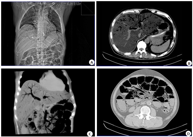
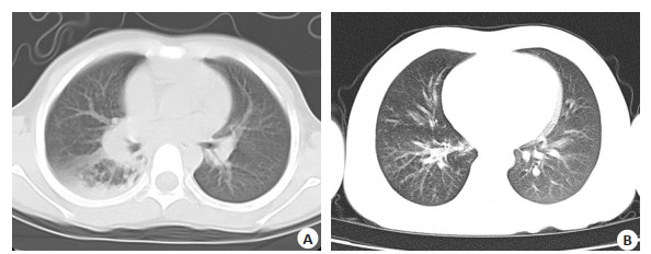
Objective To investigate the imaging and clinical features of hepatic portal vein gas (HPVG). Methods The data of 10 HPVG patients in the Second People's Hospital of Lianyungang, Zhongda Hospital Southeast University from June 2012 to April 2020 were analyzed retrospectively. The imaging and clinical features, treatment and prognosis were summarized. Results Acute gastrointestinal symptoms occurred in 7 cases and fever in 5 cases. Renal insufficiency and maintenance hemodialysis in 3 cases, 8 cases of hypertension, 6 cases of surgical history. In clinical diagnosis, 5 cases of intestinal obstruction, 6 cases of severe infection and septic shock. Only 3 of the 10 patients survived. Both patients treated with surgery were diagnosed with intestinal necrosis. The gas in portal vein was mainly distributed in the left branch of portal vein. There were 7 cases of pneumatosis of splenic vein, superior mesenteric vein and its tributaries, and 9 cases of pneumatosis of intestinal wall. There were 7 cases of local intestinal wall edema, 5 cases of intestinal obstruction and 3 cases of digestive tract perforation. Enhanced scan confirmed superior mesenteric artery thrombosis in 2 cases. Conclusion The incidence of pneumatosis cystoides intestinalis in HPVG cases is high. Clinical attention should be paid to the identification of HPVG and related imaging features, so as to intervene in patients in time.

2023, 46(4): 620-626.
doi: 10.12122/j.issn.1674-4500.2023.04.08
Abstract:
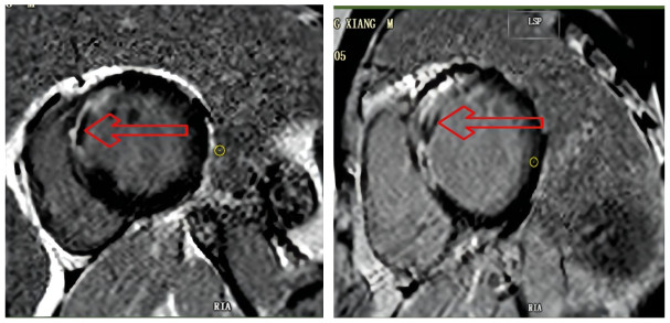
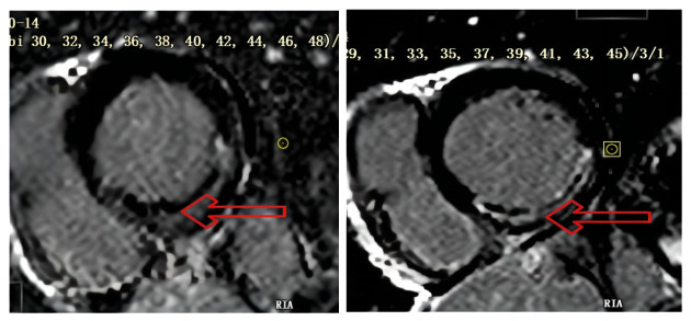
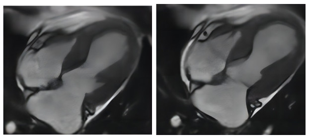
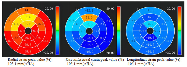
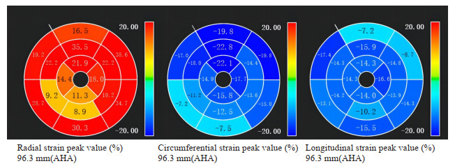
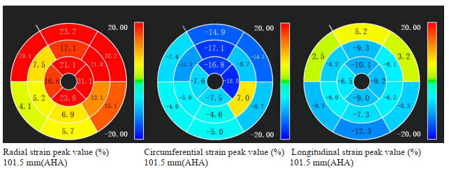
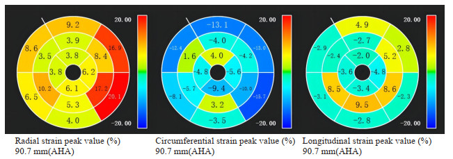
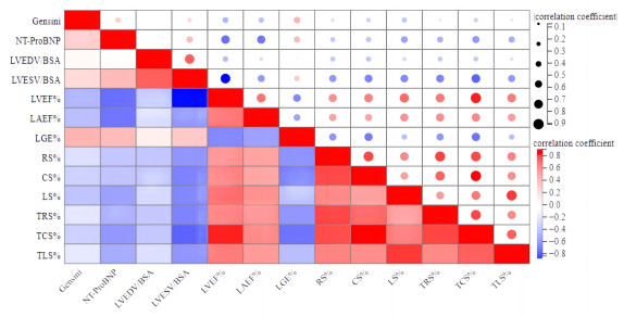
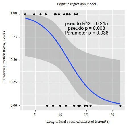
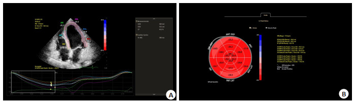
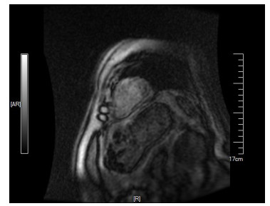
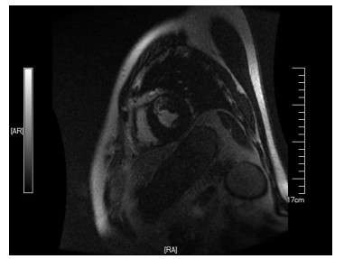
Objective To explore the value of cardiac magnetic resonance imaging in evaluating early cardiac function in patients with acute myocardial infarction. Methods The study included 24 patients with acute ST-segment elevation myocardial infarction who were admitted to our department from June 2022 to December 2022, including 22 male patients and 2 female patients, with the age at 55.3±11.3 years old. All patients underwent coronary intervention followed by cardiac magnetic resonance imaging 5-7 d after the operation. Motion picture imaging was used to analyze cardiac function status, presence or absence of reverse motion and ventricular aneurysm. The strain capacity of each myocardium segment was analyzed by tissue tracking technique. The location, size and microcirculation disturbance of myocardial infarction were analyzed by delayed enhancement technique. Results Myocardial strain analysis revealed that 21 patients were found to have decreased radial strain and longitudinal strain among the 24 patients, 18 had decreased circumferential strain, and 16 showed a reduction in the three-dimensional myocardial strain. According to the overall analysis of the myocardial strain revealed that 15 patients revealed decreased radial strain, 10 had decreased circumferential strain, 20 had decreased longitudinal strain, and 9 showed a reduction in three-dimensional myocardial strain. The mean radial strain and circumferential strain of the MI segment were significantly lower than that of the average radial strain and circumferential strain of the entire myocardium (P < 0.05), but no statistically significant difference was observed in the longitudinal strain (P > 0.05). Moreover, 13 patients were observed to have myocardial reverse movement. There were 10 and 14 patients with and without decreased left ventricular ejection fraction, respectively, among which 6 had myocardial reverse movement, 6 showed a decrease in three-dimensional myocardial strain in the infarcted segment, 7 presented microcirculatory disturbance within the infarct zone, and 7 had increased level of NT-proBNP. The mean circumferential and longitudinal strains of left ventricular ejection fraction, infarct segment myocardium and global myocardium in patients with reverse motion were all smaller than those without reverse motion (P < 0.05). Correlation analysis indicated that left ventricular ejection fraction was positively correlated with the overall myocardial strain, myocardial strain in the infarcted segment and left atrial emptying fraction. It was negatively correlated with infarct size, left ventricular end-systolic volume/body surface area ratio, NT-proBNP level and Gensini score (P < 0.05). The binary Logistic regression analysis showed that the reduction in the longitudinal strain in the infarcted segments could independently predict whether patients possessed myocardial reverse movement. Conclusion Cardiac magnetic resonance imaging provides a comprehensive evaluation of the cardiac function status, which will be a reference for patients with acute myocardial infarction, offering valuable insights into the improvement of early post-operative cardiac function assessment, treatment, intervention and prognosis.
2023, 46(4): 627-631.
doi: 10.12122/j.issn.1674-4500.2023.04.09
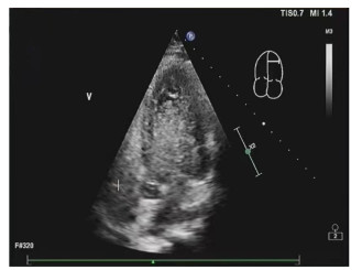
Abstract:
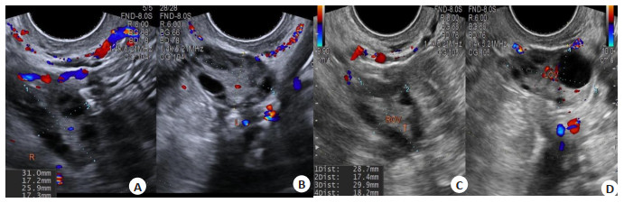
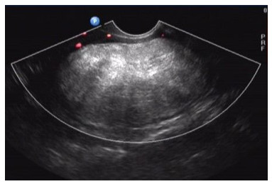
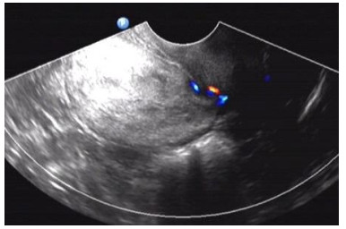
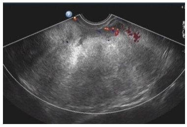
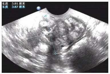
Objective The development of follicles in infertile patients with polycystic ovary syndrome was dynamically monitored by transvaginal ultrasonography, and the clinical value of combined acupuncture and drug therapy was discussed to provide further guidance for clinical treatment. Methods forty-four cases of polycystic ovary syndrome women admitted to our hospital from September 2020 to September 2022 were retrospectively studied. A total of 44 cases were divided into two groups according to the combination of medicine and acupuncture, of which 21 patients were treated with conventional western medicine (control group) and 23 patients were treated with acupuncture combined with conventional Western medicine (observation group). Ovulation, clinical pregnancy and imaging manifestations were observed in 2 groups within 3 months after treatment. Results After 3 months of treatment, the total effective rate of the control group and the observation group was 66.67% and 82.60%, the ovulation rate was 52.38% and 78.26%, the pregnancy rate was 28.57% and 43.48%, respectively. The ovarian volume, number of follicles and maximum follicle diameter in the observation group were better than those in the control group (P < 0.05). The ovulation success rate and pregnancy rate in the observation group was higher than those in the control group. The differences were statistically significant (P < 0.05). Conclusion Under the dynamic monitoring of transvaginal ultrasound, the follicular development of patients can be accurately observed, which provides guidance for clinical diagnosis and treatment of polycystic ovary syndrome infertility patients, effectively improves the pregnancy rate of patients.
2023, 46(4): 632-637.
doi: 10.12122/j.issn.1674-4500.2023.04.10
Abstract:
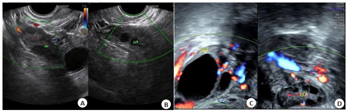
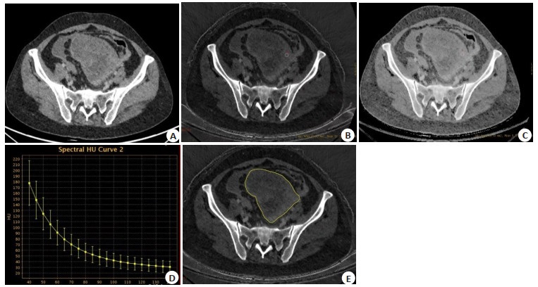
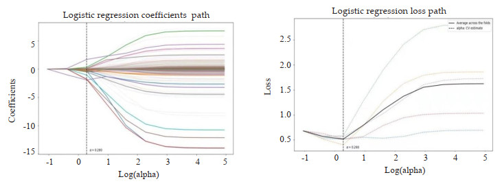
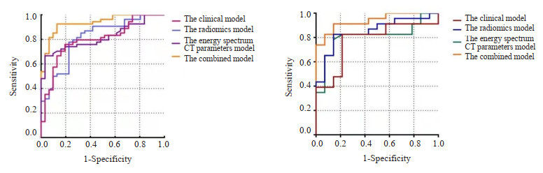
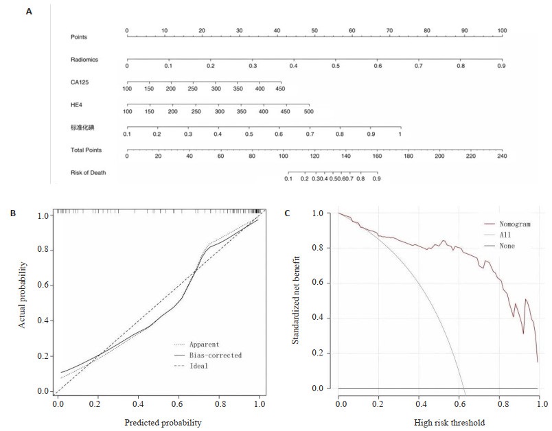
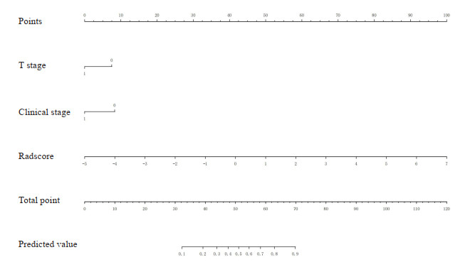
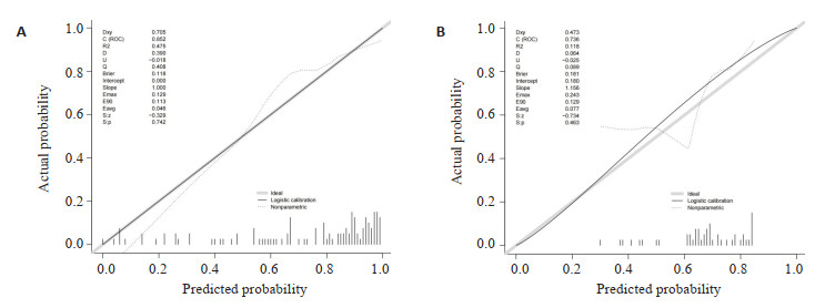
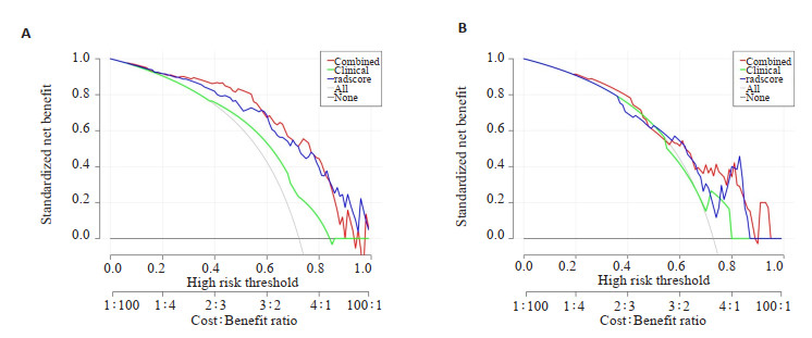
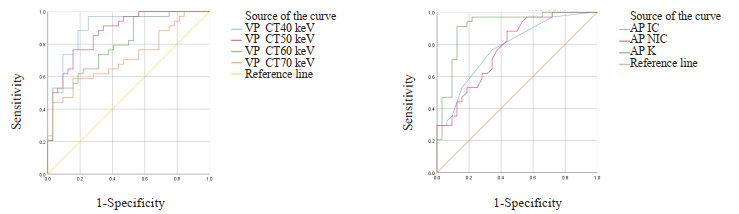
Objective To explore the value of nomogram combined with radiomics analysis from iodine overlay maps, spectral CT and clinical features in prediction of the type of epithelial ovarian cancer. Methods We retrospectively analyzed 122 patients (including 46 patients of typeⅠ and 76 patients of typeⅡ) with epithelial ovarian cancer pathologically confirmed underwent contrast enhanced spectral CT scan. The clinical characteristic model were constructed combined with serum CA125 and HE4. Iodine concentration (IC), normalized iodine concentration (NIC), slope of spectrum energy curve (λ), effective-Z (Zeff), normalized effective-Z (NZeff) of the region of interest under the venous phase image were compared. The significant energy spectrum parameters were selected by Logistic regression analysis, and then constructed the energy spectrum CT parameters model. Radiomics features were extracted from iodine overlay maps in the arteriovenous phase. Patients were randomized devided into training group (n=85) and test group (n=37) set in a ratio of 7:3. After dimensionality reduction of the data set, effective features were screened out and then construct the radiomics model. The diagnostic efficiency of the models were evaluated by using the ROC curve and the area under the curve (AUC). The clinical application value of normograph was evaluated by using the decision curve analysis and correction curve. Results Serum CA125 and HE4 were significantly different between two groups. A clinical model was established by combining the two methods, and the areas under the curve were respectively 0.797(95% CI: 0.700-0.895) for the training group 0.776(95% CI: 0.620-0.933) for the test group. λ40-70 keV, λ40-100 keV, IC, NIC, Zeff, NZeff were significantly different (P < 0.05). Binary Logistic regression analysis suggested that NIC was an independent factor (P=0.008). The AUC of NIC was 0.813(95% CI: 0.723-0.902) for the training group and 0.837(95% CI: 0.707-0.966) for the test group. Eighteen features were screened by imaging omics, including six first-order features, eight gray-scale features, one shape features and three wavelet features. The AUC was 0.825(95% CI: 0.733-0.917) for the training group and 0.851(95% CI: 0.725-0.796) for the test group. The diagnostic efficiency of the combined model was higher than of the single model. The AUC was 0.935(95% CI: 0.885-0.986) for the training group and 0.938(95% CI: 0.865-1.000) for the test group. Conclusion Clinic-spectral CT-radiomics nomogram have potential value in predicting the typing of epithelial ovarian cancer.
2023, 46(4): 638-642.
doi: 10.12122/j.issn.1674-4500.2023.04.11
Abstract:
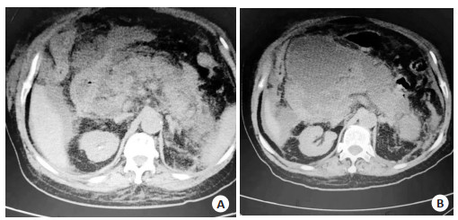
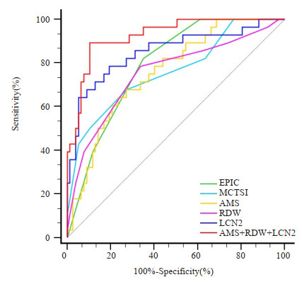
Objective To explore the relationship between CT imaging of severe pancreatitis and disease progression, serum amylase (AMS), red blood cell distribution width (RDW) and lipocain-2 (LCN2). Methods Clinical data of 105 patients with severe pancreatitis from March 2019 to December 2022 were retrospectively collected. The patients were divided into good prognosis group (n=77) and poor prognosis group (n=28) according to the clinical prognosis results of patients. CT imaging results [extra-pancreatic inflammation on CT (EPIC), modified CT severity index (MCTSI)] and serum AMS, RDW and LCN2 levels were compared between the two groups of patients. ROC curve was used to analyze the evaluated value of each indicator on prognosis of severe pancreatitis. Results The number of cases with organ failure in poor prognosis group was more than that in good prognosis group (P=0.039), and the APACHE Ⅱ score was higher than that in good prognosis group (P=0.001). EPIC score and MCTSI score were higher in poor prognosis group than those in good prognosis group (P < 0.001), and serum AMS, RDW and LCN2 levels were higher compared with those in good prognosis group (P < 0.001). Correlation analysis showed that EPIC score and MCTSI score were positively correlated with serum AMS, RDW and LCN2 levels (r=0.591, 0.668, 0.684 and 0.573, 0.637, 0.652, P < 0.001). The areas under the curves of EPIC score and MCTSI score on evaluating the prognosis of severe pancreatitis were 0.791 and 0.762, and the sensitivities were 82.14% and 67.86% and the specificities were 64.94% and 72.73%. The areas under the curves of AMS, RDW and LCN2 were 0.758, 0.754, 0.851, and the sensitivities were 64.29%, 78.57%, 78.57, the specificities were 76.62%, 66.23%, 80.52% respectively. The area under the curve, sensitivity and specificity of the combination of AMS, RDW and LCN2 on assessing the prognosis of severe pancreatitis were 0.925, 92.86% and 79.22%. Conclusion High EPIC score, MCTSI score and serum AMS, RDW and LCN2 levels in patients with severe pancreatitis are associated with poor prognosis. The above CT scores are positively correlated with serological indicators. All the indicators can be used to evaluate the poor outcomes after disease progression. The combination of serological indicators has the highest efficiency.
2023, 46(4): 643-647.
doi: 10.12122/j.issn.1674-4500.2023.04.12
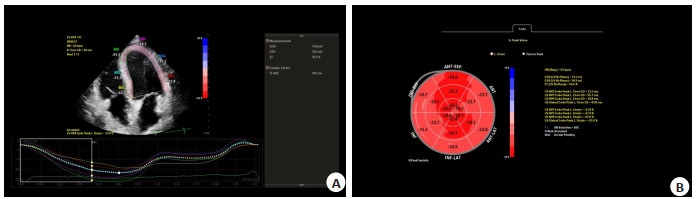
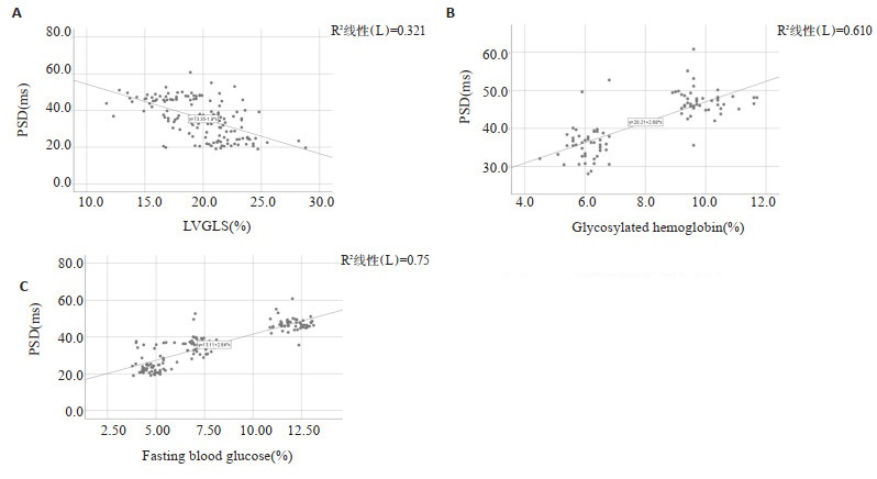
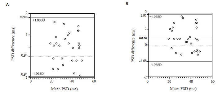
Abstract:
Objective To investigate the early changes of left ventricular systolic function in patients with type 2 diabetes mellitus using peak strain dispersion (PSD). Methods Ninety-five patients with type 2 diabetes mellitus were enrolled. They were divided into two groups based on their HbA1c levels: controlled blood glucose group (group A, HbA1c < 7%, n=45) and uncontrolled blood glucose group (group B, HbA1c≥7.0%, n=50). An additional 50 healthy subjects were selected as control group. Conventional echocardiography was performed, and parameters were obtained. The left ventricular global longitudinal strain and PSD were obtained using two-dimensional dynamic images and tracing analysis. The differences in parameters between the groups were compared. Results There was no significant difference in left ventricular global longitudinal strain between group A and the control group (P=0.08), while left ventricular global longitudinal strain in group B was significantly reduced compared with the control group (P < 0.01). Compared with the control group, PSD was significantly increased in group A and group B (P < 0.01). Correlation analysis revealed that left ventricular global longitudinal strain was negatively correlated with PSD, while glycosylated hemoglobin and fasting blood glucose were positively correlated with PSD (P < 0.01). Conclusion PSD can be used to quantitatively evaluate changes in left ventricular systolic function in patients with type 2 diabetes mellitus. PSD is an early and reliable indicator for evaluating left ventricular myocardial movement in patients with diabetes.
2023, 46(4): 648-653.
doi: 10.12122/j.issn.1674-4500.2023.04.13
Abstract:
Objective To investigate the correlation between tumor burden, metastasis and serum prostate specific antigen (PSA) in prostate cancer patients based on the dual-probe PET/CT imaging of 18F-fluorodeoxyglucose (18F-FDG) and 18F-prostate specific membrane antigen (18F-PSMA). Methods Fifty-four patients with primary prostate cancer diagnosed by pathological histology from March 1st, 2021 to November 27th, 2022 in Weifang People's Hospital. The patients underwent 18F-FDG and 18F-PSMA dual probe PET/CT. Based on the dual-probe PET/CT examination and clinical data, the patients were divided into metastasis-free group (n=16) and metastasis group (n=38), and the tumor load indexes determined by dual-probe PET/CT examination were counted in all patients. Spearman test was used to analyze the correlation between serum PSA and tumor burden index, and Mann-Whitney U test was used to compare between groups. Results The differences of serum PSA, metabolic tunour volume, volume of PSMA, total lesion glycolysis and total lesion uptake value of PSMA between the two groups were statistically significant (P < 0.05). The efficacy of detecting prostate cancer metastasis was best at serum PSA > 31.83 μg/L, with sensitivity and specificity of 84.21% and 93.75%, respectively. The tumor burden obtained by dual-probe PET/CT were significantly correlated with serum PSA (P < 0.01). Conclusion The higher the serum PSA, the greater the tumour load of prostate cancer. The tumour load of the metastatic group is higher than that of the non-metastatic group. Patients with a serum PSA > 31.83 μg/L indicates a high probability of metastatic prostate cancer and necessitated a comprehensive assessment with 18F-PSMA PET/CT imaging.
2023, 46(4): 654-660.
doi: 10.12122/j.issn.1674-4500.2023.04.14
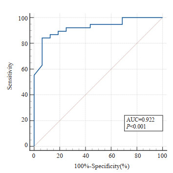
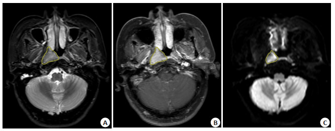
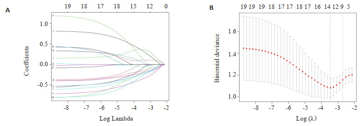
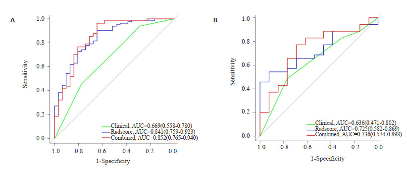
Abstract:
Objective To investigate the predictive value of nomogram based on the radiomics features of multimodal magnetic resonance combined with clinical information in the efficacy of chemoradiotherapy in advanced nasopharyngeal carcinoma. Methods The imaging and clinical data of 160 patients with pathologically confirmed nasopharyngeal carcinoma at first diagnosis were retrospectively analyzed. Patients were divided into training group (n=112) and validation group (n=48) according to a ratio of 7:3. In the training group, the radiomics features of T2-weighted fat suppression sequence, T1-weighted enhancement sequence and diffusion-weighted imaging sequence images were extracted, and the most effective features were filtered to construct the radiomics prediction model after dimensionality reduction of the LASSO data. The relevant clinical information were incorporated, and the most valuable clinical information were screened a clinical information model constructed using logistics regression. The clinical information model was combined with the radiomics feature model to construct a combined model, and the nomogram was constructed. The diagnostic efficacy of each model was assessed by the ROC curve and area under the curve. The clinical application value of the nomogram was assessed by decision curve analysis and calibration curve. Results The clinic-radiomics model constructed by combining two clinical information and nine radiomics features showed good predictive efficacy with AUCs of 0.852 (95% CI : 0.765-0.940) and 0.736(95% CI : 0.574-0.898)in the training and validation groups, respectively. Conclusion Multimodal MRI-based radiomics nomograms are feasible in predicting the efficacy of chemoradiotherapy in advanced nasopharyngeal carcinoma and have good clinical application.
2023, 46(4): 661-668.
doi: 10.12122/j.issn.1674-4500.2023.04.15
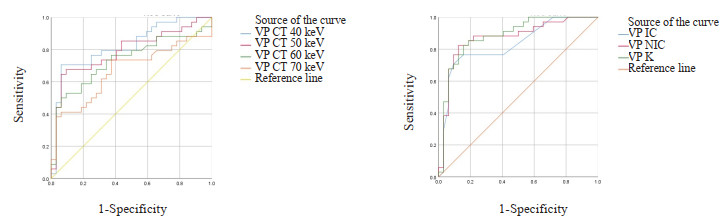
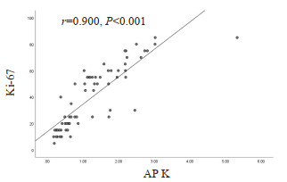
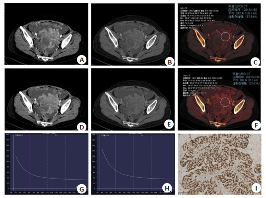
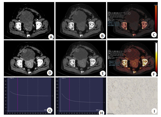
Abstract:
Objective To explore the value of non-invasive assessment for the proliferative ability of ovarian high-grade serous carcinoma tumor cells using dual-energy CT multiple quantitative parameters. Methods We retrospectively analyzed 66 cases of high-grade serous ovarian carcinoma confirmed by surgery at the First People's Hospital of Yancheng City from June 2021 to January 2023. All patients underwent spectral CT plain and enhanced scans within 2 weeks prior to surgery. After surgery, all tumor tissue pathological specimens were stained with Ki-67 immunohistochemistry. According to postoperative pathological findings, patients were classified into Ki-67 high expression group (n=34) and Ki-67 low expression group (n=32). The measurement of arterial and venous phase single-energy CT values (40-90 keV) and iodine concentration of the lesions were independently conducted by two observers. Normalized iodine concentration and energy spectrum curve slope (K40-90 keV) were calculated. Intra-group correlation coefficient was used to evaluate the consistency and correlation between the measurement parameters of the two observers. The differences in various parameters between the two groups were compared. ROC curves were calculated to evaluate the diagnostic efficacy of the related parameters with statistically significant differences. The correlation between the parameters with statistically significant differences and Ki-67 expression was analyzed. Results Two observers showed good consistency in the measurement of various parameters in the arterial and venous phases, with all the intra-group correlation coefficient was greater than 0.75. The low expression group had lower arterial and venous phase 40-70 keV single-energy CT values, iodine concentration, normalized iodine concentration and K40-90 keV than the high expression group (P < 0.05). ROC curve analysis showed that the diagnostic efficacy of arterial phase K40-90 keV was the highest, with an AUC of 0.913, sensitivity of 91.2%, specificity of 87.5%, and a threshold of 0.95. The above parameters were also correlated with Ki-67 expression, with the highest correlation coefficient observed between arterial phase K40-90keV and Ki-67 (rs=-0.900, P < 0.001). Conclusion Dual-energy CT can non-invasively evaluate the proliferative ability of high-grade serous ovarian carcinoma cells using multiple quantitative parameters.
2023, 46(4): 669-673.
doi: 10.12122/j.issn.1674-4500.2023.04.16
Abstract:
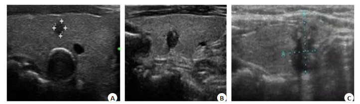
Objective To investigate the diagnostic value of multi-gene detection for thyroid nodules with unclear significance by fine needle aspiration (FNA). Methods Fifty-six cases of atypical cell lesions or follicular lesions with unclear significance diagnosed by FNA of thyroid C-TIRADS 4 nodules in the Fourth Hospital of Hebei Medical University from August 2018 to May 2020 were selected for multi-gene detection. The results of postoperative pathological diagnosis were used as the 'gold standard'. Among the 56 cases of nodules with unclear diagnostic significance of FNA, there were 42 cases of TBSRTC grade Ⅲ, 12 cases of TBSRTC grade Ⅳ and 2 cases of TBSRTC grade Ⅴ, all of which were reported by genetic test results. The sensitivity, specificity, accuracy, positive predictive value and negative predictive value of BRAF V600E mutation results and multi-gene detection results were compared. Results Among the 56 cases of FNA with unclear diagnostic significance, postoperative pathological diagnosis was benign in 2 cases (1 case of nodular goiter, 1 case of thyroid adenoma), and postoperative pathological diagnosis was malignant in 54 cases (50 cases of papillary thyroid carcinoma, 4 cases of medullary thyroid carcinoma). The diameter of the nodules was 22.5±23.3 mm in C-TIRADS 4A, including 2 cases of nodules with a diameter≤10 mm. The diameter of the nodules was 8.0±7.1 mm in C-TIRADS 4B, including 14 cases of nodules with a diameter≤10 mm. The diameter of the nodules was 7.0±6.2 mm in C-TIRADS 4C, including 36 cases of nodules with a diameter≤10 mm. The sensitivity, specificity, accuracy, positive predictive value and negative predictive value of BRAF single gene detection in the diagnosis of benign and malignant nodules with unclear significance were 60.0 %, 25.0 %, 56.7 %, 91.3 % and 3.45 %, respectively. The sensitivity, specificity, accuracy, positive predictive value and negative predictive value of multi-gene detection combined with C-TIRADS classification in the diagnosis of benign and malignant nodules with unclear significance were 80.0%, 86.4%, 75.7%, 93.3% and 65.5%, respectively. Multi-gene detection method has higher diagnostic accuracy than BRAF single gene detection. Conclusion Multi-gene detection is helpful to clarify the diagnosis of thyroid FNA nodules with unclear significance.
2023, 46(4): 674-681.
doi: 10.12122/j.issn.1674-4500.2023.04.17
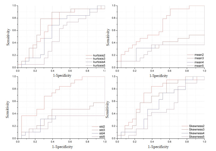
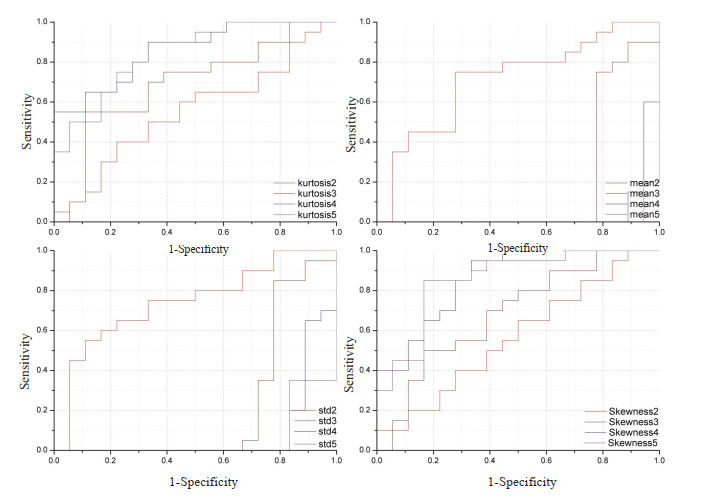
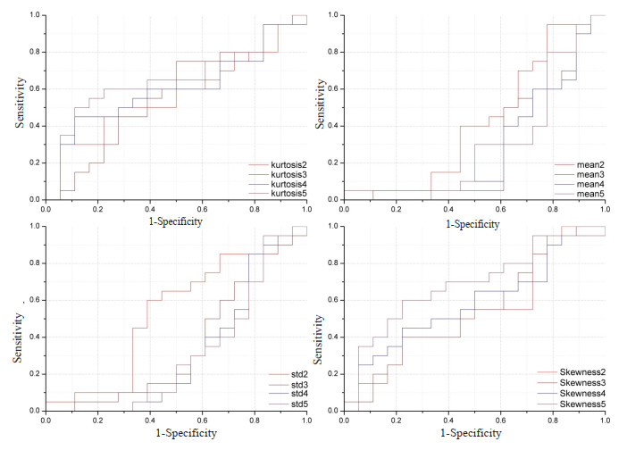
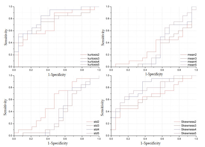
Abstract:
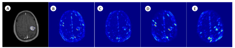
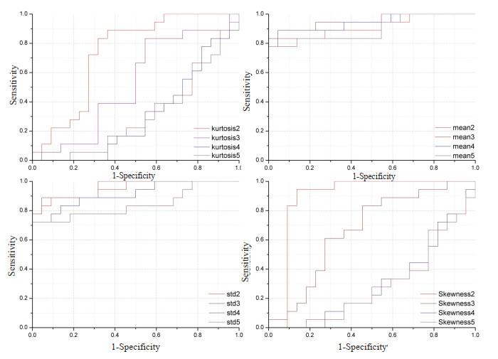
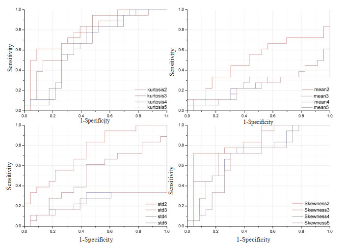
Objective To propose a histogram feature based on multi-scale multimodal magnetic resonance images and implementing a machine learning approach for the grading of gliomas. Methods sixty clinical cases of glioma were collected, including 22 cases of grade Ⅱ glioma (diffuse astrocytoma and oligodendroglioma), 18 cases of grade Ⅲ glioma (anaplastic oligoastrocytoma and anaplastic astrocytoma) and 20 cases of grade Ⅳ glioma (glioblastoma). Case image information included T2-weighted sequence, T2-weighted sequence with water pressured and fat pressured, contrast-enhanced T1-weighted sequence. Multi-scale processing was performed on the three sequence images, and texture analysis was performed on the multi-scaled images. Taking the core area of the lesion as the area of interest, the texture parameters were calculated, and the intrinsic correlation between the texture parameters and glioma was explored, ROC was used to analyze the texture parameters between grade Ⅱ and grade Ⅲ, also between grade Ⅲ and grade Ⅳ. Using the support vector machine learning, the accuracy of the texture analysis method in this paper in the grading of different grades of gliomas were obtained through the cross-validation method. Results The identification system of multi-scale and multi-modal magnetic resonance image histogram features combined with support vector machine model had an accuracy rate of 91.5% between grade Ⅱ and grade Ⅲ gliomas, and an accuracy rate of 97.9% between grade Ⅲ and grade Ⅳ gliomas. The classification accuracy rate of the overall three-category support vector machine model in the cross-validation method was 91.67%. Conclusion The histogram features of multi-scale and multi-modal magnetic resonance images combined with the identification system of support vector machine model can provide important identification information for clinical glioma tumor grade.
2023, 46(4): 682-687.
doi: 10.12122/j.issn.1674-4500.2023.04.18
Abstract:
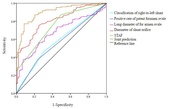
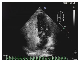
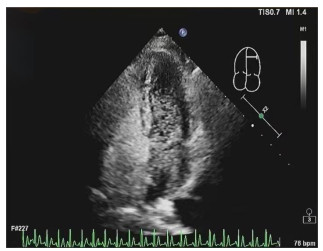
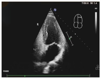
Objective To investigate the predictive value of right heart contrast echocardiography combined with the score for the targeting of atrial fibrillation (STAF) in cardiac embolism (CE). Methods We retrospectively analyzed the data of 149 patients with CE (CE group) and 93 patients with non-cardiac embolism (NCE group) who were admitted to the hospital from August 2017 to August 2021. The results of right heart contrast echocardiography and STAF scores in the two groups were compared. The predictive value of right heart contrast echocardiography combined with STAF in CE was analyzed. Patients with CE were followed up for 1 year to evaluate the prognosis. The prognostic factors in patients with CE were analyzed. Results The proportions of patients with grade 1, grade 2 and grade 3 right-to-left shunt, the positive rate of patent foramen ovale, long diameter of foramen ovale, and diameter of shunt orifice in CE group were higher/larger than those in NCE group (P < 0.05). The STAF score of CE group was higher than that of NCE group (P < 0.05). ROC curve analysis showed that the area under the curve values of right-to-left shunt grade, positive rate of patent foramen ovale, long diameter of foramen ovale, diameter of shunt orifice, STAF and their combination to predict CE were 0.582, 0.570, 0.679, 0.808, 0.750 and 0.905. Univariate analysis and multivariate logistic regression analysis found that the NIHSS score at admission, the positive rate of patent foramen ovale, long diameter of foramen ovale, and STAF were prognostic factors in patients with CE (P < 0.05). Conclusion Right heart contrast echocardiography combined with STAF is beneficial to predict CE.
2023, 46(4): 688-691.
doi: 10.12122/j.issn.1674-4500.2023.04.19
Abstract:
Objective To explore the feasibility of reducing chest CT radiation dose by adjusting tube voltage based on body mass combined with automatic tube current technology in children. Methods The chest CT images of 54 children who underwent chest CT scanning in our hospital from September to December 2019 were collected as the control group. Scanning parameters: Tube voltage: 100 kV or 120 kV, tube current using fixed value: range from 120-200 mA. The chest images of 56 children who underwent low-dose scanning from January to May 2020 were collected as the low dose group. The scanning parameters were as follows: Different tube voltages were selected according to their body mass, 80 kV for body mass ≤15 kg, 100 kV for body mass ≤40 kg, 120 kV for body mass ≥40 kg, and the tube current was automatically adjusted. The radiation dose and image quality of the two groups were compared. Results The average effective dose of chest CT in control group was 0.95±0.55 mSv, and the average effective dose of chest CT in low dose group was 0.18±0.17 mSv, the radiation dose was only 18.9% of that in the control group. The proportion of images meeting the diagnostic criteria in control group and low dose group were 98.15% and 96.43%, respectively(P=0.579). Conclusion On the premise of meeting the diagnostic requirements, choosing different tube voltages according to the body mass of children combined with automatic regulation of tube current technology can significantly reduce the radiation dose of chest CT.
2023, 46(4): 692-696.
doi: 10.12122/j.issn.1674-4500.2023.04.20
Abstract:
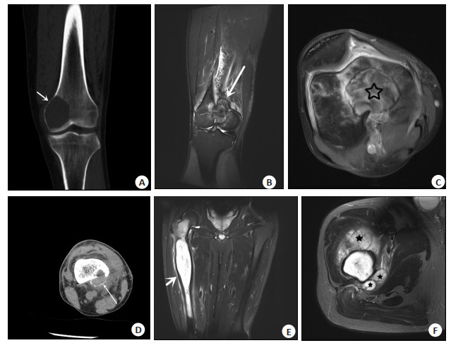
Objective To investigate the imaging differential diagnosis features of giant cell tumor of bone and chondrosarcoma occurring in the end of long bone. Methods Twenty patients with giant cell tumor of bone (giant cell tumor of bone group) and 16 patients with chondrosarcoma (chondrosarcoma group), which were confirmed by surgical pathology to be primary in the bone end of long bone in our hospital from May 2015 to May 2022 were selected for the study, and the clinical and imaging characteristics between the two groups were retrospectively analyzed using independent samples t-test and Fisher's precision probability test. Results Comparing the imaging features of the two groups, the differences were statistically significant (P < 0.05): A higher percentage of patients with giant cell tumor of bone in the end of long bone are 20 to 40 years old (60%), and there was a higher percentage of simple involvement of bone end (90%), eccentric growth (70%), clear boundary (100%), bone cortex expansion (95%), uniform cortical thinning (100%), soft tissue masses without lobulation (100%), localized growth of soft tissue masses (100%), slightly high signal predominates on T2WI (100%), with Cystic degeneration/necrosis /bleeding/liquid-liquid level (60%). While the percentage of patients with chondrosarcoma in the end of long bone greater than 40 years was 87.5%, and there was a higher percentage of the lesions involved both the bone end and the diaphysis (81.2%), non-eccentric growth (87.5%), unclear boundary (56.2%), without bone cortex expansion (87.5%), uneven cortical thickness/roughness (100%), lobulated soft tissue mass (100%), soft tissue mass surrounding the diaphysis/infiltrating surrounding tissue (100%), high signal or Significantly high signal on T2WI (100%), without Cystic degeneration/necrosis/bleeding/liquid-liquid level (100%). There was no statistically significant difference between the two groups of gender, presence or absence of soft tissue mass, bone marrow edema, soft tissue exudation and MR edge low signal ring (P > 0.05). Conclusion There are some similarities in age of onset and imaging manifestations between giant cell tumor of bone and chondrosarcoma occurring in the end of long bone. Comprehensive assessment of onset age, onset location, eccentric growth or not, clear boundary or not, abnormal changes of the bone cortex or not, features of the soft tissue mass, and internal signal characteristics of the lesion on MR can help in the differential diagnosis of the two deases.
2023, 46(4): 697-700.
doi: 10.12122/j.issn.1674-4500.2023.04.21
Abstract:
Objective To assess left ventricular function in patients with grade 1 essential hypertension combined with overweight and obesity through echocardiographic techniques. Methods Sixty patients diagnosed with grade 1 essential hypertension in our hospital were classified as the case group, including 35 hypertensives in the group with normal BMI (case A group) and 25 hypertensives in the group with combined overweight and obesity (case B group). Fifty healthy people were selected as the control group. The conventional ultrasound indices: interventricular septal thickness, left ventricular posterior wall thickness, left ventricular ejection fraction, left ventricular myocardial function Tei index, overall left ventricular global longitudinal strain, and myocardial work done indices, including global work index, global constructive work, global wasted work and global work efficiency were compared among the three groups. Results There were no significant differences in interventricular septal thickness, left ventricular posterior wall thickness and left ventricular ejection fraction among three groups (P > 0.05). Compared with the control group, the Tei index increased and global longitudinal strain decreased (P < 0.05), and there was no significant difference in the case groups (P > 0.05). Compared with the control group, there were no significant differences in the heart rate and myocardial work parameters global work index, global constructive work, global wasted work and global work efficiency in case group A (P > 0.05). The heart rate and global wasted work of case B increased, while the global work efficiency decreased (P < 0.05), and no statistical differences were found in global work index and global constructive work (P > 0.05). Conclusion In patients with grade 1 essential hypertension combined with overweight and obese patients, the global left ventricular function and global longitudinal strain was decreased, global wasted work was increased, and global work efficiency was reduced. The combined application of multiple parameters of echocardiography can more objectively and comprehensively evaluate left ventricular function in patients with grade 1 essential hypertension complicated with overweight and obesity.
2023, 46(4): 701-705.
doi: 10.12122/j.issn.1674-4500.2023.04.22
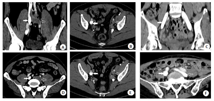
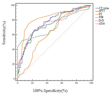
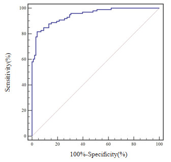
Abstract:
Objective To analyze the diagnostic value of CT value combined with coagulation and fibrinolysis index in the diagnosis of lower extremity deep vein thrombosis (DTV). Methods A total of 98 patients with lower extremity DTV admitted to our hospital from January 2019 to May 2022 were selected as the research group, and 98 patients without lower extremity DTV in our hospital during the same period were selected as the control group. All patients underwent CT examination and determination of coagulation and fibrinolysis indexes [activated partial prothrombin time (aPTT), prothrombin time (PT), fibrinogen (FIB), D-dimer (D-D), antithrombin Ⅲ (ATⅢ), Logistic regression was used to analyze the correlation between CT value, coagulation and fibrinolysis index and lower extremity DTV, and ROC curve was used to analyze the diagnostic value of CT values and coagulation and fibrinolysis indexes for lower extremity DTV, and the area under the curve was calculated. Results CT images showed that the venous lumen of DTV in lower limbs was not filled with contrast agent, showing low density; compared with the control group, the CT value, FIB, and D-D of the study group were significantly higher (P < 0.05), aPTT, PT and ATⅢ were significantly lower (P < 0.05); Logistic regression results showed that CT value, aPTT, PT, FIB, D-D, ATⅢ were significantly correlated with lower extremity DTV (P < 0.05). ROC curve results showed that the area under the curve of the combined diagnosis was 0.946, the sensitivity was 81.63%, and the specificity was 95.92%. Conclusion CT value, coagulation and fibrinolysis index are correlated with lower extremity DTV, and their combined diagnosis has high application value in patients with lower extremity DTV.
2023, 46(4): 706-710.
doi: 10.12122/j.issn.1674-4500.2023.04.23
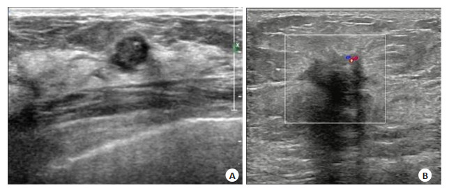
Abstract:
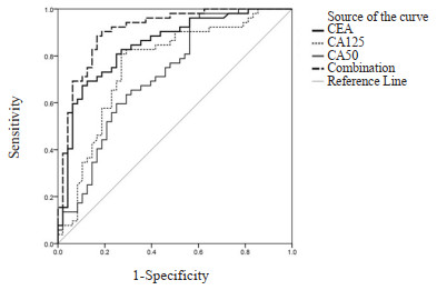
Objective To analyze the correlation between ultrasonic image manifestations of breast cancer and serum carcinoembryonic antigen (CEA), carbohydrate antigen (CA) 125, CA50. Methods A retrospective analysis was performed on the case data of 100 patients with suspected breast cancer admitted to the hospital from June 2020 to September 2022. All patients underwent ultrasound examination. According to the results of pathological examination, they were divided into malignant group (n=52) and benign group (n=48). The characteristics of ultrasonic images in malignant group were analyzed. The levels of serum CEA, CA125 and CA50 were detected by chemiluminescence immunoassay. The changes of CEA, CA125 and CA50 in patients with different ultrasonic image characteristics were compared, and their correlation was analyzed. The levels of serum CEA, CA125 and CA50 in malignant group and benign group were compared, and their diagnostic value for breast cancer was analyzed. Results The proportions of cases with tumor ≥2 cm, irregular morphology, marginal spiculation, aspect ratio > 1, internal low/mixed echo, posterior echo attenuation, micro-calcification and abundant blood supply of lesions in malignant group were higher than those in benign group (P < 0.05). There were significant differences in levels of serum CEA, CA125 and CA50 among patients with different ultrasonic image characteristics [tumor size (≥2 cm vs < 2 cm), aspect ratio (≤1 vs > 1), internal echo (low/mixed echo vs high/equal echo), posterior echo (attenuation vs non-attenuation), micro-calcification (yes vs no), blood flow distribution (abundant blood supply vs lack of blood supply] (P < 0.05). The levels of serum CEA and CA50 in patients with irregular morphology of breast cancer were higher than those with regular morphology (P < 0.05). The levels of serum CA125 and CA50 in patients with marginal spiculation were higher than those without marginal spiculation (P < 0.05). The levels of serum CEA, CA125 and CA50 were positively correlated with aspect ratio > 1, micro-calcification and abundant blood supply of lesions (raspect ratio > 1=0.415, 0.341, 0.424; rmicro-calcification=0.374, 0.394, 0.311; rabundant blood supply of lesions=0.419, 0.533, 0.461, P < 0.05). The levels of serum CEA, CA125 and CA50 in malignant group were 9.36±2.16 ng/mL, 46.84±7.66 U/mL and 42.61±6.48 μg/L, higher than those in benign group (6.9±2.04 ng/mL, 38.73±8.82 U/mL, 36.70±6.87 μg/L, P < 0.05). AUC values of serum CEA, CA125, CA50 and combined detection were 0.845, 0.760, 0.721 and 0.910, respectively. AUC of combined detection was greater than that of single index (ZCEA/combination=2.109, P=0.035; ZCA125/combination=3.232, P=0.001; ZCA50/combination=3.821, P < 0.001). Conclusion The serum CEA, CA125 and CA50 are closely related to ultrasonic image characteristics of breast cancer (tumor size, micro- calcification, abundant blood supply of lesions), and their combined detection has auxiliary diagnosis value for breast cancer.
2023, 46(4): 711-718.
doi: 10.12122/j.issn.1674-4500.2023.04.24
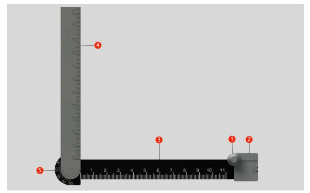
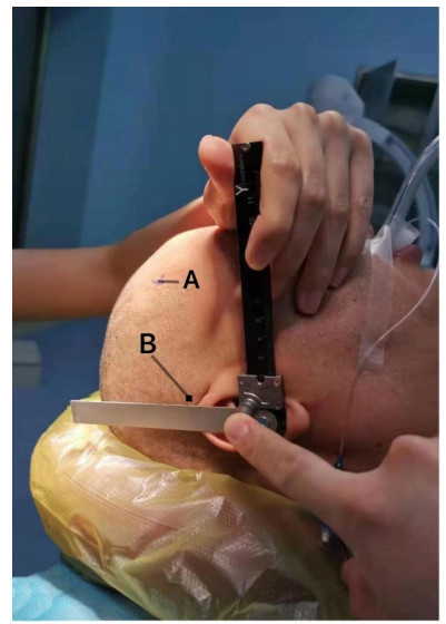
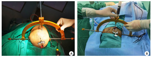
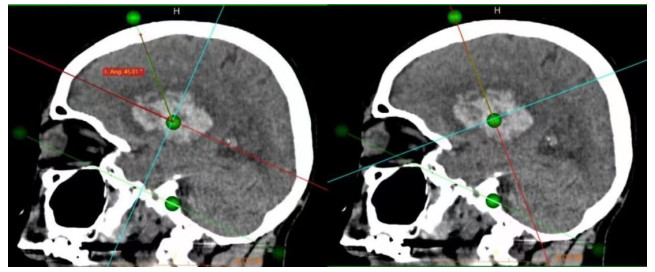
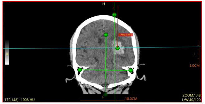
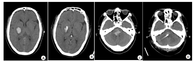
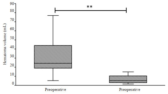
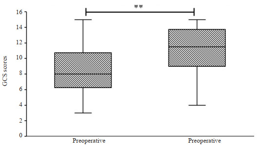
Abstract:
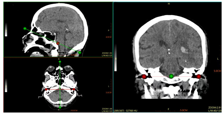
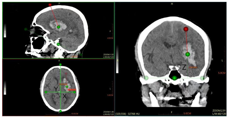
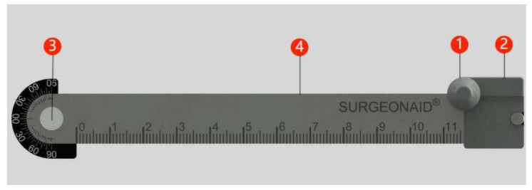
Objective To evaluate the application value of the novel stereotactic puncture technique in hypertensive intracerebral hemorrhage drainage. Methods Clinical data of 24 patients who underwent hematoma puncture and drainage using this technique at our hospital from January 2017 to January 2021 were retrospectively analyzed. The puncture accuracy, hematoma clearance rate, pre- and postoperative Glasgow coma scale score, and the incidence of complications were observed to further investigate the clinical value of this technique. Results In all 24 cases, postoperative cranial CT scans showed good placement of the drainage tube, all along the long axis of the hematoma with the tip inside the hematoma. The offset distance from the preoperative target point was 4.53±3.40 mm, all≤10 mm. The hematoma clearance was satisfactory postoperatively and there were no instances of re-bleeding or catheter-related infections. Conclusion This stereotactic puncture technique is simple to operate and accurately positioned, which can save surgery and preoperative preparation time, ensure the success rate and accuracy of hematoma puncture, and reduce surgical trauma and postoperative complications, with a low risk of re-bleeding. It is suitable for emergency cases of cerebral hemorrhage.
2023, 46(4): 719-723.
doi: 10.12122/j.issn.1674-4500.2023.04.25
Abstract:
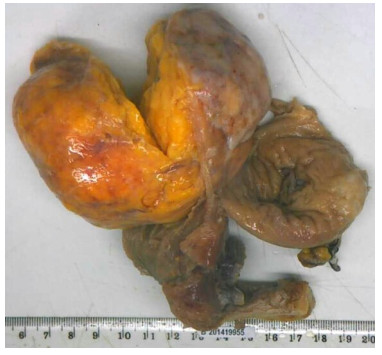
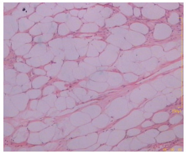
Objective To analyze the clinical manifestations and ultrasonic characteristics of uterine lipoleiomyomas (LL) at different sites. Methods A retrospective analysis was performed on the clinical data of 60 patients with LL confirmed by surgical pathology in the hospital from September 2019 to September 2022. The clinical information, laboratory examinations, characteristics of ultrasonic images and pathological results were analyzed. Results In the 60 patients with LL, there were 41 cases without clinical manifestations, 7 cases with abdominal distension, 6 cases with increased menstruation, 3 cases with postmenopausal vaginal bleeding, 1 case with dyspareunia, 1 case with anal distension and 1 case with frequent and urgent urination. There was no significant difference in clinical manifestations among patients with LL at different sites (P > 0.05). There were 41 cases with lesions located in the intermuscular wall of uterine body or subserous membrane (5 cases in broad ligament), 13 cases in the cervix (1 case in submucosa of cervical canal) and 6 cases in uterine submucosa. In ultrasound detection, LL were mostly round or quasi-round, there were false envelope-like echoes, most boundaries were clear. There were solid and slightly hyperechoic lumps, relatively homogeneous internal echoes and blood flow signals around tumors. Preoperative ultrasound showed that there were 30 misdiagnosed cases, with misdiagnosis rate of 50%. There was no significant difference in misdiagnosis rate among patients with LL at different sites (P > 0.05). Conclusion There are certain characteristics of ultrasonic manifestations in LL. Most of them are round or quasi-round, with solid and slightly hyperechoic lumps. Preoperative ultrasound can preliminarily determine LL, but the misdiagnosis rate is high.
2023, 46(4): 724-730.
doi: 10.12122/j.issn.1674-4500.2023.04.26
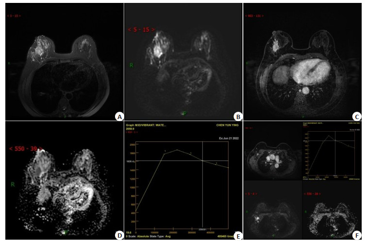
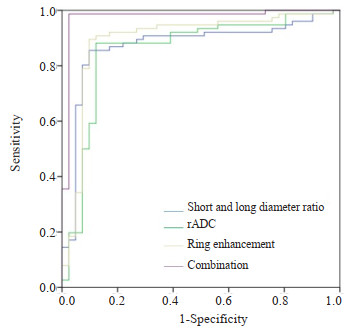
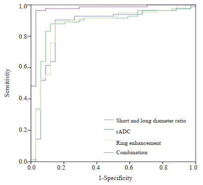
Abstract:
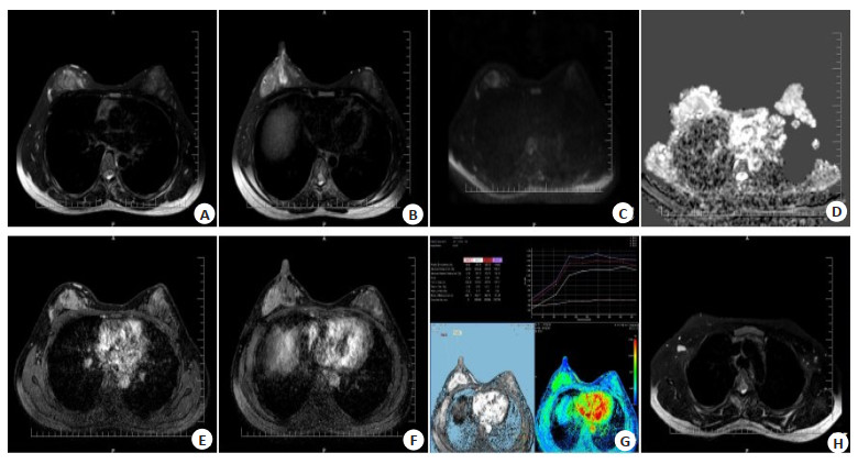
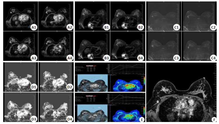
Objective To explore the MRI imaging features of breast cancer and its relationship with sentinel lymph node and axillary lymph node metastasis. Methods A total of 117 patients with breast cancer admitted to our hospital from May 2019 to May 2022 were selected as the study objects, including 41 patients with sentinel lymph node metastasis, 34 with axillary lymph node metastasis and 42 without metastasis. The patients underwent MRI scan for breast cancer. ROC curve was drawn to analyze the diagnostic value of axillary lymph node metastasis and sentinel lymph node metastasis. Results Breast cancer patients with sentinel lymph node metastasis and axillary lymph node metastasis had significantly lower ratio of ratio of short diameter to long diameter in MRI imaging than those without metastasis. The relative apparent diffusion coefficient (rADC) value was significantly higher than that without metastasis(P < 0.05). There were significant differences in ring enhancement between patients with sentinel lymph node metastasis, axillary lymph node metastasis and those without metastasis (P < 0.05). The prediction model of sentinel lymph node by combined application of MRI features of breast cancer was Log(P)=-0.602×ratio of short diameter to long diameter+0.675×rADC-0.754×ring enhancement+0.895. The combined application of MRI features to predict axillary lymph node metastasis was Log(P)=-0.685×ratio of short diameter to long diameter+0.712×rADC-0.695×ring enhancement+0.794. The combined application of ratio of short diameter to long diameter, rADC and ring enhancement in predicting the AUC of sentinel lymph node metastasis was significantly higher than that applied alone, and the difference was statistically significant (P < 0.05). Combined application of ratio of short diameter to long diameter, rADC and ring enhancement was significantly higher in predicting axillary lymph node metastasis AUC than that applied alone, and the difference was statistically significant (P < 0.05). Conclusion MRI imaging features of breast cancer are highly correlated with sentinel and axillary lymph node metastasis. The combined application of MRI imaging features can significantly and effectively predict sentinel lymph node metastasis and axillary lymph node metastasis.
2023, 46(4): 731-735.
doi: 10.12122/j.issn.1674-4500.2023.04.27
Abstract:
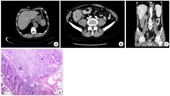
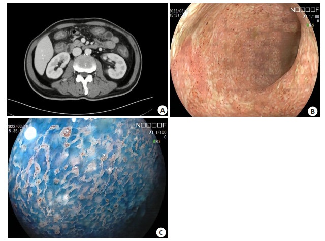
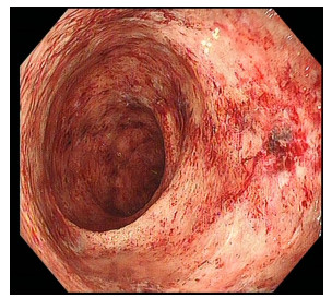
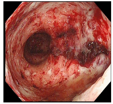
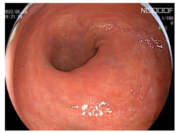
Immune checkpoint inhibitor-induced colitis is one of the most common immune-related adverse events, which clinical feature is diarrhea. The endoscopic feature of immune checkpoint inhibitor-induced colitis are erythema (93.5%), brittleness (58.6%), congestion (48.2%) and ulcer (37.9%). More than 98% of immune checkpoint inhibitor-induced colitis could affect the rectum. Here we report a case of death due to severe immune checkpoint inhibitor-induced colitis.The main clinical manifestations of the patient were abdominal pain and diarrhea. The endoscopy showed that the mucosa was rough and edematous, and the vascular texture disappeared. The patient had previously received immune checkpoint inhibitor. After treatment with glucocorticoid and infliximab, although the endoscopic performance of the patient improved, thrombocytopenia, massive gastrointestinal bleeding and myocardial damage occurred. Death due to severe immune checkpoint inhibitor-induced colitis are rare in clinical practice. In this case, glucocorticoid and infliximab were effective, but they still failed to reverse the progress of the patient, resulting in clinical death. The diagnosis and treatment of immune-related adverse events needs more clinical exploration.
Immune checkpoint inhibitor-induced colitis is one of the most common immune-related adverse events, which clinical feature is diarrhea. The endoscopic feature of immune checkpoint inhibitor-induced colitis are erythema (93.5%), brittleness (58.6%), congestion (48.2%) and ulcer (37.9%). More than 98% of immune checkpoint inhibitor-induced colitis could affect the rectum. Here we report a case of death due to severe immune checkpoint inhibitor-induced colitis.The main clinical manifestations of the patient were abdominal pain and diarrhea. The endoscopy showed that the mucosa was rough and edematous, and the vascular texture disappeared. The patient had previously received immune checkpoint inhibitor. After treatment with glucocorticoid and infliximab, although the endoscopic performance of the patient improved, thrombocytopenia, massive gastrointestinal bleeding and myocardial damage occurred. Death due to severe immune checkpoint inhibitor-induced colitis are rare in clinical practice. In this case, glucocorticoid and infliximab were effective, but they still failed to reverse the progress of the patient, resulting in clinical death. The diagnosis and treatment of immune-related adverse events needs more clinical exploration.
2023, 46(4): 736-740.
doi: 10.12122/j.issn.1674-4500.2023.04.28
Abstract:
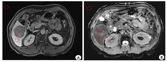
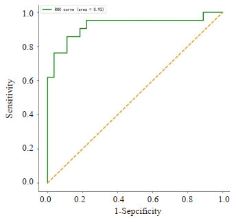
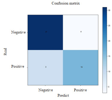
Objective To explore the effectiveness of a machine learning model based on Gd-EOB-DTPA enhanced MRI in predicting microvascular invasion (MVI) of hepatocellular carcinoma. Methods We retrospectively analyzed MRI images and clinical data from 59 patients who underwent Gd-EOB-DTPA enhanced imaging and were pathologically confirmed with hepatocellular carcinoma from January 2017 to December 2020. Based on histopathological results, the patients were divided into MVI-positive group and MVI-negative group. The signal noise ratio and contrast to noise ratio in the hepatobiliary-specific phase and apparent dispersion coefficient images were measured. Principal component analysis was used for feature dimension reduction, and a support vector machine model was developed. The diagnostic performance of the model was evaluated using ROC curves and a confusion matrix. Results The AUC value of the support vector machine model was 0.86 (95% CI: 0.83-0.95). The accuracy, sensitivity and specificity were 0.80, 0.64 and 0.91, respectively. Conclusion The machine learning model based on Gd-EOB-DTPA enhanced MRI has potential in predicting MVI before hepatocellular carcinoma surgery.
2023, 46(4): 741-745.
doi: 10.12122/j.issn.1674-4500.2023.04.29
Abstract:
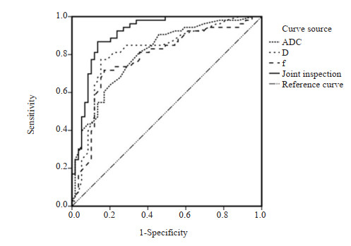
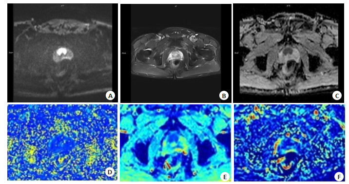
Objective To investigate the value of MRI texture analysis and intravoxel incoherent motion (IVIM) sequences based on zoomed imaging with parallel transmission technique (ZOOMit) for the differential diagnosis of benign and malignant prostate nodules. Methods Ninety-five patients with prostate nodules in our hospital between March 2021 and August 2022 were selected. A total of 112 nodules were divided into benign group (n=59) and malignant group (n=53). All patients underwent MRI including T2* mapping and ZOOMit IVIM sequence scans before surgery, then the MRI plain scanning images, diffusion weighted imaging (DWI) images, parameters including apparent diffusion coefficient (ADC), diffusion coefficient of pure diffusion (D), pseudo-diffusion coefficient (D*) and perfusion fraction (f) were obtained. The misjudgment rates of the two scanning sequences were compared. ROC curve was plotted to evaluate the diagnostic value of each parameter for benign and malignant prostate nodules. Results There was no significant difference in the misjudgment rate between benign and malignant prostate nodules diagnosed by MRI texture analysis and ZOOMit IVIM sequence scan alone and in combination (P > 0.05). Compared with benign group, malignant group had remarkably decreased ADCMean, variance, D value and f value (P < 0.05), notably increased skewness, kurtosis and entropy (P < 0.05), and slightly increased D* value (P > 0.05). Among the parameters in the differential diagnosis of benign and malignant prostate nodules, ADC value had high sensitivity and D value had high specificity, while the diagnostic efficacy of ADC combined with D and f values was the highest. Conclusion MRI texture analysis and ZOOMit IVIM sequence have high value in differentiating benign and malignant prostate nodules.
2023, 46(4): 746-750.
doi: 10.12122/j.issn.1674-4500.2023.04.30
Abstract:
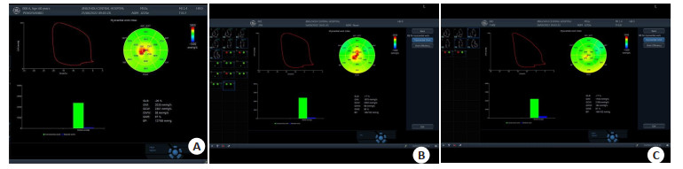
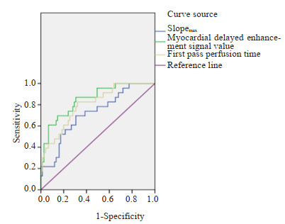
Objective To explore the clinical value of cardiac magnetic resonance (CMR) myocardial perfusion imaging in coronary artery disease and microcirculation obstruction (MVO). Methods 106 patients with coronary heart disease admitted to our hospital from January 2020 to December 2022 were selected to undergo CMR myocardial perfusion imaging before and after percutaneous coronary intervention. Using the diagnostic results of coronary angiography as the gold standard, the parameters of delayed enhanced transmural regional wall abnormal motion and CMR myocardial perfusion imaging in the diseased and normal myocardial areas in CMR myocardial perfusion imaging before percutaneous coronary intervention were compared. To analyze the diagnostic efficacy of CMR myocardial perfusion imaging in coronary artery disease. After percutaneous coronary intervention, the patients were divided into MVO group (n=29)and non-MVO group (n=77)according to whether there was MVO. The parameters of CMR myocardial perfusion imaging were compared between the two groups, and the diagnostic value of CMR myocardial perfusion imaging parameters to MVO was analyzed by ROC curve. Results 1802 myocardial segments in 106 patients were included in the evaluation. CMR myocardial perfusion imaging showed that there were 147 myocardial segments with delayed enhancement in 106 patients (at least one myocardial segment with delayed enhancement in each patient), including 68 transmural enhancement and 79 non-transmural enhancement. There were significant differences in the first pass perfusion time, the maximum slope of first pass perfusion (Slopemax) and the delayed enhancement signal value between the diseased myocardium and the normal myocardium (P < 0.05). Based on the results of coronary angiography as the gold standard, the sensitivity of CMR myocardial perfusion imaging in the diagnosis of left anterior descending artery, right coronary artery and left circumflex artery lesions was 94.12%, 88.64% and 88.89% respectively, the specificity was 95.00%, 96.55% and 94.74% respectively, and the accuracy was 94.66%, 93.89% and 93.13% respectively. After percutaneous coronary intervention, there were 29 cases with MVO and 77 cases without MVO. In MVO group, the first perfusion time, Slopemax and delayed myocardial enhancement signal value were significantly different from those without MVO group (P < 0.05). The ROC curve analysis showed that AUC of the first perfusion time, Slopemax, and delayed myocardial enhancement signal value of CMR myocardial perfusion imaging parameters for the diagnosis of MVO in patients with coronary heart disease were 0.803, 0.718 and 0.851, respectively, with sensitivity of 82.76%, 72.41% and 86.21%, specificity of 66.23%, 68.83%, and 67.53%, and accuracy of 70.75%, 69.81% and 72.64%. Conclusion CMR myocardial perfusion imaging can display the changes of the cardiovascular disease area of coronary heart disease, and has high diagnostic value for diagnosing coronary artery disease and MVO.
2023, 46(4): 751-758.
doi: 10.12122/j.issn.1674-4500.2023.04.31
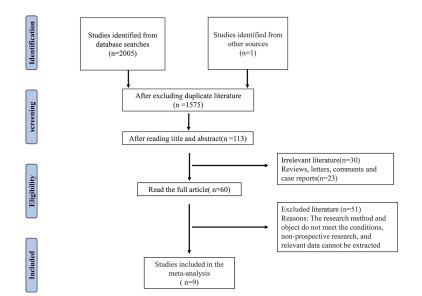
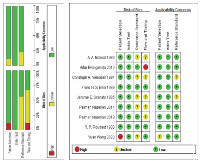
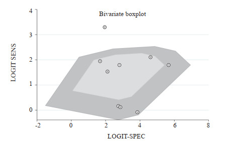
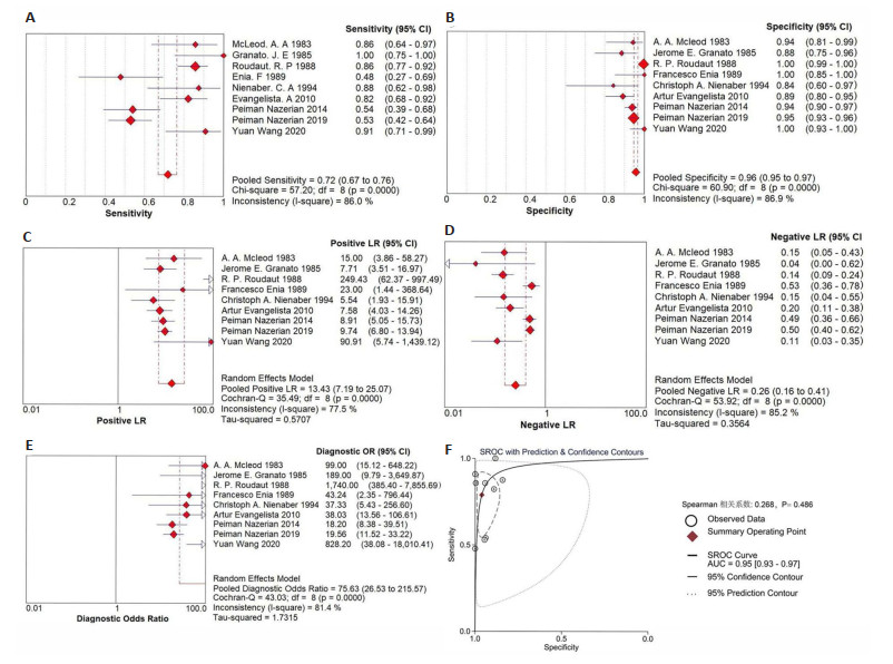
Abstract:
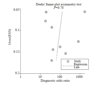
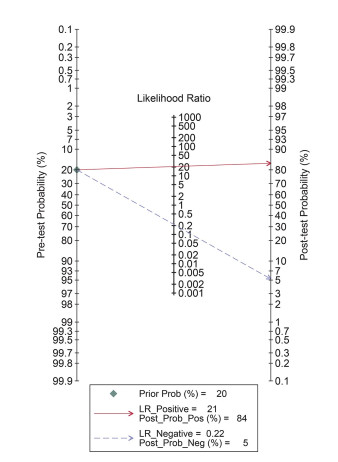
Objective Meta-analysis was used to systematically review the diagnostic efficacy of transthoracic echocardiography on type A aortic dissection (AAD). Methods The Meta-disc1.4 and Stata 16.0 software was used to analyze 9 literatures as well as 9 groups of data including true positive, false positive, true negative and false negative values, and estimated the combined effect values of sensitivity, specificity, positive likelihood ratio, negative likelihood ratio and diagnostic odds ratio were estimated. The area under curve of the summary ROC curve and the Fagan nomogram were summarized to estimate the diagnostic accuracy of transthoracic echocardiography. The sources of heterogeneity were analyzed by Meta-regression analysis. Deeks' funnel diagram asymmetry test was used to test publication bias. Results The combined value of sensitivity was 0.72 (95%CI: 0.67-0.76), specificity was 0.96 (95% CI: 0.95-0.97), positive likelihood ratio was 13.43 (95%CI: 7.19-25.07), negative likelihood ratio was 0.26 (95%CI: 0.16-0.41)and the area under curve was 0.95 (95%CI: 0.93-0.97). The post-test probability increased from 20% to 84% in Fagan nomogram, all indicating a high diagnostic accuracy. Sensitivity and specificity Q test were less than 0.01, which indicated some heterogeneity. Meta-regression showed that sample size, age of population, ultrasound technology, operator population, selection of ultrasound diagnostic criteria and selection of gold standard were not the sources of heterogeneity. However, Subgroup analysis showed that the strict setting of ultrasound positive criteria and the lack of experience in the operator population made the diagnostic sensitivity too low, which may be the source of heterogeneity in our study. Deeks' funnel plot showed that there was no publication bias for the 9 included literatures (P=0.78). Conclusion Transthoracic echocardiography has high diagnostic efficacy for AAD and can be used as a preliminary imaging evaluation method for AAD. However, it may be necessary to adjust the setting of ultrasound positive criteria and strengthen the relevant training of operators to reduce the missed diagnosis of AAD and further improve the diagnostic sensitivity.
2023, 46(4): 759-764.
doi: 10.12122/j.issn.1674-4500.2023.04.32
Abstract:
As the prevalence of low-dose CT scans for the lungs continues to rise, coupled with the enhancement of public health awareness, there has been a significant surge in the detection rate of pulmonary nodules. However, discerning between benign and malignant cases remains a contemporary challenge and hot topic in radiology. It is crucial to establish an early definitive diagnosis for the proper selection of treatment plans and to anticipate prognosis outcomes. The current investigative focus in radiology concerning pulmonary nodules has evolved from morphology to molecular imaging. Molecular imaging is capable of conducting qualitative, quantitative, and dynamic studies at the cellular and molecular level in vivo, revealing its important value and potential in diagnosing pulmonary nodules. This paper provides an overview of the pathological progression of pulmonary nodules of different pathological types and reviews the latest research developments of several different types of nuclear medicine molecular imaging agents in pulmonary nodules.
As the prevalence of low-dose CT scans for the lungs continues to rise, coupled with the enhancement of public health awareness, there has been a significant surge in the detection rate of pulmonary nodules. However, discerning between benign and malignant cases remains a contemporary challenge and hot topic in radiology. It is crucial to establish an early definitive diagnosis for the proper selection of treatment plans and to anticipate prognosis outcomes. The current investigative focus in radiology concerning pulmonary nodules has evolved from morphology to molecular imaging. Molecular imaging is capable of conducting qualitative, quantitative, and dynamic studies at the cellular and molecular level in vivo, revealing its important value and potential in diagnosing pulmonary nodules. This paper provides an overview of the pathological progression of pulmonary nodules of different pathological types and reviews the latest research developments of several different types of nuclear medicine molecular imaging agents in pulmonary nodules.
2023, 46(4): 765-768.
doi: 10.12122/j.issn.1674-4500.2023.04.33
Abstract:
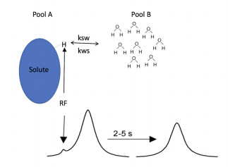
Gliomas originate from the central nervous system, with a high degree of malignancy and a poor prognosis. At present, the diagnosis of gliomas is mostly based on magnetic resonance examination in addition to surgical pathology. Traditional magnetic resonance imaging presents information in the form of images, with tumor enhancement scope and peritumoral edema as the main description objects, and tumor properties are judged based on human eye recognition and clinical experience. The distinction between gliomas without enhancement and treatment-related changes is largely limited. amide proton transfer magnetic resonance imaging is an emerging autologous imaging technique, which expands the traditional anatomical imaging to pH imaging and biochemical metabolism imaging. The current technique is still under investigation. This review provides a comprehensive overview of the fundamental principles, commonly employed quantitative methods, applications in glioma imaging using amide proton transfer imaging, as well as the limitations and future prospects of this technology.
Gliomas originate from the central nervous system, with a high degree of malignancy and a poor prognosis. At present, the diagnosis of gliomas is mostly based on magnetic resonance examination in addition to surgical pathology. Traditional magnetic resonance imaging presents information in the form of images, with tumor enhancement scope and peritumoral edema as the main description objects, and tumor properties are judged based on human eye recognition and clinical experience. The distinction between gliomas without enhancement and treatment-related changes is largely limited. amide proton transfer magnetic resonance imaging is an emerging autologous imaging technique, which expands the traditional anatomical imaging to pH imaging and biochemical metabolism imaging. The current technique is still under investigation. This review provides a comprehensive overview of the fundamental principles, commonly employed quantitative methods, applications in glioma imaging using amide proton transfer imaging, as well as the limitations and future prospects of this technology.
2023, 46(4): 769-773.
doi: 10.12122/j.issn.1674-4500.2023.04.34
Abstract:
Near-infrared has better penetration depth and biocompatibility for living tissues, which minimizes the scattering and attenuation of irradiation in tissues. In recent years, it has been used in many aspects such as in vivo imaging, 3D image visualization, photothermal therapy, drug release and in vivo optogenetics. Near-infrared is now increasingly used in clinical medicine, combining with various molecular imaging modalities and using nano-probes to play a photoacoustic imaging role; developing photothermal agents with different properties to achieve near-infrared light therapy through the conversion of light and heat energy; combining with 3D printing, an emerging technology, to personalize stents or implants, using the spatio-temporal tunability of near-infrared light to enable simultaneous diagnosis and treatment, and achieving precision medicine.
Near-infrared has better penetration depth and biocompatibility for living tissues, which minimizes the scattering and attenuation of irradiation in tissues. In recent years, it has been used in many aspects such as in vivo imaging, 3D image visualization, photothermal therapy, drug release and in vivo optogenetics. Near-infrared is now increasingly used in clinical medicine, combining with various molecular imaging modalities and using nano-probes to play a photoacoustic imaging role; developing photothermal agents with different properties to achieve near-infrared light therapy through the conversion of light and heat energy; combining with 3D printing, an emerging technology, to personalize stents or implants, using the spatio-temporal tunability of near-infrared light to enable simultaneous diagnosis and treatment, and achieving precision medicine.
2023, 46(4): 774-778.
doi: 10.12122/j.issn.1674-4500.2023.04.35
Abstract:
MRI has the advantages of radiation-free, high soft tissue resolution, multi-parameter and multi-sequence imaging, and is widely used in all systems of the whole body.In the past, due to the low content of hydrogen protons in the lung, respiratory motion artifacts and magnetic susceptibility artifacts, MRI was considered not to be used in lung scanning. With the rapid development of MRI technology, such as parallel acquisition technology, respiratory gating technology, and development of new sequences and artificial intelligence, lung MRI technology is gradually improved. With the increase of people's attention to radiation dose and pulmonary nodules, the clinical demand of pulmonary MRI is also increasing. This article will review the new technology of MRI from two aspects: nodule detection and benign and malignant differentiation, including star volume interpolated breath-hold examination sequence, ultrashort echo time, zero echo time, compression sensing volume interpolated breath-hold examination sequence, longitudinal relaxation time quantitative imaging, chemical exchange saturation transfer and other new MRI techniques.
MRI has the advantages of radiation-free, high soft tissue resolution, multi-parameter and multi-sequence imaging, and is widely used in all systems of the whole body.In the past, due to the low content of hydrogen protons in the lung, respiratory motion artifacts and magnetic susceptibility artifacts, MRI was considered not to be used in lung scanning. With the rapid development of MRI technology, such as parallel acquisition technology, respiratory gating technology, and development of new sequences and artificial intelligence, lung MRI technology is gradually improved. With the increase of people's attention to radiation dose and pulmonary nodules, the clinical demand of pulmonary MRI is also increasing. This article will review the new technology of MRI from two aspects: nodule detection and benign and malignant differentiation, including star volume interpolated breath-hold examination sequence, ultrashort echo time, zero echo time, compression sensing volume interpolated breath-hold examination sequence, longitudinal relaxation time quantitative imaging, chemical exchange saturation transfer and other new MRI techniques.