


High-resolution magnetic resonance vessel wall imaging can evaluate recurrence of cerebral infarction tiggered by carotid plaques
-
摘要:
目的 探究高分辨磁共振血管壁成像对颈动脉斑块引发脑梗死再发的评估价值。 方法 选取我院2021年6月~2022年6月收治的脑梗死再发患者作为研究组(n=60), 另选取初发脑梗死患者作为对照组(n=60)。两组研究对象均进行高分辨磁共振血管壁成像检查, 比较两组研究对象临床特征资料、颈动脉斑块负荷、颈动脉斑块成分、颈动脉斑块成分面积。 结果 研究组的管腔面积高于对照组(P < 0.05), 血管总面积值和管壁标准化指数均高于对照组(P < 0.05), 但管壁面积值和管腔狭窄率的差异无统计学意义(P > 0.05)。研究组钙化、脂质坏死核心、斑块内出血和纤维帽破裂占斑块成分的比例高于对照组(P < 0.05), 但斑块内钙化及管腔狭窄率 > 50%的比例的差异无统计学意义(P > 0.05)。研究组患者的脂质坏死核心面积、斑块内钙化面积及斑块内出血面积均大于对照组(P < 0.001)。 结论 高分辨磁共振血管壁成像不仅能显示血管壁的狭窄程度, 同时可以有效显示颈动脉斑块的成分及面积, 对颈动脉斑块引发的脑梗死预防提供可靠依据, 有效降低脑梗死的再发率。 -
关键词:
- 高分辨磁共振血管壁成像 /
- 脑梗死 /
- 动脉斑块 /
- 评估价值 /
- 管腔狭窄
Abstract:Objective To evaluate the value of high-resolution magnetic resonance vessel wall imaging for recurrence of cerebral infarction triggered by carotid plaques. Methods Patients with recurrent cerebral infarction and patients with initial cerebral infraction in our hospital from June 2021 to June 2022 were enrolled as study group (n=60) and control group (n=60).All patients received high-resolution magnetic resonance vessel wall imaging.Then comparison was conducted on the clinical characteristics, carotid plaque burden and compositional features. Results Study group had notably increased luminal area and larger total vessel area and vessel wall normalization index than control group (P < 0.05), while no statistical difference was found in vessel wall area and luminal stenosis rate between two groups (P > 0.05).The proportions of carotid plaques of calcification, lipoid-rich necrotic core, intraplaque hemorrhage and fibrous cap rupture were higher in study group than in control group (P < 0.05), while the proportion of intraplaque calcification and luminal stenosis > 50% demonstrated no statistical difference between two groups (P > 0.05).The area of lipoid-rich necrotic core, intraplaque calcification and intraplaque hemorrhage were significantly larger in study group than in control group (P < 0.001). Conclusion Application of high-resolution magnetic resonance vessel wall imaging can effectively display the stenosis degree of the vessel wall, carotid plaque burden and compositional features.It provide a basis for the prevention of cerebral infarction triggered by carotid plaques, thus effectively reducing the recurrence rate of cerebral infarction. -

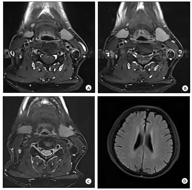
图 1 脑梗死再发患者的HR-MRI图像
Figure 1. HR-MRI of patients with recurrent cerebral infarction. A: T1WI enhanced imaging; B: T1WI imaging; C: PDWI proton imaging; D: Brain T2 FLAIR imaging.
表 1 两组研究对象颈动脉斑块负荷比较
Table 1. Comparison of carotid plaque load between the two groups (n=60, Mean±SD)
Group LA(mm2) WA(mm2) TVA(mm2) NWI(mm2) Luminal stenosis rate(%) Study group 31.84±10.86 72.63±20.41 113.09±31.22 72.23±18.76 52.75±15.68 Control group 40.57±12.32 70.87±19.54 101.47±29.65 60.85±16.34 48.33±15.31 t 4.1175 0.4825 2.0905 3.5432 1.5623 P 0.0001 0.6304 0.0387 0.0006 0.1209 LA: Luminal area; WA: Vessel wall area; TVA: Total vessel area; NWI: Vessel wall normalization index.  下载: 导出CSV
表 2 两组颈动脉斑块成分及管腔狭窄率比较
Table 2. Comparison of carotid plaque components and lumen stenosis rate between the two groups [n=60, n(%)]
Group LRNC Intraplaque calcification IPH Fibrous cap rupture Luminal stenosis > 50% Study group 42(70.00) 31(51.67) 25(41.67) 21(35.00) 30(50.00) Control group 29(48.33) 28(46.67) 9(15.00) 3(5.00) 27(45.00) χ2 5.8293 0.3001 10.5062 16.8750 0.3008 P 0.0158 0.5838 0.0012 < 0.001 0.5834 LRNC: Lipoid-rich necrotic core; IPH: Intraplaque hemorrhage.
下载: 导出CSV
表 3 两组颈动脉斑块成分面积比较
Table 3. Comparison of carotid plaque component area between the two groups (n=60, Mean±SD)
Group LRNC(mm2) Intraplaque calcification(mm2) IPH(mm2) Study group 25.41±6.43 3.86±1.14 12.33±3.29 Control group 3.57±0.88 1.57±0.42 0.42±0.09 t 26.0668 14.6005 28.0304 P < 0.001 < 0.001 < 0.001
下载: 导出CSV
-
[1] 李耀志, 谢艺才. 磁共振颅颈一体化高分辨血管壁成像评价颈动脉斑块负荷与脑卒中的关系[J]. 中国医学物理学杂志, 2022, 39(3): 328-32. https://www.cnki.com.cn/Article/CJFDTOTAL-YXWZ202203012.htm [2] 赵楠, 李俊林. 磁共振血管壁成像检测颈动脉斑块内出血的应用进展[J]. 内蒙古医学杂志, 2021, 53(9): 1084-6. https://www.cnki.com.cn/Article/CJFDTOTAL-NMYZ202109019.htm [3] Jia Y, Liu X, Zhang L, et al. Integrated head and neck imaging of symptomatic patients with stroke using simultaneous non-contrast cardiovascular magnetic resonance angiography and intraplaque hemorrhage imaging as compared with digital subtraction angiography[J]. J Cardiovasc Magn Reson, 2022, 24(1): 19. doi: 10.1186/s12968-022-00849-1 [4] 张明宇, 陈红燕, 马丁. 高分辨率磁共振血管壁成像评估椎-基底动脉硬化血管壁对急性脑梗死患者预后的评估价值[J]. 卒中与神经疾病, 2020, 27(3): 323-6. doi: 10.3969/j.issn.1007-0478.2020.03.009 [5] 秦海松, 林中青, 梅凯, 等. 磁共振血管壁成像在评价脑卒中患者颈动脉斑块负荷中的意义[J]. 吉林医学, 2022, 43(5): 1370-2. doi: 10.3969/j.issn.1004-0412.2022.05.083 [6] 巴建, 苗红, 张艾红. 磁共振高分辨技术在脑梗死复发患者颈动脉斑块评估中的应用观察[J]. 癫痫与神经电生理学杂志, 2020, 29(1): 17-20. https://www.cnki.com.cn/Article/CJFDTOTAL-ZGQX202214032.htm [7] 刘良进, 毕俊英, 曾艳妮, 等. 能谱CT成像对颈动脉粥样硬化斑块性质评估及其与脑卒中发生率的关系[J]. 中国CT和MRI杂志, 2021, 19(1): 44-6. doi: 10.3969/j.issn.1672-5131.2021.01.015 [8] Li J, Xiong JD, Chen KX, et al. Comparison of magnetic resonance imaging versus computed tomography-based thrombolysis treatment in patients with acute ischemic stroke[J]. J Clin Ultrasound, 2022, 50(2): 176-81. doi: 10.1002/jcu.23126 [9] 梁汉祥, 李新春. 高分辨磁共振成像检测轻度大脑中动脉粥样斑块情况对梗死类型的评估价值[J]. 中西医结合心脑血管病杂志, 2020, 18(21): 3672-5. doi: 10.12102/j.issn.1672-1349.2020.21.042 [10] 闫力永, 武刚. 血清C反应蛋白联合颈动脉血管壁磁共振高分辨率成像检查对脑梗死复发的预测价值[J]. 检验医学与临床, 2022, 19(13): 1781-4, 1790. https://www.cnki.com.cn/Article/CJFDTOTAL-JYYL202213014.htm [11] 孙逢春, 尤精武, 张雅静. 3D高分辨率磁共振血管壁成像技术对脑血管病变的评估价值[J]. 影像研究与医学应用, 2021, 5(5): 31-2. doi: 10.3969/j.issn.2096-3807.2021.05.015 [12] Zhang DF, Wang M, Wu LL, et al. Assessing the characteristics and diagnostic value of plaques for patients with acute stroke using high-resolution magnetic resonance imaging[J]. Quant Imaging Med Surg, 2022, 12(2): 1529-38. [13] 刘永志, 何国华, 廖俊杰. 高分辨磁共振血管壁成像对颅内动脉瘤的评估诊断价值[J]. 临床医学工程, 2020, 27(6): 701-2. https://www.cnki.com.cn/Article/CJFDTOTAL-YBQJ202006010.htm [14] Danish K, Patel Himanshu A, Zhang Tony J, et al. Abstract TP105: optimizing the utilization of cardiac magnetic resonance imaging among patients with ischemic stroke[J]. Stroke, 2022, 53(Suppl_1): 105-6. [15] 刘春霞, 王丽亚, 陈玉红, 等. 高分辨率磁共振血管壁成像技术对缺血性脑卒中患者的评估价值[J]. 菏泽医学专科学校学报, 2021, 33(4): 71-5. https://www.cnki.com.cn/Article/CJFDTOTAL-HZYZ202104022.htm [16] 刘一江, 张雪峰, 彭昌勇, 等. 评估颈动脉易损斑块核心类型高分辨率MRI的价值[J]. 分子影像学杂志, 2021, 44(2): 355-8. doi: 10.12122/j.issn.1674-4500.2021.02.28 [17] 赵富强, 郑红伟, 彭晓博, 等. 磁共振血管成像对急性脑梗死血管病变的评估价值[J]. 中国CT和MRI杂志, 2020, 18(6): 15-7. https://www.cnki.com.cn/Article/CJFDTOTAL-CTMR202006005.htm [18] Zhao JJ, Lu Y, Cui JY, et al. Characteristics of symptomatic plaque on high-resolution magnetic resonance imaging and its relationship with the occurrence and recurrence of ischemic stroke[J]. Neurol Sci, 2021, 42(9): 3605-13. [19] 杨帅, 王小宜, 胡平, 等. 头颈动脉高分辨率MR血管壁成像在隐匿性卒中病因诊断的临床价值[J]. 临床放射学杂志, 2020, 39(10): 1936-40. https://www.cnki.com.cn/Article/CJFDTOTAL-LCFS202010009.htm [20] 吴经纬, 吴水仙, 陈斌. 高分辨率磁共振评估颈动脉粥样硬化斑块与脑梗死患者血浆Lp-PLA2水平相关性[J]. 分子影像学杂志, 2022, 45(3): 348-52. doi: 10.12122/j.issn.1674-4500.2022.03.08 [21] 凌秀梅, 苏寿红, 曾阳东. 快速三维MR血管壁成像评估急性脑梗死及颈动脉粥样硬化的研究[J]. 影像科学与光化学, 2022, 40(1): 105-8. https://www.cnki.com.cn/Article/CJFDTOTAL-GKGH202201020.htm -

 点击查看大图
计量
- 文章访问数: 205
- HTML全文浏览量: 56
- PDF下载量: 4
- 被引次数: 0