


Value of quantitative multi- parameter assessment of arterial enhancement fraction and extracellular volume based on the iodine maps on spectral CT in the diagnosis of colorectal cancer
-
摘要:
目的 探讨光谱CT定量增强参数对结直肠癌的诊断价值。 方法 对46例行腹盆部光谱CT三期增强的结直肠癌患者的能谱基数据进行处理,生成无水碘图、动脉增强分数(AEF)图及细胞外容积(ECV)图,分别测量肿瘤及正常肠壁的碘浓度(IC)、AEF及ECV值。采用独立样本t检验及Mann-Whitney U检验进行组间比较,并采用ROC曲线评价其诊断效能。 结果 结直肠癌病变的AEF及IC值大于正常肠壁AEF值及IC值(0.338±0.122 vs 0.225±0.072,1.007±0.402 mg/mL vs 0.599±0.229 mg/mL,P<0.0001),ECV在结直肠癌病变与正常肠壁间的差异无统计学意义(P>0.999)。AEF、IC、ECV及三者联合诊断结直肠癌的ROC曲线下面积分别为0.806、0.797、0.671及0.842,其约登指数分别为0.565、0.565、0.326及0.609。三者联合诊断结直肠癌的截断值为0.660,敏感度为63.04%,特异性为97.83%,其逻辑回归方程为Y=13.75×AEF+16.30×ECV-6.70。 结论 光谱CT定量增强参数AEF、ECV及IC联合诊断对结直肠癌的诊断具有一定价值。 Abstract:Objective To explore the diagnostic value of quantitative contrast-enhanced parameters of spectral CT in colorectal cancer. Methods Spectrum base images of 46 patients with colorectal cancer who underwent three- phase enhancement of abdominal and pelvic spectral CT were processed to generate the water-free iodine images, arterial enhancement fraction (AEF) and extracellular volume (ECV) images, and the iodine concentration (IC) value, AEF value and ECV value were measured on the colorectal lesions and the normal colorectal walls. The independent sample t test and Mann-Whitney U test were applied for inter-group comparisons, and ROC curve was used to evaluate the diagnostic efficacy. Results The AEF value and IC value presented significantly highly in the patients than that in normal intestinal wall (0.338±0.122 vs 0.225±0.072, 1.007±0.402 mg/mL vs 0.599±0.229 mg/mL, P < 0.0001). ECV showed no significant difference between colorectal lesion and the normal intestinal wall(P > 0.999). The areas under the ROC curve of AEF, IC, ECV and three indicators combination in the diagnosis of colorectal cancer were 0.806, 0.797, 0.671 and 0.842, respectively, and the Jordan index were 0.565, 0.565, 0.326 and 0.609, respectively. The cutoff value of three indicator combination in the diagnosis of colorectal cancer was 0.660 with the sensitivity of 63.04% and the specificity of 97.83%, and the logistic regression equation was Y=13.75×AEF+16.30×ECV-6.70. Conclusion The combined diagnostic model of spectral CT quantitative contrast-enhanced parameter of AEF, ECV and IC has certain value in the diagnosis of colorectal cancer. -

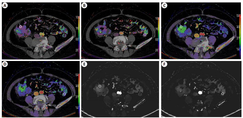
图 1 经结直肠癌癌灶及正常肠壁层面,取感兴趣区(白色圆圈)
Figure 1. The regions of interest were placed on the cancer foci and normal intestinal wall level (White circles). A: AEF on the cancer lesion level; B: AEF on normal intestinal wall level; C: ECV on the cancer lesion level; D: ECV on the normal intestinal wall level; E: Iodine no water on the cancer lesion level; F: Iodine no water on the normal intestinal wall level.

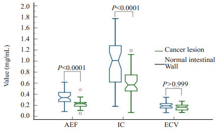
图 2 结直肠癌光谱CT定量增强参数AEF、IC及ECV组间比较
Figure 2. The comparison of spectral CT arterial enhancement fraction, arterial phase iodine concentration and extracellular volume between the colorectal cancer and the normal intestinal wall.

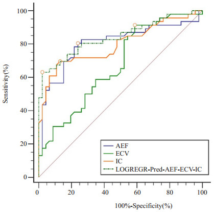
图 3 结直肠癌光谱CT定量增强参数AEF、IC、ECV及三者联合ROC曲线
Figure 3. The ROC analysis of spectral CT arterial enhancement fraction, arterial phase iodine concentration, extracellular volume and three indicators combination in colorectal cancer.
表 1 结直肠癌光谱CT动脉增强函数、动脉期碘浓度及细胞外容积组间比较
Table 1. Comparison of arterial enhancement fraction, arterial phase iodine concentration and extracellular volume of spectral CT between colorectal cancer and the normal intestinal wall (n=46, Mean±SD).
Index Cancer lesion Normal intestinal wall t/U P AEF 0.338±0.122 0.225±0.072 5.433 <0.0001 IC (mg/mL) 1.007±0.402 0.599±0.229 5.983 <0.0001 ECV 0.197±0.059 0.169(0.070, 0.274)* 1058.000 >0.999 AEF: Arterial enhancement fraction; IC: Iodine concentration; ECV: Extracellular volume; *M(Min, Max).  下载: 导出CSV
表 2 结直肠癌光谱CT动脉增强函数、动脉期碘浓度及细胞外容积的ROC曲线分析
Table 2. ROC analysis of arterial enhancement fraction, arterial phase iodine concentration and extracellular volume of spectral CT in colorectal cancer
Index AUC Youden index Sensitivity (%) Specificity (%) Cut-Off AEF 0.806(0.710, 0.881) 0.565 80.43 76.09 0.252 IC 0.797(0.700, 0.874) 0.565 69.57 86.96 0.790 ECV 0.671(0.566, 0.766) 0.326 91.30 41.30 0.128 AEF+IC+ECV 0.842(0.751, 0.909) 0.609 63.04 97.83 0.660
下载: 导出CSV
-
[1] Tang JWC, Lam WWT, Ma ASY, et al. Dietary changes adopted by Chinese colorectal cancer patients: a qualitative study[J]. Eur J Cancer Care, 2019, 28(6): e13159. [2] 田剑波, 温艳, 杨卓煜, 等. 全球结直肠癌筛查指南及共识质量评价[J]. 中华流行病学杂志, 2021, 42(2): 248-57. https://www.cnki.com.cn/Article/CJFDTOTAL-BYDB201903029.htm [3] 李江, 要鹏韬, 牛军强, 等. 结直肠癌筛查领域指南方法学质量的系统评价[J]. 中华预防医学杂志, 2019, 53(4): 398-404. doi: 10.3760/cma.j.issn.0253-9624.2019.04.013 [4] Kontovounisios C, Tan E, Pawa N, et al. The selection process can improve the outcome in locally advanced and recurrent colorectal cancer: activity and results of a dedicated multidisciplinary colorectal cancer centre[J]. Colorectal Dis, 2017, 19(4): 331-8. doi: 10.1111/codi.13517 [5] McCollough CH, Leng S, Yu LF, et al. Dual- and multi-energy CT: principles, technical approaches, and clinical applications[J]. Radiology, 2015, 276(3): 637-53. doi: 10.1148/radiol.2015142631 [6] Ananthakrishnan L, Rajiah P, Ahn R, et al. Spectral detector CT-derived virtual non-contrast images: comparison of attenuation values with unenhanced CT[J]. Abdom Radiol, 2017, 42(3): 702-9. doi: 10.1007/s00261-016-1036-9 [7] Zhuo SQ, Ma HL, Liu ZL, et al. Evaluation of a second-generation monoenergetic reconstruction algorithm for lesion contrast and venous invasion in pancreatic ductal adenocarcinomas[J]. 2019, 23 (21): 9341-50. [8] Lu XM, Lu ZM, Yin JD, et al. Effects of radiation dose levels and spectral iterative reconstruction levels on the accuracy of iodine quantification and virtual monochromatic CT numbers in duallayer spectral detector CT: an iodine phantom study[J]. Quant Imaging Med Surg, 2019, 9(2): 188-200. doi: 10.21037/qims.2018.11.12 [9] Nagayama Y, Iyama A, Oda S, et al. Dual-layer dual-energy computed tomography for the assessment of hypovascular hepatic metastases: impact of closing k-edge on image quality and lesion detectability[J]. Eur Radiol, 2019, 29(6): 2837-47. doi: 10.1007/s00330-018-5789-0 [10] Brun AM, Dyveke E, Jesper T, et al. Impact of spectral body imaging in patients suspected for occult cancer: a prospective study of 503 patients[J]. Eur Radiol, 2020, 30(10): 1-12. [11] Liu JJ, Liu W, Jin ZY, et al. Improved visualization of gastric cancer and increased diagnostic performance in lesion depiction and depth identification using monoenergetic reconstructions from a novel dual-layer spectral detector CT[J]. Acad Radiol, 2020, 27(6): e140-7. doi: 10.1016/j.acra.2019.09.004 [12] Sauter AP, Kössinger A, Beck S, et al. Dual-energy CT parameters in correlation to MRI-based apparent diffusion coefficient: evaluation in rectal cancer after radiochemotherapy[J]. Acta Radiol Open, 2020, 9(9). [13] Longarino Friderike K, Thomas T, Stewart M, et al. Dual-layer spectral CT for proton, helium, and carbon ion beam therapy planning of brain tumors[J]. J Appl Clin Med Phys, 2021, 23(1): e13465. [14] Johnson TRC. Dual-energy CT: general principles[J]. Am J Roentgenol, 2012, 199(5): S3-8. [15] 中华放射学杂志双层探测器光谱CT临床应用协作组. 双层探测器光谱CT临床应用中国专家共识(第一版)[J]. 中华放射学杂志, 2020, 54(7): 635-43. [16] 谭晶文, 朱兰, 王兰, 等. 新型双层探测器光谱CT在直肠癌术前T分期中的价值[J]. 中华放射学杂志, 2020, 54(7): 671-6. [17] 杨琰昭, 徐嘉旭, 李若坤, 等. 双层探测器光谱CT能谱图像在胰腺神经内分泌肿瘤检出中的应用价值[J]. 中华放射学杂志, 2020, 54(6): 534-8. [18] Mao XN, Guo Y, Lu ZM, et al. Enhanced CT textures derived from computer mathematic distribution analysis enables arterial enhancement fraction being an imaging biomarker option of hepatocellular carcinoma[J]. Front Oncol, 2020, 10: 1337. doi: 10.3389/fonc.2020.01337 [19] Kim KW, Lee JM, Klotz E, et al. Quantitative CT color mapping of the arterial enhancement fraction of the liver to detect hepatocellular carcinoma[J]. Radiology, 2009, 250(2): 425-34. doi: 10.1148/radiol.2501072196 [20] 容鹏飞, 冯智超, 郭睿, 等. CT动脉增强分数评估肝硬化患者肝功能水平[J]. 中南大学学报: 医学版, 2019, 44(5): 469-76. https://www.cnki.com.cn/Article/CJFDTOTAL-HNYD201905002.htm [21] 冯智超, 朱文卫, 刘倩云, 等. 动脉增强分数定量彩图对肝脏局灶性结节增生与肝细胞癌的鉴别诊断价值[J]. 临床放射学杂志, 2017, 36 (2): 231-5. https://www.cnki.com.cn/Article/CJFDTOTAL-LCFS201702020.htm [22] Mao XN, Guo Y, Wen F, et al. Applying arterial enhancement fraction (AEF) texture features to predict the tumor response in hepatocellular carcinoma (HCC) treated with Transarterial chemoembolization (TACE)[J]. Cancer Imaging, 2021, 21(1): 49. [23] 刘洋, 李洪燕, 楚琳, 等. 螺旋CT动脉增强分数在结直肠癌分期中的诊断价值[J]. 肿瘤研究与临床, 2022, 34(9): 674-8. [24] 王警建, 李娜, 王龙龙, 等. 能谱CT鉴别肺部炎性病变和肺癌的临床价值[J]. 中国医学物理学杂志, 2018, 35(10): 1164-8. https://www.cnki.com.cn/Article/CJFDTOTAL-YXWZ201810010.htm [25] 杨峰峰, 董杰, 闫晓龙, 等. 能谱CT定量参数: 术前诊断肺癌转移性淋巴结的价值[J]. 中国肺癌杂志, 2016, 19(11): 738-45. https://www.cnki.com.cn/Article/CJFDTOTAL-FAIZ201611006.htm [26] 王宝玲, 周连新. 螺旋CT、能谱CT和MRI诊断原发性肝癌的临床价值比较[J]. 实用肝脏病杂志, 2016, 19(4): 467-70. https://www.cnki.com.cn/Article/CJFDTOTAL-GBSY201604020.htm [27] 赵云松, 张慧滔, 赵星, 等. 双能谱CT的迭代重建模型及重建方法[J]. 电子学报, 2014, 42(4): 666-71. https://www.cnki.com.cn/Article/CJFDTOTAL-DZXU201404007.htm [28] Sauter AP, Kopp FK, Münzel D, et al. Accuracy of iodine quantification in dual-layer spectral CT: influence of iterative reconstruction, patient habitus and tube parameters[J]. Eur J Radiol, 2018, 102: 83-8. [29] Ehn S, Sellerer T, Muenzel D, et al. Assessment of quantification accuracy and image quality of a full-body dual-layer spectral CT system[J]. J Appl Clin Med Phys, 2018, 19(1): 204-17. [30] Sadoughi N, Krishna S, MacDonald DB, et al. Diagnostic accuracy of attenuation difference and iodine concentration thresholds at rapid-kilovoltage-switching dual-energy CT for detection of enhancement in renal masses[J]. Am J Roentgenol, 2019, 213(3): 619-25. [31] Lourenco PDM, Rawski R, Mohammed MF, et al. Dual-energy CT iodine mapping and 40-keV monoenergetic applications in the diagnosis of acute bowel ischemia[J]. Am J Roentgenol, 2018, 211(3): 564-70. [32] Weidman EK, Plodkowski AJ, Halpenny DF, et al. Dual-energy CT angiography for detection of pulmonary emboli: incremental benefit of iodine maps[J]. Radiology, 2018, 289(2): 546-53. [33] Thaiss WM, Haberland U, Kaufmann S, et al. Dose optimization of perfusion-derived response assessment in hepatocellular carcinoma treated with transarterial chemoembolization: comparison of volume perfusion CT and iodine concentration[J]. Acad Radiol, 2019, 26(9): 1154-63. [34] 孙雯雯, 王哲海. 表皮生长因子受体单克隆抗体疗效预测因素的研究进展[J]. 中华临床医师杂志: 电子版, 2013, 7(18): 183-4. https://www.cnki.com.cn/Article/CJFDTOTAL-ZLYD201318071.htm [35] 哈力木拉提·吾布力卡斯木, 吾买尔江·买买提, 段绍斌. 癌相关成纤维细胞异质性的研究进展[J]. 肿瘤学杂志, 2021, 27(3): 222-5. https://www.cnki.com.cn/Article/CJFDTOTAL-XHON202103014.htm [36] 郑文霞, 王莉莉, 陈杏彪, 等. 光谱CT量化的细胞外容积评估结直肠癌神经、血管及淋巴管浸润[J]. 中国医学影像学杂志, 2022, 30(9): 896-902. https://www.cnki.com.cn/Article/CJFDTOTAL-ZYYZ202209018.htm [37] Bandula S, Punwani S, Rosenberg WM, et al. Equilibrium contrastenhanced CT imaging to evaluate hepatic fibrosis: initial validation by comparison with histopathologic sampling[J]. Radiology, 2015, 275(1): 136-43. -

 点击查看大图
计量
- 文章访问数: 104
- HTML全文浏览量: 50
- PDF下载量: 6
- 被引次数: 0