


Application value of MRI in the treatment of ectopic pregnancy in uterus by high-intensity focused ultrasound ablation
-
摘要:
目的 探讨MRI在高强度聚焦超声消融(HIFU)治疗子宫内异位妊娠中的应用价值。 方法 回顾性分析2016年12月~2021年12月在佛山市妇幼保健院经HIFU治疗的5例子宫内异位妊娠患者资料, 中位年龄35岁, 所有患者在术前及术后均行MRI检查。观察术前、术后MRI图像, 了解妊娠囊部位、大小、形态、生长方式、信号特点、妊娠囊附着处子宫肌层改变、强化及其HIFU术后MRI表现。 结果 HIFU术前MRI示4例为瘢痕妊娠(CSP), 1例为宫颈妊娠; 2例妊娠囊信号均匀, 3例妊娠囊信号不均; CSP患者子宫前壁肌层明显变薄, 厚度1~2 mm。强化: 1例CSP患者妊娠囊未见强化, 但宫旁可见多发增粗、迂曲强化血管影; 余3例CSP患者妊娠囊内可见条索状、斑片状明显强化; 宫颈妊娠患者见妊娠囊壁均匀强化。HIFU术后MRI示: 4例妊娠囊均较前缩小, 1例妊娠囊较前无明显改变; 5例病灶信号均较前升高/不均匀; 1例CSP妊娠囊术前强化灶未见显示; 1例CSP患者术前无强化, 但术后宫旁增粗、迂曲强化血管较前减少, 强化程度减弱。另2例CSP妊娠囊仍见少许条索状、斑片状强化, 但强化范围较前缩小、部分坏死改变。宫颈妊娠患者妊娠囊壁未见强化。 结论 MRI术前检查能明确妊娠囊位置及信号情况, 有助于HIFU术前准确定位, 且能显示妊娠囊与周围组织的关系; 术后动态增强扫描可判断HIFU治疗疗效。 Abstract:Objective To explore the application value of MRI in high-intensity focused ultrasound ablation (HIFU) ablation of intrauterine ectopic pregnancy. Methods The data of five patients with intrauterine ectopic pregnancy treated by HIFU ablation in Foshan Women and Children Hospital from December 2016 to December 2021 were analyzed retrospectively.All the patients had median age of 35 years old and underwent MRI before and after operation.We observed the preoperative and postoperative MRI images with location, size, shape, growth mode and signal intensity characteristics of gestational sacs, the changes and enhancement of myometrium at the attachment of gestational sacs, and MRI findings after HIFU. Results MRI before HIFU showed that 4 cases were cesareanscar pregnancy (CSP) and 1 case was cervical pregnancy.2 cases gestational sac signal were homogeneous and 3 cases were inhomogeneous.In CSP patients, the anterior uterine wall muscle layer was significantly thinner, with a thickness of 1-2 mm.In 1 CSP patient, there was no enhancement of pregnancy sac, but multiple thickening and tortuous enhanced vascular shadow were seen near the uterus.In the other 3 CSP patients, there was obvious enhancement of strip-like and patchy lesions in the pregnancy sac.In cervical pregnancy, the wall of the pregnancy sac was uniformly strengthened.MRI after HIFU showed that 4 cases of pregnancy sac were smaller than before, and 1 case had no significant change.The signal intensity of 5 cases ware higher/inhomogeneous than before.No enhancement lesion was found in 1 case of CSP pregnancy sac before operation.1 case of CSP patient had no enhancement before operation, but the periuterine thickening and tortuous enhancement vessels decreased after operation, and the enhancement degree was weakened.In the other 2 cases of CSP gestational sac, there were still some cord-like and patchy enhancement, but the enhancement range was smaller than before, and some necrosis changes were found.There was no enhancement of gestational sac wall in patients with cervical pregnancy. Conclusion MRI preoperative examination can clearly determine the position and signal of gestational sac, which is helpful to accurately locate HIFU before operation, and can show the relationship between gestational sac and surrounding tissues.Postoperative dynamic contrast-enhanced scanning can judge the therapeutic effect of HIFU. -

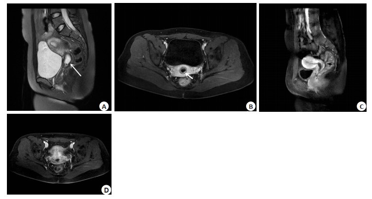
图 1 41岁女性CSP患者
Figure 1. A 41-year-old female patient with CSP. T2WI showed mixed signal masses in the scar of lower uterine segment (arrow in A, B), the lesion showed cord-like and patchy enhancement, and the periuterine vessels were thickened and tortuous (arrow in C). After HIFU treatment, the enhanced scan showed that there was no enhanced focus in the gestational sac, and the enhanced focus was necrotic before HIFU treatment (D).

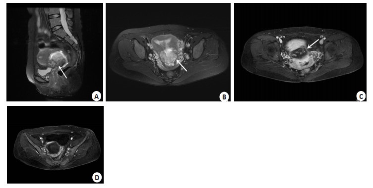
图 2 23岁女性,宫颈妊娠
Figure 2. Female, 23 years old, cervical pregnancy. Preoperative T2WI sagittal, cervical cystic mass (arrow in A); Preoperative contrast-enhanced scan, ring-like homogeneous enhancement of the gestational sac wall (B). Postoperative HIFU, in contrast-enhanced sagittal view, the gestational sac was significantly smaller than that before HIFU (C). After HIFU, the gestational sac wall was not enhanced on contrast-enhanced scan (D).
-
[1] 朱颖, 王霄英. 异位妊娠的MRI诊断及鉴别诊断[J]. 实用放射学杂志, 2016, 32(3): 388-91. doi: 10.3969/j.issn.1002-1671.2016.03.016 [2] 靳金岩, 邢健, 杨景堯, 等. 3.0T磁共振在异位妊娠诊断的临床价值分析[[J]. 中国CT和MRI杂志, 2020, 18(10): 112-5. https://www.cnki.com.cn/Article/CJFDTOTAL-CTMR202010033.htm [3] 贺成英. 高龄产妇不同部位异位妊娠的MRI影像特点及诊断准确度分析[J]. 湖南师范大学学报: 医学版, 2019, 16(4): 147-51. https://www.cnki.com.cn/Article/CJFDTOTAL-HNYG201904048.htm [4] 董天发, 吴美仙, 陈永露, 等. 子宫内异位妊娠MRI表现及临床意义[J]. 南昌大学学报: 医学版, 2017, 57(3): 31-5. https://www.cnki.com.cn/Article/CJFDTOTAL-JXYB201703009.htm [5] 孙丽, 高毅. DCE-MRI在高强度聚焦子宫肌瘤消融术疗效评估中的应用价值[J]. 中国CT和MRI杂志, 2021, 19(3): 107-9. https://www.cnki.com.cn/Article/CJFDTOTAL-CTMR202103037.htm [6] 张玉婷, 罗欣, 明雪, 等. 高强度聚焦超声联合术后清宫治疗胎盘植入的有效性研究[J]. 中国医药导报, 2019, 16(11): 69-72, 76. https://www.cnki.com.cn/Article/CJFDTOTAL-YYCY201911018.htm [7] 冯敏清, 张婧, 陈向东, 等. MRI在高强度聚焦超声消融治疗产后胎盘植入中的应用价值[J]. 实用放射学杂志, 2023, 39(2): 255-8. [8] 吴娜, 严凤, 李全香. 海扶刀联合宫腔镜治疗瘢痕型妊娠的疗效及安全性[J]. 中国性科学, 2020, 29(3): 102-5. https://www.cnki.com.cn/Article/CJFDTOTAL-XKXZ202003032.htm [9] 张晓荣, 马艳伟, 董渠龙, 等. 高强度聚焦超声联合清宫术治疗宫颈妊娠一例[J]. 国际妇产科学杂志, 2021, 48(5): 525-7. https://www.cnki.com.cn/Article/CJFDTOTAL-GWVC202105012.htm [10] Uysal F, Uysal A, Adam G. Cesarean scar pregnancy[J]. J Ultrasound Med, 2013, 32(7): 1295-300. [11] 孙婷, 许永红. 超声诊断少见类型异位妊娠的相关影像学特征分析[J]. 中国计划生育学杂志, 2019, 27(1): 111-3. https://www.cnki.com.cn/Article/CJFDTOTAL-JHSY201901035.htm [12] 徐嘉璐, 孙明华, 朱家樑, 等. 磁共振检查在宫角妊娠诊断及鉴别诊断中的价值[J]. 中国医学计算机成像杂志, 2017, 23(6): 535-8. https://www.cnki.com.cn/Article/CJFDTOTAL-YJTY201706010.htm [13] Marques A, Andres MP, Kho RM, et al. Is high-intensity focused ultrasound effective for the treatment of adenomyosis? A systematic review and meta-analysis[J]. J Minim Invasive Gynecol, 2020, 27(2): 332-43. [14] 朱颖, 王岑. 磁共振检查在妊娠期的应用[J]. 中华围产医学杂志, 2020, 23(3): 150-4. https://www.cnki.com.cn/Article/CJFDTOTAL-JXYY202109066.htm [15] 潘志立, 吕维富, 刘影. 子宫切口妊娠的MRI表现[J]. 医学影像学杂志, 2014, 24(7): 1203-6. https://www.cnki.com.cn/Article/CJFDTOTAL-XYXZ201407037.htm [16] 张向群, 许乙凯, 罗小琴. 子宫切口瘢痕内妊娠的MR影像分析[J]. 中华放射学杂志, 2012, 46(9): 812-5. [17] 何慧, 顾晗, 何玲, 等. 磁共振对剖宫产瘢痕妊娠的诊断及临床价值[J]. 磁共振成像, 2020, 11(3): 207-10. https://www.cnki.com.cn/Article/CJFDTOTAL-CGZC202003010.htm [18] 张伟. 剖宫产后子宫瘢痕妊娠异常供血的数字减影血管造影表现及相关研究[J]. 实用放射学杂志, 2020, 36(7): 1117-20, 1128. [19] 陈丽平, 吕发金, 郑伊能, 等. 高强度聚焦超声治疗子宫肌瘤疗效的影响因素研究[J]. 磁共振成像, 2020, 11(11): 1019-22. https://www.cnki.com.cn/Article/CJFDTOTAL-CGZC202011013.htm [20] 陆安伟, 秦娟. 宫颈妊娠诊治策略[J]. 中国实用妇科与产科杂志, 2017, 33(9): 900-3. https://www.cnki.com.cn/Article/CJFDTOTAL-ZGSF201709007.htm -

 下载:
下载:
 点击查看大图
计量
- 文章访问数: 65
- HTML全文浏览量: 126
- PDF下载量: 5
- 被引次数: 0