


Dynamic and static low-frequency amplitude combined functional connectivity in primary insomnia
-
摘要:
目的 采用动态和静态低频振幅联合功能链接方法探讨原发性失眠(PI)患者静息状态下自发脑功能活动特征。 方法 收集20例原发性失眠患者(失眠组)、20例健康志愿者(对照组)的临床量表(匹兹堡睡眠质量指数量表、抑郁自评量表、焦虑自评量表,失眠患者同时完成失眠严重指数量表)评分结果和功能性磁共振成像数据,采用动态和静态低频振幅(ALFF)方法分析两组间自发脑功能活动的差异,进一步分析差异脑区的信号值与失眠的相关性。再基于动态ALFF差异脑区与全脑其余体素进行基于种子的功能连接分析。 结果 与对照组相比,PI组左侧额上回动态ALFF值增加,提示该区大脑内在活动的时间变异性升高,且其与失眠严重指数量表间呈负相关关系(r=-0.463,P=0.04)。与对照组相比,PI组右侧颞中回、左侧额上回静态ALFF值增加,左侧扣带回、左侧中央前回、右侧额叶补充运动区静态ALFF值减少。以左侧额上回为种子点进行与全脑体素的相关性分析,相对于对照组,PI组功能连接上升的脑区主要有:左侧眶部额中回、右侧尾状核、右侧眶部额中回、左侧背外侧额上回。 结论 PI患者在动态ALFF方面存在局部脑活动的改变且与失眠指数具有相关性,联合功能连接可以更全面地显示PI患者的异常脑功能活动,提示功能性磁共振成像可在一定程度上评价PI患者自发脑神经功能活动改变的情况。 Abstract:Objective To investigate the characteristics of spontaneous brain function activity in the resting state of primary insomnia (PI) patients using a combined dynamic and static low-frequency amplitude functional linkage method. Methods The scores and fMRI data of clinical scales (Pittsburgh sleep quality index scale, depression self-rating scale, anxiety self-rating scale, insomnia patients also completed the insomnia severity index scale) were collected from 20 PI patients (PI group) and 20 healthy volunteers (control group). The differences in spontaneous brain functional activity between the two groups were analyzed using dynamic and static low frequency amplitude (ALFF) methods, further analysing the correlation between the signal values of the differential brain regions and insomnia. A seed-based functional connectivity analysis was performed between the differential brain regions based on dynamic ALFF and the remaining voxels of the whole brain. Results Compared to normal controls, the PI group had increased dynamic ALFF values in the left superior frontal gyrus, suggesting increased temporal variability in intrinsic brain activity in this region. A significant negative correlation was found with insomnia severity index (r=-0.463, P=0.04). Compared to the control group, the PI group showed increased static ALFF values in the right middle temporal gyrus and left superior frontal gyrus and decreased static ALFF values in the left cingulate gyrus, left precentral gyrus and right frontal supplementary motor area. Correlation analysis with whole brain voxels was performed using the left superior frontal gyrus as the seed point, and the main brain regions with increased functional connectivity in the PI group relative to the HC group were: left orbital middle frontal gyrus, right caudate nucleus, right orbital middle frontal gyrus, and left dorsolateral superior frontal gyrus. Conclusion The presence of localised changes in brain activity in PI patients in terms of dynamic low-frequency amplitude correlates with the insomnia index, and combined functional connectivity can provide a more comprehensive picture of abnormal brain function activity in PI patients. It is suggested that fMRI can evaluate to some extent the altered spontaneous neurological activity in PI patients. -

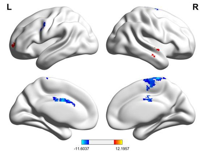
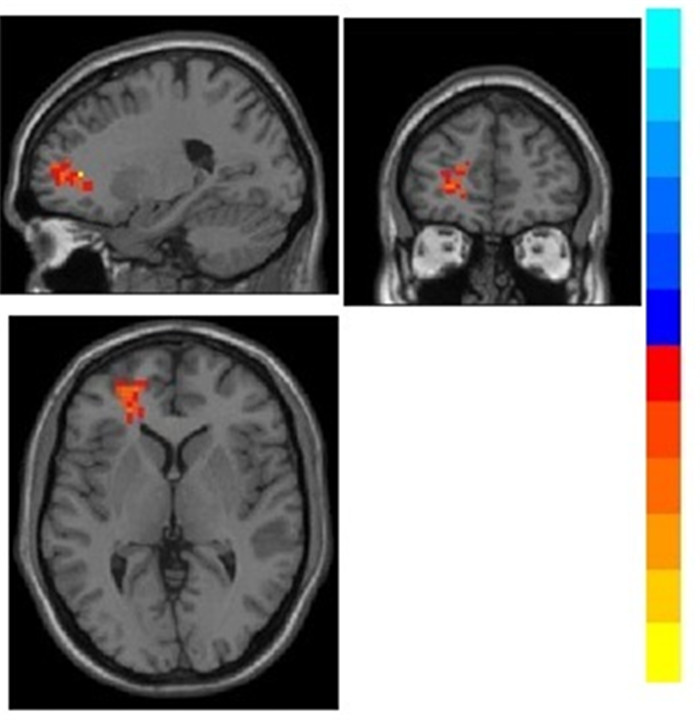
图 1 动态ALFF时间变异性有显著差异的脑区
Figure 1. Brain regions with significant differences in dynamic ALFF temporal variability.
表 1 两组临床资料比较
Table 1. Comparison of clinical data between the two groups (n=20)
Index PI group HC group P Age (years, Mean±SD) 49.10±11.37 50± 11.44 0.15 Gender(Male/Female, n) 6/14 7/13 0.736 Years of education (years, Mean±SD) 12±3 15±2.5 0.20 Course (month) 19.65 - - ISI (Mean±SD) 14.55±2.625 - - PSQI (Mean±SD) 15.73±2.70 3.05±1.15 0.004 SAS[M(P25, P75)] 40(31, 50) 31.65±5.50 0.001 SDS[M(P25, P75)] 47(35, 55) 35±7.42 0.017 PI: Primary insomnia; ISI: Insomnia severity index; PSQI: Pittsburgh sleep quality index; SAS: Self-rating anxiety scale; SDS: Self-rating depression scale.  下载: 导出CSV
表 2 PI组与HC组的差异脑区
Table 2. Differential brain areas in the PI and HC groups
Area Serial Position T X Y Z Middle Temporal Gyrus-R 46 60 -6 -6 11.6257 Frontal Lobe-R 36 -21 51 3 9.6073 Limbic Lobe-L 72 -3 0 33 -10.6933 Precentral_L (aal) 36 -54 9 39 -10.3717 Supp_Motor_Area_R (aal) 175 6 -30 75 -10.8611
下载: 导出CSV
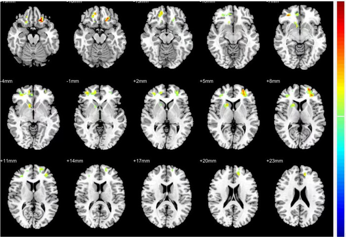
表 3 以左侧额上回为种子点时PI组功能连接的异常脑区
Table 3. Abnormal brain areas in the PI group functional connectivity when the left superior frontal gyrus was used as the seed point.
Area Serial Position T X Y Z Frontal_Mid_Orb_L (aal) 36 -18 27 -18 4.9294 Caudate_R (aal) 49 9 13 -3 4.0569 Middle Frontal Gyrus 135 33 42 -6 4.2781 Frontal_Sup_L (aal) 141 -24 57 9 4.3894
下载: 导出CSV
-
[1] Association AP. Diagnostic and Statistical Manual of Mental Disorders, Fourth Edition, Text Revision (DSM-IV-TR)[M]. Arlington, VA: American Psychiatric Association, 2000. [2] Covassin N, de Zambotti M, Sarlo M, et al. Cognitive performance and cardiovascular markers of hyperarousal in primary insomnia[J]. Int J Psychophysiol, 2011, 80(1): 79-86. doi: 10.1016/j.ijpsycho.2011.02.005 [3] Damaraju E, Allen EA, Belger A, et al. Dynamic functional connectivity analysis reveals transient states of dysconnectivity in schizophrenia[J]. Neuroimage Clin, 2014, 5: 298-308. doi: 10.1016/j.nicl.2014.07.003 [4] Zang YF, He Y, Zhu CZ, et al. Altered baseline brain activity in children with ADHD revealed by resting-state functional MRI[J]. Brain Dev, 2007, 29(2): 83-91. doi: 10.1016/j.braindev.2006.07.002 [5] Hou YN, Wu XM, Hallett M, et al. Frequency-dependent neural activity in Parkinson's disease[J]. Hum Brain Mapp, 2014, 35(12): 5815-33. doi: 10.1002/hbm.22587 [6] Drevets WC, Videen TO, Price JL, et al. A functional anatomical study of unipolar depression[J]. J Neurosci, 1992, 12(9): 3628-41. doi: 10.1523/JNEUROSCI.12-09-03628.1992 [7] Stephan KE, Marshall JC, Penny WD, et al. Interhemispheric integration of visual processing during task-driven lateralization[J]. J Neurosci, 2007, 27(13): 3512-22. doi: 10.1523/JNEUROSCI.4766-06.2007 [8] Li J, Duan XJ, Cui Q, et al. More than just statics: temporal dynamics of intrinsic brain activity predicts the suicidal ideation in depressed patients[J]. Psychol Med, 2018, 49(5): 852-60. [9] Fu ZN, Tu YH, Di X, et al. Characterizing dynamic amplitude of low-frequency fluctuation and its relationship with dynamic functional connectivity: an application to schizophrenia[J]. Neuroimage, 2018, 180(Pt B): 619-31. [10] Friston KJ. Functional and effective connectivity: a review[J]. Brain Connect, 2011, 1(1): 13-36. doi: 10.1089/brain.2011.0008 [11] Van Dijk KRA, Hedden T, Venkataraman A, et al. Intrinsic functional connectivity as a tool for human connectomics: theory, properties, and optimization[J]. J Neurophysiol, 2010, 103(1): 297-321. doi: 10.1152/jn.00783.2009 [12] Gonzalez-Castillo J, Hoy CW, Handwerker DA, et al. Tracking ongoing cognition in individuals using brief, whole-brain functional connectivity patterns[J]. Proc Natl Acad Sci USA, 2015, 112(28): 8762-7. doi: 10.1073/pnas.1501242112 [13] 聂晓, 彭德昌, 李海军, 等. 原发性失眠的不同频段低频振幅静息态功能磁共振研究[J]. 中国医学影像技术, 2016, 32(2): 204-8. doi: 10.13929/J.1003-3289.2016.02.011 [14] Briggs RG, Khan AB, Chakraborty AR, et al. Anatomy and white matter connections of the superior frontal gyrus[J]. Clin Anat, 2020, 33(6): 823-32. doi: 10.1002/ca.23523 [15] 谭志, 骆俊佳, 罗树存, 等. 低频振幅联合功能连接对原发性失眠的研究[J]. 中国CT和MRI杂志, 2022(2): 1-4. https://www.cnki.com.cn/Article/CJFDTOTAL-CTMR202202001.htm [16] 王钰凯, 李铁, 莽靖, 等. 基于静息态功能磁共振的健康人与原发性失眠患者的影像学研究[J]. 中国实验诊断学, 2018, 22(11): 1976-80. https://www.cnki.com.cn/Article/CJFDTOTAL-ZSZD201811036.htm [17] 黄海军, 王勇. 基于全脑静息态fMRI评价针刺对原发性失眠病人脑区功能的影响[J]. 中西医结合心脑血管病杂志, 2019, 17(19): 3040-3. doi: 10.12102/j.issn.1672-1349.2019.19.047 [18] 姚慷, 周群, 陈丽君. 原发性失眠患者工作记忆的研究[J]. 精神医学杂志, 2016, 29(3): 168-9. doi: 10.3969/j.issn.2095-9346.2016.03.003 [19] 窦社伟, 王恩锋, 张红菊, 等. 空间工作记忆任务态fMRI在原发性失眠患者认知功能障碍评价中的应用[J]. 中华医学杂志, 2015, 95(21): 1677-80. doi: 10.3760/cma.j.issn.0376-2491.2015.21.014 [20] 张亮, 李则宣, 路晓文, 等. 左侧额上回网络节点高效率值与抑郁症症状特征的关系[J]. 中南大学学报: 医学版, 2022, 47(3): 289-300. https://www.cnki.com.cn/Article/CJFDTOTAL-HNYD202203003.htm [21] 何全兴, 李曼, 林琳, 等. 儿童期虐待对状态焦虑的影响: 左额上回的中介作用[C]//中国心理学会第二十四届全国心理学学术会议摘要集, 2022: 1389-90. [22] Wang TY, Li SM, Jiang GH, et al. Regional homogeneity changes in patients with primary insomnia[J]. Eur Radiol, 2016, 26(5): 1292-300. doi: 10.1007/s00330-015-3960-4 [23] 曾少庆, 黎程, 江桂华, 等. 原发性失眠局部脑区功能变化的静息态功能磁共振成像研究[J]. 功能与分子医学影像学杂志: 电子版, 2015, 4(4): 7-12. https://www.cnki.com.cn/Article/CJFDTOTAL-GNFZ201504002.htm [24] Killgore WDS, Schwab ZJ, Kipman M, et al. Insomnia-related complaints correlate with functional connectivity between sensory-motor regions[J]. NeuroReport, 2013, 24(5): 233-40. doi: 10.1097/WNR.0b013e32835edbdd [25] Kessler RC, Gruber M, Hettema JM, et al. Co-morbid major depression and generalized anxiety disorders in the National Comorbidity Survey follow-up[J]. Psychol Med, 2008, 38(3): 365-74. doi: 10.1017/S0033291707002012 [26] Jiang GH, Li C, Ma XF, et al. Abnormal spontaneous regional brain activity in primary insomnia: a resting-state functional magnetic resonance imaging study[J]. Neuropsychiatr Dis Treat, 2016: 1371. doi: 10.2147/NDT.S109633 [27] Dai XJ, Gong HH, Wang YX, et al. Gender differences in brain regional homogeneity of healthy subjects after normal sleep and after sleep deprivation: a resting-state fMRI study[J]. Sleep Med, 2012, 13(6): 720-7. doi: 10.1016/j.sleep.2011.09.019 [28] 杨旭, 赵建农. 轻微肝性脑病患者默认模式网络功能连接研究[J]. 中国CT和MRI杂志, 2018, 16(9): 54-6, 72. https://www.cnki.com.cn/Article/CJFDTOTAL-CTMR201809017.htm [29] 张晓琦. 原发性失眠患者脑结构及静息态脑功能连接的磁共振研究[D]. 郑州: 郑州大学, 2014. [30] Smith EE, Jonides J. Storage and executive processes in the frontal lobes[J]. Science, 1999, 283(5408): 1657-61. [31] 张红菊, 姜晓锋, 王恩峰, 等. 原发性失眠患者前额叶腹外侧区静息态功能连接[J]. 中国临床医生杂志, 2014, 42(10): 51-4. https://www.cnki.com.cn/Article/CJFDTOTAL-ZLYS201410020.htm [32] 蔡万野. 原发性失眠患者白质通路及纹状体功能连接异常的影像学研究[D]. 西安: 西安电子科技大学, 2020. [33] 江琦, 侯璐璐, 邱江, 等. 尾状核-眶部内侧前额叶的功能连接与反应性攻击的关系: 基于静息态功能磁共振研究[J]. 心理学报, 2018, 50(6): 655-66. https://www.cnki.com.cn/Article/CJFDTOTAL-XLXB201806007.htm [34] 姜雨, 程敬亮, 郑瑞平, 等. 伴自杀观念抑郁障碍患者动态和静态低频振幅fMRI研究[J]. 放射学实践, 2022, 37(8): 934-40. https://www.cnki.com.cn/Article/CJFDTOTAL-FSXS202208001.htm -

 点击查看大图
计量
- 文章访问数: 89
- HTML全文浏览量: 46
- PDF下载量: 9
- 被引次数: 0