

 Find Duplicates
 Check Document
 Submission(new)
 Experts Office
 Editorial Office
2023 Vol. 46, No. 6
column
Display Method:
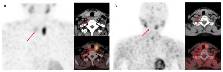
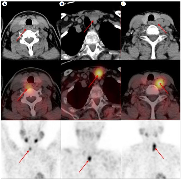
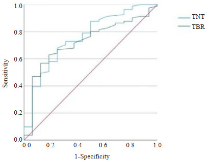
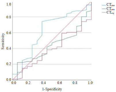
2023, 46(6): 957-963.
doi: 10.12122/j.issn.1674-4500.2023.06.01
Abstract:
Objective To investigate the clinical value of SPECT/CT and CT imaging features in primary hyperparathyroidism (PHPT) with inaccurate imaging location. Methods A total of 90 patients with PHPT confirmed by surgery and pathology were selected and divided into a missed diagnosis group (n=12) and a correct diagnosis group (n=78) according to pathological results, and a parathyroid lesion group (n=81) and a thyroid lesion group (n=29) according to pathological results. The same size areas of interest were delineated in the focal areas of parathyroid, thyroid and contralateral normal areas of parathyroid lesions on the SPECT/CT fusion images, and the radioactive counts of these different sites were recorded, and target-to-nontarget ratio (T/NT) and target-to- non-target ratio (TBR) were calculated. The mean values of maximum CT density (CTmax), minimum CT density (CTmin) and mean CT density (CTavg) were collected at three consecutive layers. The data of each group were compared between groups, the working curve of subjects was drawn, and the diagnostic efficacy of each data was evaluated. Results On 99mTc-MIBI SPECT/CT images, the T/NT, TBR, volume, and diameter of the lesions in the missed group were smaller than those in the correctly diagnosed group, and the difference in T/NT between the two groups was statistically significant (P=0.002) and the diagnostic efficiency was the best, while there was no significant difference in TBR, volume, and diameter between the two groups (P>0.05). There was no significant difference in blood Ca and blood P between the parathyroid lesion group and the thyroid lesion group(P>0.05). The parathyroid hormone, TBR, CTmax, CTmin and CTavg in the thyroid lesion group were all lower than those in the parathyroid lesion group (P < 0.05). The diagnostic efficiency of CTmax was the best, with AUC of 0.623, sensitivity of 74.10% and specificity of 62.50%. Conclusion SPECT/CT has better diagnostic efficacy than TBR in preoperative localization of PHPT, which can provide more valuable reference for preoperative localization of PHPT. It has good clinical value to treat TBR and CTmax with thyroid lesions.
2023, 46(6): 964-969.
doi: 10.12122/j.issn.1674-4500.2023.06.02
Abstract:
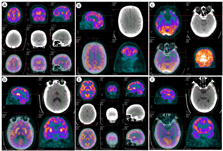
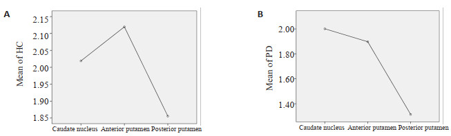
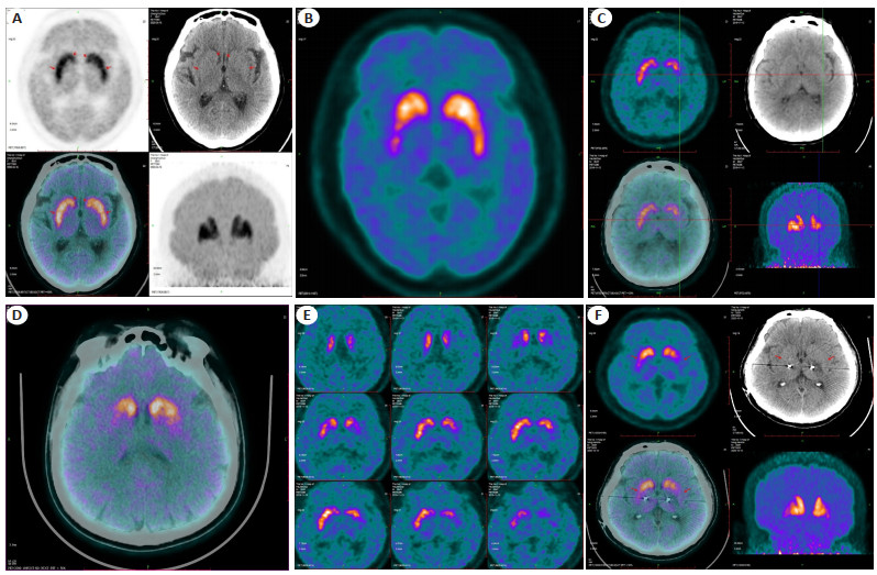
Objective To study the application of 18F-FDG and 11C-CFT PET imaging in the combined diagnosis, evaluation, and differential diagnosis of Parkinson's disease (PD). Methods A total of 107 patients admitted to our hospital from September 2018 to March 2022 who were initially diagnosed as PD according to clinical symptoms and the effectiveness of oral metoba were selected, and 43 healthy subjects were selected as the control group. Both groups underwent 18F-FDG and 11C-CFT PET imaging. 11C- CFT analysis: The PD group was divided into two levels: H&Y 1- 2 (early) and H&Y 3- 5 (late). The 11C- CFT standard uptake values of the caudate nucleus, anterior putamen nucleus, and posterior putamen nucleus were measured in the PD group and the control group. Independent sample t-test was used to compare the PD group and the control group. The caudate nucleus, anterior putamen nucleus, and posterior putamen nucleus were then divided into three groups, Using oneway ANOVA for inter group comparison; 18F-FDG analysis: Analyze the metabolic changes in the putamen, thalamus, caudate nucleus, and frontal parietal temporal occipital lobe based on the patient's motor symptoms. Results 11C-CFT PET imaging: At the early stage of H&Y 1-2, the contralateral posterior putamen of the affected limb showed low metabolism (P < 0.05). Compared with the control group, some patients showed low metabolism in the contralateral anterior putamen and ipsilateral posterior putamen of the affected limb, and the results were statistically significant (P < 0.05), while the ipsilateral anterior putamen and caudate nuclei of the affected limb were basically normal. In H&Y 3- 5 (late stage), the metabolism of the posterior putamen on the opposite side of the affected limb was lower (P < 0.05). The opposite and ipsilateral posterior putamen and anterior putamen of the affected limb exhibited uneven hypometabolism, and the results were statistically significant (P < 0.05); Comparing the caudate nucleus, anterior putamen nucleus, and posterior putamen nucleus, there were differences between the contralateral and ipsilateral groups of the affected limb in the early and late stages (P < 0.05), providing additional evidence that the lesion originated in the posterior putamen nucleus; 18F-FDG PET imaging: 107 Parkinson's patients showed 81.31% increase in putamen metabolism, 54.20% increase in thalamus metabolism, and 26.17% increase in caudate nucleus metabolism. The metabolic reduced area of the cerebral cortex showed 89.72% decrease in parietal lobe metabolism, 57.94% decrease in frontal lobe metabolism, 68.22% decrease in temporal lobe metabolism, and 14.02% decrease in occipital lobe metabolism. Conclusion 18F-FDG combined with 11C-CFT PET binuclear imaging provide objective data for clinical diagnosis, differential diagnosis, and evaluation of PD.
2023, 46(6): 970-977.
doi: 10.12122/j.issn.1674-4500.2023.06.03
Abstract:
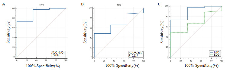
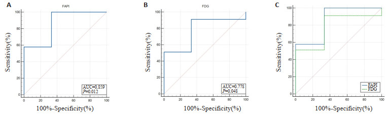
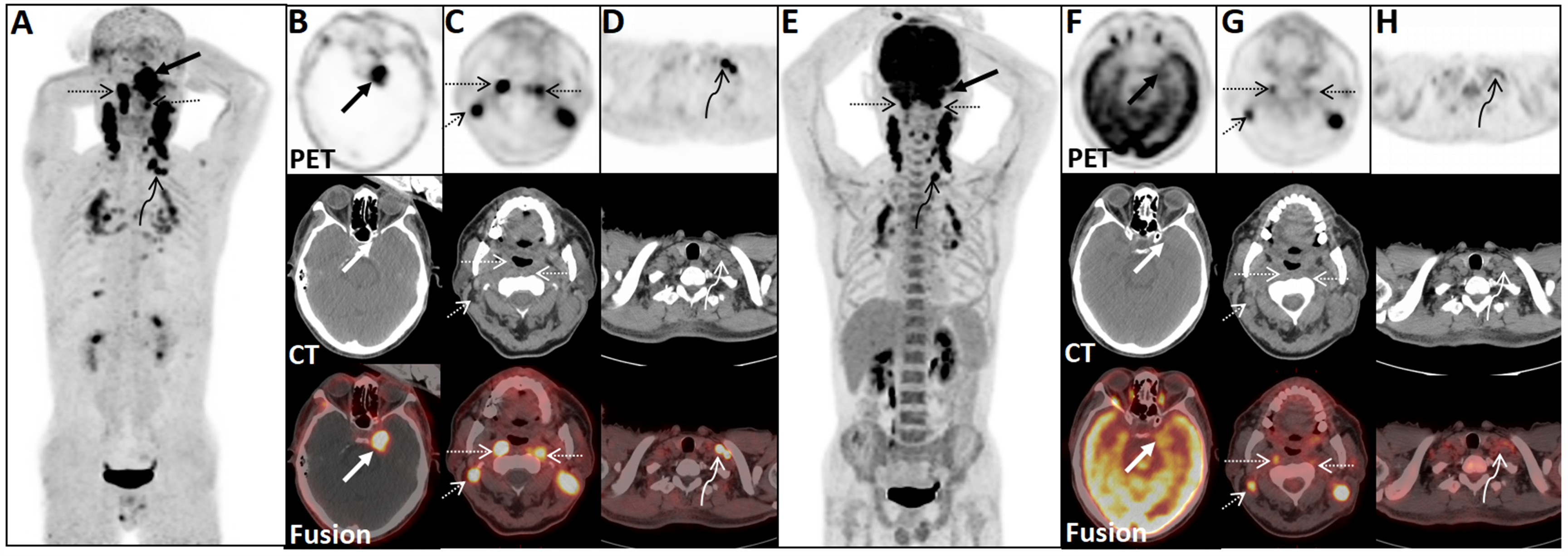
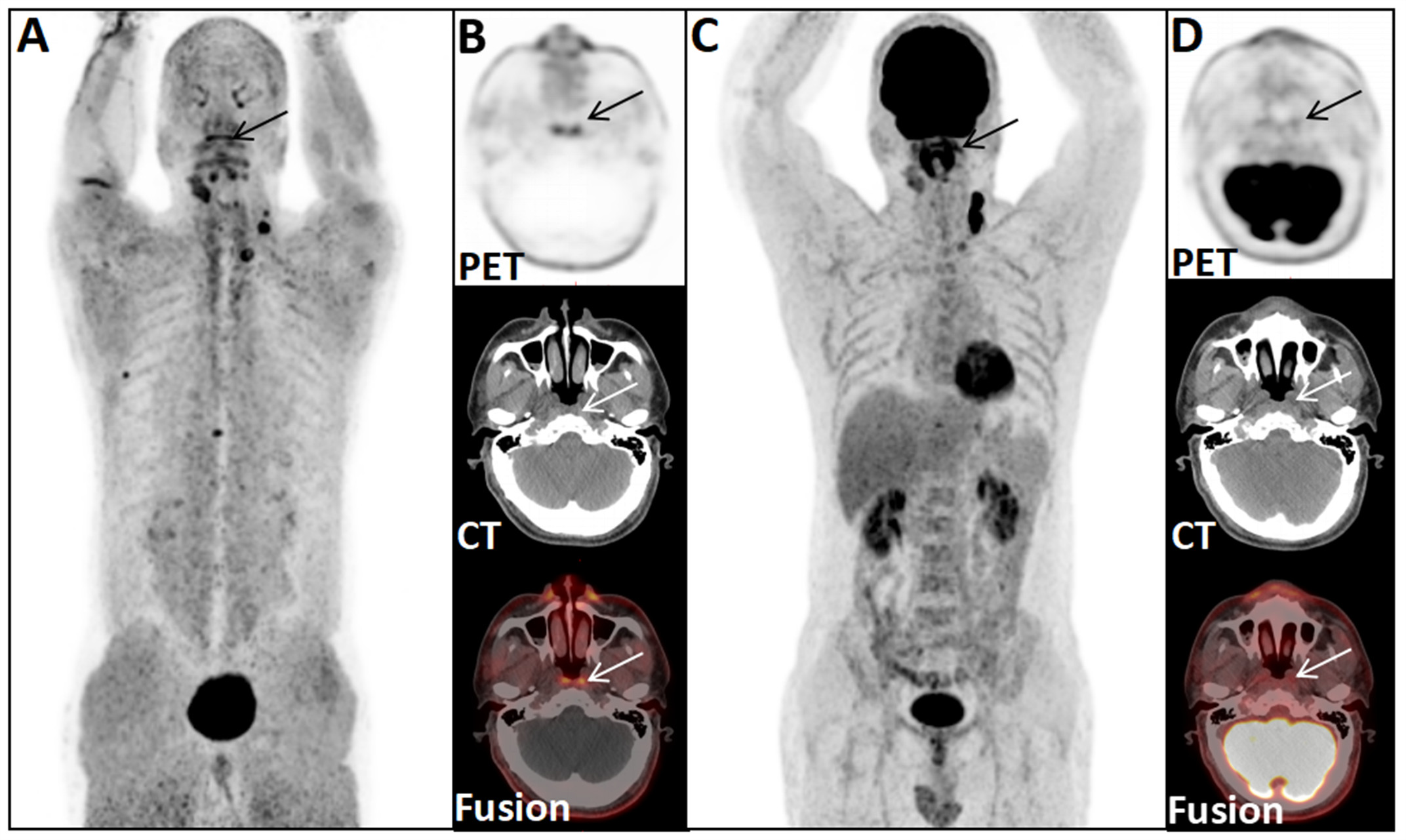
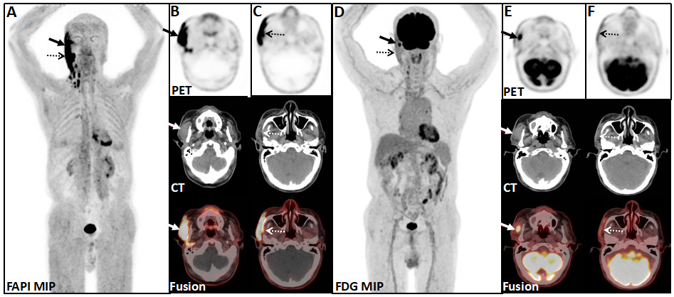
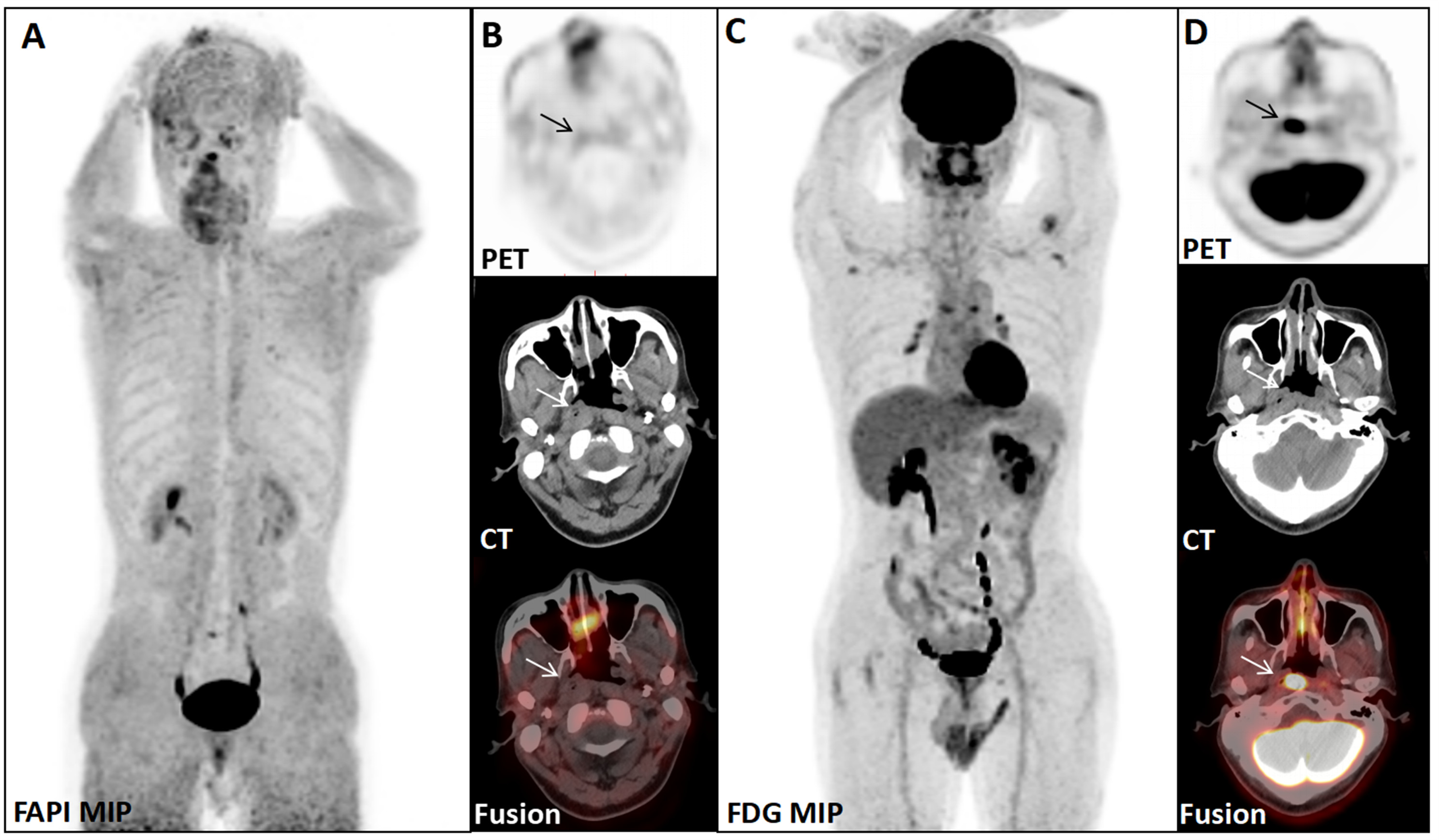
Objective To compare the value of 68Ga-FAPI PET/CT and 18F-FDG PET/CT in the diagnosis and staging of head and neck tumors. Methods Fourty-five patients with head and neck tumors who underwent 68Ga-FAPI and 18F-FDG PET/CT dual imaging in our department were collected from October 2020 to June 2022. All patients were initially staged with two imaging methods. The differences in detection rates of primary lesions and metastatic lesions between 18F-FDG PET/CT and 68Ga-FAPI PET/CT were compared. The differences in maximum standard uptake value (SUVmax) and target to background ratio (TBR) values between the two examination methods were compared. Results The detection rates of primary tumor, lymph node metastasis and distant metastasis by 68Ga-FAPI and 18F-FDG PET/CT were 97.78% and 97.78%, 90.48% and 96.54%, 75% and 87.5%, respectively. The FAPI-SUVmax and FDG-SUVmax of primary tumors were 10.90 and 10.45, respectively, and the difference was not statistically significant (P=0.551). The FAPI-TBR and FDG-TBR were 11.50 and 9.80, respectively, and the difference between two imaging in TBR was statistically significant (P=0.007). 68Ga- FAPI PET/CT upstaged 6 cases of T staging, but underestimated 2 cases of T staging, 1 case of N staging and 1 case of M staging. 18F-FDG PET/CT underestimated 1 case of N staging and 1 case of M staging. 18F-FDG PET/CT overestimated 7 cases of N staging and 1 case of M staging. Conclusion The detection ability of 68Ga-FAPI PET/CT for head and neck tumors was comparable to that of 18F-FDG PET/CT. The detection rates of lymph node metastasis and distant metastasis were lower than those of 18F- FDG PET/CT. However, 68Ga- FAPI PET/CT reduced the detection of false-positive lymph nodes. 68Ga-FAPI PET/CT has a higher accuracy for T-staging, N-staging and Mstaging comparing to 18F-FDG PET/CT.
2023, 46(6): 978-982.
doi: 10.12122/j.issn.1674-4500.2023.06.04
Abstract:
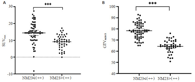
Objective To analyze the influence of NM23 gene expression status on the biological behavior of non-small cell lung cancer and on radiotherapy planning. Methods A total of 107 patients with non-small cell lung cancer in the Cancer Hospital of Xinjiang Medical University were taken as the study subjects, and the radiotherapy protocols were formulated under PET/CT images, the general target area volume outlined under PET/CT images, and the amount of critical organs irradiated, which were divided into high NM23 expression group(n=43) and low NM23 expression group(n=64) according to the results of immunohistochemistry, and the differences in the maximal standardized uptake value of metabolic parameters of PET/CT, the PET/CT images, the volume of the radiotherapy macroscopic target area outlined under the images, and the differences in the amount of critical organs irradiated were compared. Results The maximum standardized uptake value of metabolic parameters measured in PET/CT in the NM23 high-expression group was smaller than that of the low-expression group, and the difference was statistically significant (P < 0.05). In the radiotherapy plans formulated by the NM23 low-expression group and the NM23 high- expression group, the volume of the macroscopic target area, and the volume of the organs at risk of radiation were smaller than that of the low-expression group in the NM23 high-expression group, and the differences were statistically significant (P < 0.05). Conclusion In-depth understanding of NM23 gene expression and its biological behavior in NSCLC patients, and the use of PET/CT in the development of radiotherapy plans can provide more reference information for the precise sculpting of radiotherapy targets in non-small cell lung cancer patients.
2023, 46(6): 983-988.
doi: 10.12122/j.issn.1674-4500.2023.06.05
Abstract:
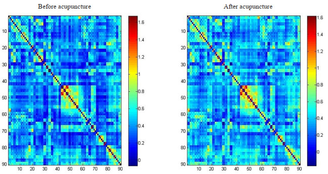
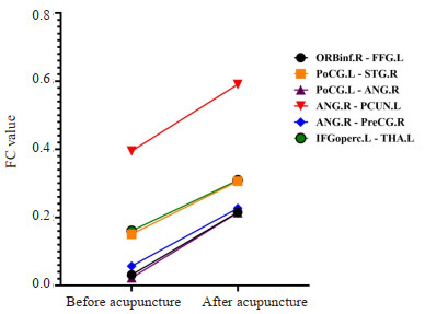
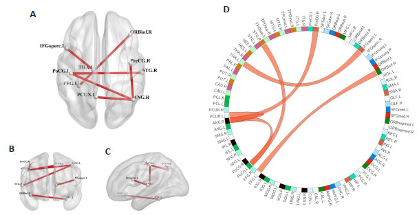
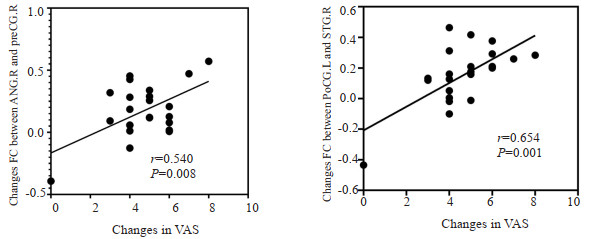
Objective To explore the changes of the rest brain at rest functional connectivity in patients with knee osteoarthritis (KOA) after acupuncture treatment, so as to provide neuroimaging basis for clinical acupuncture treatment of KOA. Methods Twenty-five patients with KOA were enrolled in the study and treated with acupuncture for 3 weeks. The points of Dubi, Xuehai, Yanglingquan, Zusanli, Sanyinjiao, Neixiyan and Liangqiu group were selected for compatibility, 5 times a week, 30 minutes each time. The data of visual analogue scale (VAS), knee joint function scale (KSS) and resting state functional MRI of KOA patients before and after acupuncture were collected, and the changes of functional connectivity of various brain regions in KOA patients after acupuncture were described with the method of edge linking analysis, and the correlation analysis was made with the clinical scores. Results After acupuncture treatment, VAS score of KOA patients decreased significantly and KSS score increased significantly (P < 0.001). Edge linking analysis showed that after acupuncture, functional connectivity of the right orbital inferior frontal gyrus and the left fusiform gyrus, the right angular gyrus and the left anterior cuneiform lobe, the right anterior central gyrus, the left insular inferior frontal gyrus and the left thalamus, the left posterior central gyrus and the right angular gyrus, and the right superior temporal gyrus were enhanced in KOA patients (P < 0.001). Correlation analysis showed that the changes of functional connectivity in right angular gyrus and right anterior central gyrus (r=0.540, P=0.008), left posterior central gyrus and right superior temporal gyrus (r=0.654, P=0.001) were positively correlated with the changes of VAS. Conclusion Acupuncture may relieve pain and improve knee joint movement disorder in KOA patients by positively activating functional connections in some brain regions and strengthening connections between central analgesia regions.These brain regions are mainly distributed in the default mode network, sensorimotor network and cingulate island network.
2023, 46(6): 989-993.
doi: 10.12122/j.issn.1674-4500.2023.06.06
Abstract:
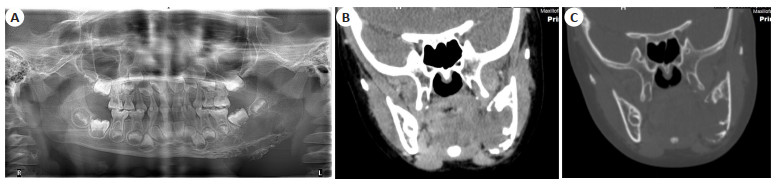
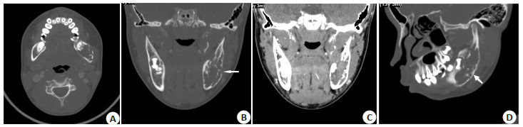
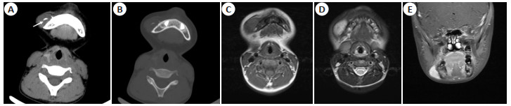
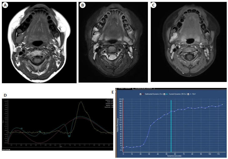
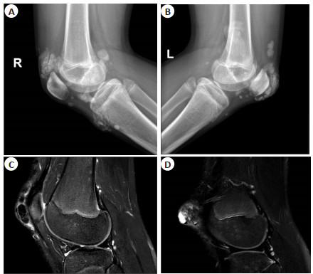
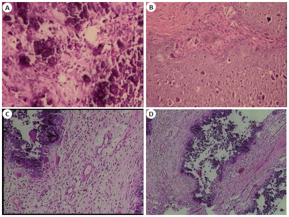
Objective To analyze the clinical manifestations and radiographic features of desmoplastic fibroma (DF). Methods The clinical and imaging findings of 17 patients with DF were retrospectively analyzed, including 11 male patients and 6 female patients, with the age ranged from 4-64 years old and a mean age of 20.27 years old. 17 patients underwent CT examination (enhanced scan for 16 cases, non-enhanced CT for 1 case). 5 patients underwent enhanced MR examination. Results The main clinical manifestations were painless mandibular mass and restricted mouth opening among 17 patients. All cases were in the mandibles, mainly located in the corner of the mandibles. CT images revealed expansile osteolytic lesions and partial compression bone destruction, central or eccentric growth, with soft tissue masses. All cases had "tree root-like" bone ridges. The lesions were divided into 2 types based on the different patterns of bone destruction: osteolytic type (n=12) and parosteal type (n=5). Lesions showed moderate/marked enhancement after contrast-enhanced scanning. All cases have no calcification and periosteal reaction. MR showed isointense or hypointense on T1 weighted images, hyperintense on T2 weighted images. The mean ADC value was 1.2×10⁻³ mm2/s. The time-signal intensity curve type of 3 cases was type Ⅰ, and 2 cases with type Ⅱ indicating benign tumor. Conclusion DF is invasive and prone to recurrence. By combining CT, MR and functional examinations, it is possible to reflect the tissue composition of the lesions, and provide important evidence for the accurate preoperative diagnosis.
2023, 46(6): 994-1000.
doi: 10.12122/j.issn.1674-4500.2023.06.07
Abstract:
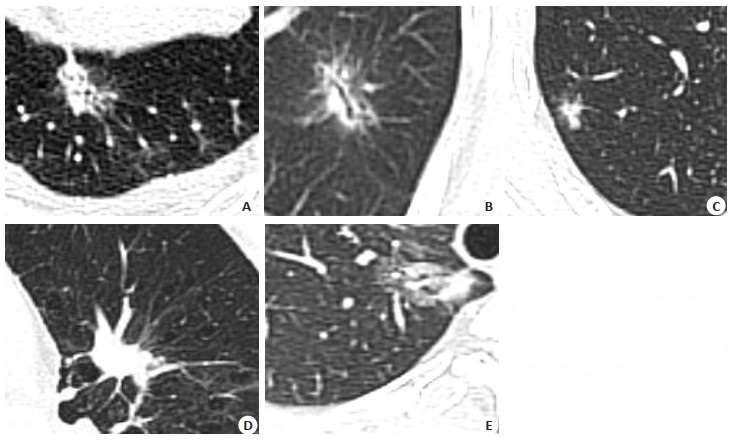
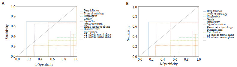
Objective To explore the relationship between CT imaging features of non-small cell lung cancer (NSCLC) and the expression of Ki- 67 and vascular endothelial growth factor (VEGF), providing a theoretical basis for the clinical diagnosis, treatment, and prognosis of NSCLC. Methods A retrospective analysis of 112 patients diagnosed with NSCLC in our hospital from January 2018 to January 2023. Philips Brilliance 256iCT was used to examine CT imaging features, and the expression of Ki-67 and VEGF in NSCLC tissues was detected. The relationship between imaging features, clinical data (pathological type, gender, age, smoking history), Ki- 67 and VEGF expression levels was analyzed. Results In the relationship between CT features and Ki-67 expression, the presence of spiculation sign, deep lobulation sign, arterial phase enhancement CT value ≥20 Hu, venous phase enhancement CT value ≥20 Hu, enlargement of mediastinal lymph nodes and diameter ≥3 cm was associated with a high Ki- 67 positive expression rate (P < 0.05). The presence of calcification and pleural traction sign was associated with a low Ki- 67 positive expression rate (P < 0.05). Other indicators showed no statistical significance. In the relationship between CT features and VEGF expression, the presence of vessel convergence sign, significant enhancement in the arterial phase ≥20 Hu, diameter ≥3 cm, and enlargement of mediastinal lymph nodes was associated with a high VEGF positive expression rate (P < 0.05). The presence of air bronchogram sign, deep lobulation sign, and diameter < 3 cm was associated with a low VEGF positive expression rate (P < 0.05). In the relationship between clinical data and Ki-67 and VEGF expression, males, those with a smoking history, and a high Ki-67 positive expression rate in patients, and a low Ki-67 positive expression rate in squamous cell carcinoma, were statistically significant (P < 0.05). There was no statistical difference in VEGF expression between different clinical data groups (P>0.05). Conclusion CT imaging features in NSCLC patients are closely related to the expression of Ki-67 and VEGF, providing a certain predictive value for immunohistochemical marker expression.
2023, 46(6): 1001-1008.
doi: 10.12122/j.issn.1674-4500.2023.06.08
Abstract:
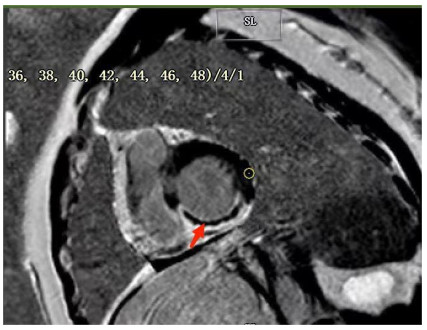
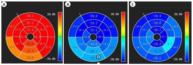
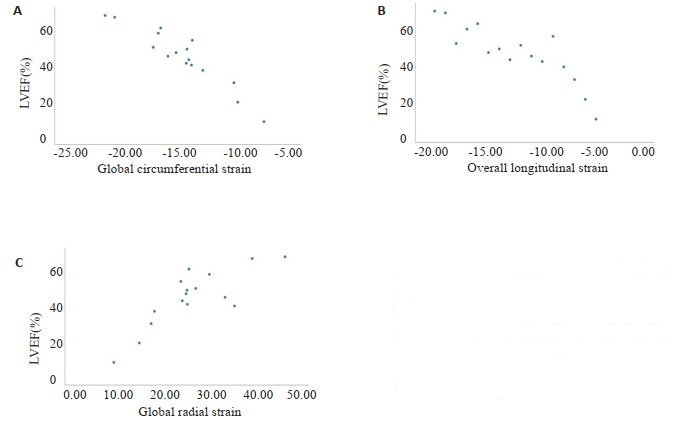
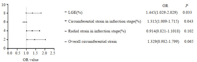
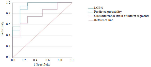
Objective To explore the correlation analysis between myocardial MRI assessment of microcirculation disorders (MVO), infarct size, myocardial strain and clinical prognosis in patients with acute myocardial infarction. Methods A total of 24 patients treated with percutaneous coronary intervention for ST segment elevation myocardial infarction (STEMI) at the First Affiliated Hospital of Bengbu Medical College from June 2022 to December 2022 were selected as the study subjects, including 22 male and 2 female patients, with an average age of 55.3±11.3 years old. All patients underwent cardiac magnetic resonance imaging 5-7 d after surgery. Based on the presence and absence of microcirculation disorders, the 24 patients were divided into MVO group (n=16) and non MVO group (n=8), then compared the baseline data, cardiac function, myocardial infarction area (LGE%) and myocardial strain between the two groups. The discharged STEMI patients were followed up for an average of 6 months in the outpatient clinic or by telephone to record the occurrence of adverse cardiovascular events (MACE events), and compare the differences in the MACE events between the two groups. The MACE events defined in this study include recurrent chest pain, heart failure, stroke, recurrent myocardial infarction, bleeding, revascularization, in-stent thrombosis, in-stent restenosis and death. Results The radial strain, circumferential strain, and overall circumferential strain of the infarcted segment in the MVO group were lower than those in the non MVO group (P < 0.05); The myocardial infarction area in the MVO group was larger than that in the non MVO group (25.18%±10.51% vs 9.93%±5.96%). The left ventricular ejection fraction in the MVO group was highly correlated with radial and circumferential strain [r=0.815(0.536-0.934), P < 0.001; r=-0.938(-0.978—-0.852), P < 0.001], and strongly correlated with longitudinal strain [r=-0.767(-0.915—-0.437), P < 0.001]. LGE% and circumferential strain of the infarcted segment in binary regression analysis are independent risk factors for MVO in STEMI patients. In univariate analysis, ROC curve showed that LGE% could assist in the diagnosis of MVO. The AUC was 0.922 (0.796-1.000), with an optimal cutoff point of 14.92%, sensitivity of 87.5% and specificity of 87.5% (P < 0.05). The circumferential strain of the infarcted segment also has diagnostic value for MVO, with an AUC of 0.781 (0.591- 0.971), an optimal cutoff point of 10.58%, sensitivity of 62.5%, and specificity of 87.5% (P < 0.05). After LGE% was combined with the circumferential strain of the infarcted segment, its AUC and sensitivity for MVO diagnosis increased, with an AUC of 0.938 (0.827-1.000), a sensitivity of 93.8%, and a specificity of 87.5%(P < 0.05). MACE events occurred in a total of 10 STEMI patients followed up in the study, accounting for 41.7% of the total number. There was no statistically significant difference in the occurrence of MACEs events between the two groups of patients (P=0.558). Conclusion LGE% and circumferential strain of myocardial infarction segment are independent risk factors for MVO after percutaneous coronary intervention in STEMI patients, and they also have high diagnostic value for MVO. The combination of the two has higher diagnostic value for MVO. There is a strong correlation between radial strain, circumferential strain, and longitudinal strain and left ventricular ejection fraction in the MVO group.
2023, 46(6): 1009-1014.
doi: 10.12122/j.issn.1674-4500.2023.06.09
Abstract:
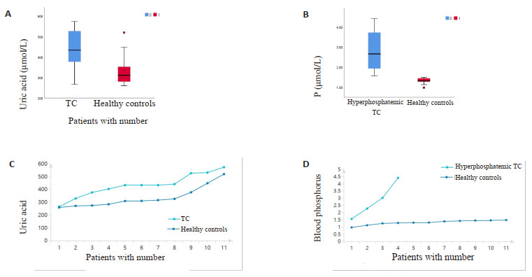
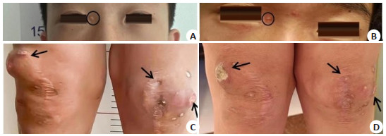
Objective To investigate the clinical characteristics of patients with tumor-like calcinosis (TC) in rare diseases and analyze the associated risk factors. Methods We collected clinical data and laboratory data on 25 patients with TC at Nanfang Hospital from January 2001 to August 2021. All the patients were diagnosed by surgical pathology. The clinical, imaging and pathological features were analyzed. Eleven hospitalized patients with complete clinical data and eleven age- and gender-matched healthy controls were enrolled into the study. The clinical and biochemical characteristics of TC patients were analyzed and summarized to explore the potential risk factors associated with the development of TC. Results X-ray of patients often showed high-density calcifications in soft tissues and MRI showed short T1 and long T2 signal shadows. The tumor exhibited uneven signal intensity, with clear boundary, and the local areas may be connected to the synovial capsule. Compared with healthy people, the serum uric acid levels were significantly increased in TC patients (P=0.038). The serum uric acid levels were positively correlated with the prevalence of TC, and the risk of TC increased by threefold (RR=3, 95% CI: 1.041-8.646). In addition, the product of serum phosphorus and calcium- phosphorus ratio in TC patients with hyperphosphatemia was increased significantly to the healthy population P < 0.05). Hyperphosphatemia increased the risk of TC by 3.2-fold (RR=3.2, 95% CI: 1.547-6.619). Conclusion High serum phosphate and uric acid increased the risk of TC. Therefore, it was important to make a differential diagnosis between TC patients and patients with hyperuricemia or hyperphosphatemia. A comprehensive consideration, including age of disease onset, blood phosphorus concentration, past medical history, imaging, and pathological diagnosis could help to reduce the rate of misdiagnosis.
2023, 46(6): 1015-1020.
doi: 10.12122/j.issn.1674-4500.2023.06.10
Abstract:
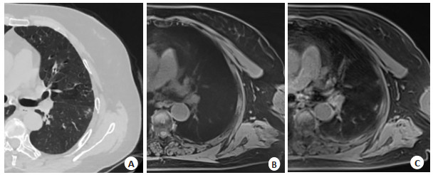
Objective To compare the image quality and the ability of showing nodules and morphology of lung nodules in MRI free breathing sequence: star volumetric interpolated breath-hold examination (star VIBE) sequence and spiral ultrashort echo time sequence (spiral UTE). Methods Patients with pulmonary nodules detected by chest CT and examined by chest MRI from November 2019 to September 2022 were collected. The interval between CT and MRI was 48 h. MRI examination was performed with star VIBE sequence and spiral UTE sequence. Two radiologists used the 5-point method to independently evaluate the image quality of MRI sequences, and compared the detection rate of pulmonary nodules and the ability to display morphological signs. Results In terms of image quality evaluation, the vascular sharpness score of spiral UTE image was higher than that of star VIBE sequence (P < 0.05), while the motion artifact score of star VIBE image was lower than that of spiral UTE image. In terms of nodule detection ability, the ground glass nodule detection rate of spiral UTE sequence was higher than that of star VIBE sequence (P < 0.05), and the total detection rate of nodules in the middle and lower lobe was higher than that of star VIBE sequence (P < 0.05). The detection rates of lobulation, burr, spinous process, vacuole and cavity in Spiral UTE sequence (90.90%, 85.71%, 88.88%, 100%, 100%) were higher than those in star VIBE sequence (86.36%, 71.42%, 77.77%, 50%, 83.33%), but the difference was not statistically significant (P>0.05). Conclusion Star VIBE sequence and spiral UTE sequence have their own advantages. Star VIBE sequence is not easily affected by respiration and cardiac movement. Spiral UTE sequence can better detect middle and lower lobe nodules, especially ground glass nodules, and spiral UTE sequence can better clarify pulmonary vessels. Therefore, star VIBE sequence scan is recommended for patients with large and irregular breathing, and spiral UTE sequence is recommended for patients with ground glass nodules and middle and lower lobe nodules.
2023, 46(6): 1021-1027.
doi: 10.12122/j.issn.1674-4500.2023.06.11
Abstract:
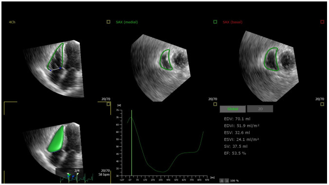
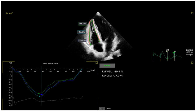
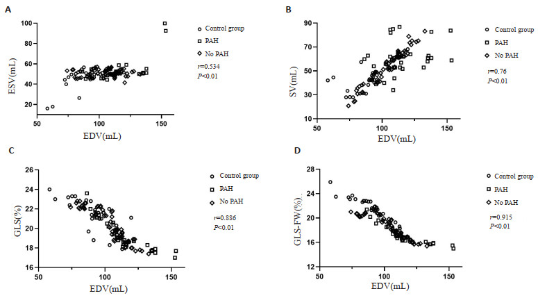
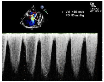
Objective To explore real-time three-dimensional (RT-3DE) combined with speckle tracking technology (2D-STI) to quantitatively assess right heart function in patients with hyperthyroidism. Methods Eighty patients with hyperthyroidism were selected among the patients attending our hospital, of which 30 patients with pulmonary artery systolic pressure>35 mmHg were included in the hyperthyroidism pulmonary hypertension group, and the remaining 50 were included in the hyperthyroidism without pulmonary hypertension group, while 40 healthy people with physical examination in the same period were selected as the control group. Parameters were obtained by conventional ultrasound techniques: right ventricular basal segment, right ventricular mid-segment, right ventricular long-axis, right atrial upper and lower diameters, right atrial left and right diameters, right atrial systolic area, right ventricular field of change, tricuspid annulus systolic displacement, tricuspid annulus systolic peak lateral wall velocity, right ventricular myocardial work index, capillary wedge pressure, and parameters obtained by RT-3DE technique: right ventricular end-diastolic volume (RVEDV), right ventricular end-diastolic volume index (RVEDVi), right ventricular end-systolic volume (RVESV), right ventricular end-systolic volume index (RVESVi), right ventricular output (RVSV), right ventricular ejection fraction (RVEF), applying 2D-STI technique to obtain the parameters: right ventricular free wall longitudinal strain (GLS-FW), right ventricular global longitudinal strain (GLS), and comparing the differences in the above mentioned results. Pearson's analysis was performed to obtain the correlation between the parameters. Results In the comparison among the three groups, right ventricular basal segment, right ventricular midsegment, right ventricular long-axis, right atrial upper and lower diameters, right atrial left and right diameters, right atrial systolic area, capillary wedge pressure, tricuspid annulus systolic displacement, tricuspid annulus systolic peak lateral wall velocity, and right ventricular myocardial work index increased in the control group, hyperthyroidism without pulmonary hypertension group, and hyperthyroidism with pulmonary hypertension group, and right ventricular field of change decreased in the control group, hyperthyroidism without pulmonary hypertension group and hyperthyroidism with pulmonary hypertension group (P < 0.05). RVEDV, RVESV, RVEDVi, RVESVi, RVSV increased sequentially, and there was a statistically significant difference by pairwise comparison (P < 0.05), but the difference was not statistically significant compared to the RVEF of the three groups (P>0.05). GLS-FW and GLS decreased sequentially in the control group, hyperthyroidism without pulmonary hypertension group and hyperthyroidism with pulmonary hypertension group, and there was a statistically significant difference by pairwise comparison (P < 0.05). Correlation test analyses showed that RVEDV was positively correlated with RVESV and SV (r=0.534, 0.760, P < 0.01), and RVEDV was negatively correlated with GLS-FW and GLS (r=-0.915, -0.886, P < 0.01). Conclusion The results of RT-3DE and 2D-STI parameters showed that patients with hyperthyroidism had increased volume load and decreased right heart function compared with the control group, and that the combination of both pathologies, pulmonary hypertension and hyperthyroidism, would lead to a further increase in volume load and a further decrease in right heart function. Therefore, RT-3DE combined with 2D-STI technology can accurately assess the status of right heart function, provide technical support for clinical assessment of patients conditions, and provide important theoretical support for further development of diagnostic and therapeutic programmes.
2023, 46(6): 1028-1034.
doi: 10.12122/j.issn.1674-4500.2023.06.12
Abstract:
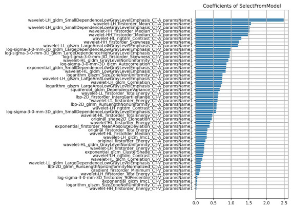
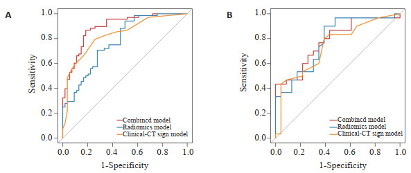
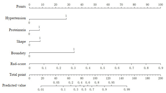
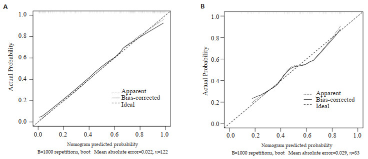
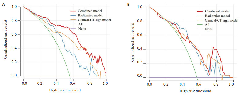
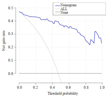
Objective To investigate the value of enhanced CT imaging nomogram in preoperative prediction of muscular infiltration in urinary tract carcinoma of bladder. Methods A retrospective analysis was performed on 175 patients diagnosed with bladder urothelial carcinoma from August 2018 to April 2023 in the First Affiliated Hospital of Bengbu Medical College. All cases were randomly divided into training group (n=122) and verification group (n=53) according to 7:3. The region of interest was manually defined and the image omics features were extracted from the multi-phase enhanced CT images. The dimensionality was reduced by minimum absolute contraction and selection operator, and machine learning was performed on the extracted features using support vector machine classifier to screen out the optimal image omics features and construct the image omics scoring model. The independent predictors of muscular infiltration of bladder urothelial carcinoma were screened by univariate analysis and multivariate binary logistic regression analysis, and the clinical-CT signs model was constructed. Combining the imaging omics model with the clinical-CT sign model, the combined model was constructed. The ROC curve is plotted, the area under the curve (AUC), sensitivity, and specificity were calculated to evaluate the predictive efficacy of different models, and then the best model was visualized to construct a nomogram. Results The diagnostic efficacy of the combined model was the highest (AUC=0.891), which was higher than that of the imaging model (AUC=0.777) and the clinical-CT sign model (AUC=0.829). Decision curve analysis and correction curve confirm that the nomogram has high predictive performance. Conclusion Enhanced CT image nomogram has a high value in predicting myoinfiltration of bladder urothelial carcinoma before operation.
2023, 46(6): 1035-1042.
doi: 10.12122/j.issn.1674-4500.2023.06.13
Abstract:
Objective To explore the clinical application value of multiple quantitative dual-energy CT parameters combined with conventional CT signs to identify benign and malignant primary epithelial tumors of the ovary. Methods A retrospective study was conducted in 166 cases with primary epithelial tumors of the ovary confirmed by pathology findings from January 2021 to June 2023 continuously at the First People's Hospital of Yancheng City. All patients underwent contrast-enhanced dualenergy CT scans within 2 weeks prior to surgery. Patients were devided into benign group (n=74) and malignant group (n=92) according to pathology. Common clinical characteristics and conventional CT signs were recorded. Two independent observers measured the lesion-related quantitative dual-energy CT parameters: iodine concentrations of arterial and venous phases, standardized iodine concentrations, and monoenergetic CT values at 40, 50, 60, 70, 80, 90 keV levels. The slope of energy spectrum curve (K40-90 keV) during both arterial and venous phase were calculated. Intra-group correlation coefficient was used to evaluate the consistency of the parameters obtained by the 2 observers. Differences in parameters between the two groups were compared. Variables with a P-value less than 0.1 were selected as input variables for constructing stepwise binary logistic regression models based on conventional CT signs, quantitative dual-energy CT parameters and the combination of both. The efficacy of the three models in the differential diagnosis of benign and malignant ovarian tumors were evaluated with ROC curve. The areas under these curves were compared using the DeLong test. Results Parameters of the arterial and venous phase obtained by the two observers showed consistency in the two groups. The intra-group correlation coefficients were all above 0.75. Significant differences were found in general clinical characteristics (age, clinical symptoms), eight conventional CT signs (tumor location, morphology, boundary, density, ascites, metastases to peritoneum/omentum, supradiaphragmatic lymph nodes, and other organs or not), and various quantitative dual-energy CT parameters (CT40keV, CT50keV, CT60keV, CT70keV, CT80keV, CT90keV, K40-90keV, iodine concentration and standardized iodine concentration) in benign and malignant groups (P < 0.05). The area under ROC curve of binary logistic regression model based on conventional CT signs, quantitative dualenergy CT parameters and their combination were 0.760, 0.764, 0.883 respectively. According to the DeLong test, no significant difference was found in the area under the curve between the model based on conventional CT signs and the quantitative dual-energy CT parameters model (Z=-0.659, P=0.510). Significant differences were observed between the models based on conventional CT signs and the combined model (Z=-4.007, P < 0.001), and between the quantitative dual-energy CT parameters model and the combined model (Z=-3.870, P=0.001). Conclusion Compared with conventional CT signs or multiple quantitative dual-energy CT parameters, the combination of them has better diagnostic performance in differentiating benign from malignant primary epithelial tumors of the ovary.
2023, 46(6): 1043-1049.
doi: 10.12122/j.issn.1674-4500.2023.06.14
Abstract:
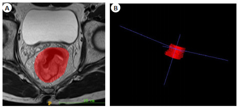
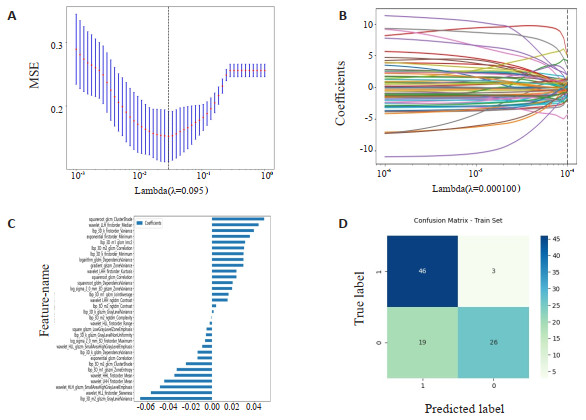
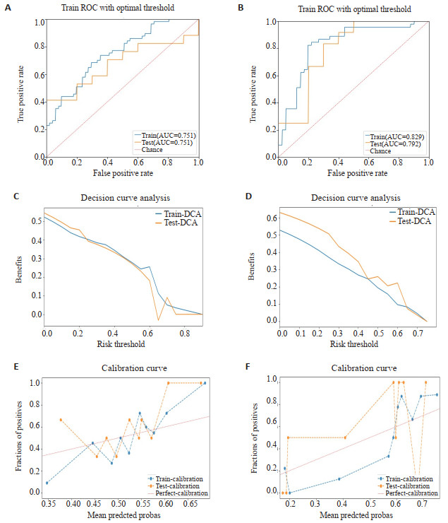
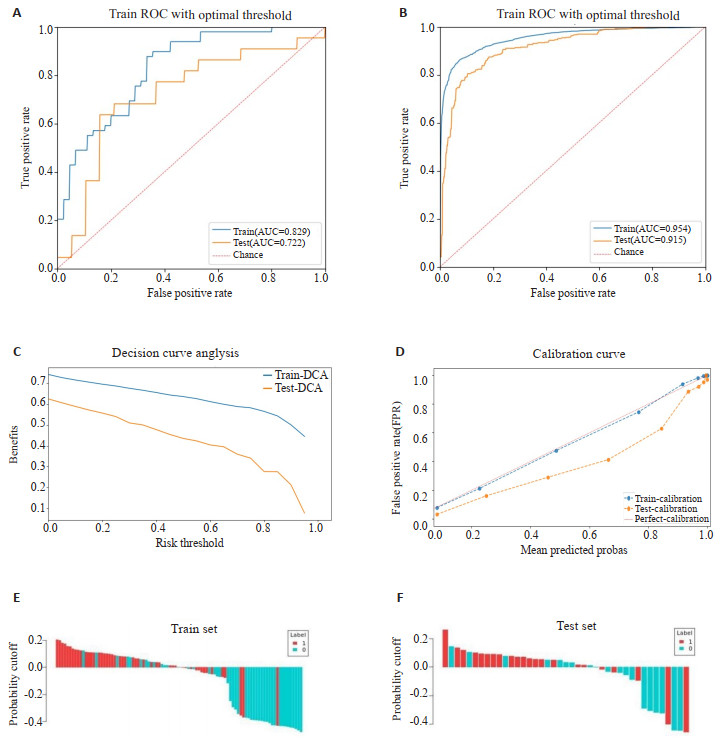
Objective To evaluate the effectiveness of combining MRI balanced steady-state free precession (b SSFP) sequence imaging histology based on clinical factors for diagnosing extra-mural vascular invasion (EMVI) in rectal cancer. Methods A total of 135 patients were included in the study whose diagnosed with rectal cancer and treated at the Lujiang Branch of Dongfang Hospital, affiliated with Tongji University. 71 cases were EMVI-positive, and 64 cases were EMVI-negative among the patients, all of which were confirmed by postoperative pathologic macroscopic specimens. Firstly, the imaging histological features were extracted from the conventional T2WI sequences and b SSFP sequences. The dataset was randomly divided into a training set (n=94) and a validation set (n=41) at a ratio of 7∶3. The imaging histological features and clinical features were analyzed, and the imaging histological model were constructed, by the t-test/Wilcoxon rank sum test, and least absolute shrinkage, and selection operator algorithms were utilized for analysis, and Spearman correlation analysis and logistic regression model. Subsequently, ROC curves were plotted, and the area under the ROC curve (AUC), specificity, and sensitivity were calculated. In order to assess and compare the diagnostic efficacy of the two sequential imaging histologies, it was imperative to quantify the discriminatory performance of the model for both the training and validation groups. Subsequently, the clinical factors were integrated to construct a joint clinical factor and b SSFP sequential imaging histology model for the assessment and validation of diagnostic efficacy. Results The imaging histology models displayed strong discriminatory ability in both the training and validation sets. Specifically, the training set of T2WI sequences demonstrated a diagnostic AUC of 0.751, a specificity of 0.746, and a sensitivity of 0.825, while the b SSFP sequences yielded an AUC of 0.829, a specificity of 0.778, and a sensitivity of 0.939. The validation set T2WI sequence imaging group demonstrated a pre-diagnostic AUC of 0.676, a specificity of 0.650, and a sensitivity of 0.706. In contrast, the b SSFP sequence exhibited an AUC of 0.792, a specificity of 0.716, and a sensitivity of 0.909. Comparison between the two indicates the latter's improved diagnostic efficacy and tangible clinical applicability. The combined clinical factors with b SSFP sequence imaging histology joint model exhibited an AUC of 0.954, specificity of 0.891, and sensitivity of 0.930 for the training set. For the validation set, the AUC was 0.915, specificity was 0.803, and sensitivity was 0.936. Both results were deemed statistically significant (P < 0.05). Conclusion The combination of clinical factors and b SSFP sequence imaging histology demonstrates strong diagnostic efficacy for rectal cancer EMVI. This facilitates imaging physicians and clinicians in making accurate differential diagnoses and formulating appropriate treatment strategies.
2023, 46(6): 1050-1054.
doi: 10.12122/j.issn.1674-4500.2023.06.15
Abstract:
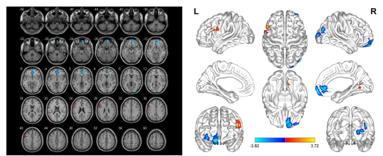
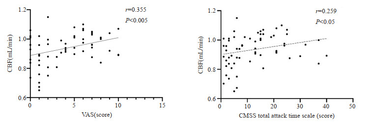
Objective To explore the changes of cerebral blood flow in patients with primary dysmenorrhea (PDM) and analyze the correlation between them and clinical scale scores by using voxel-based morphological methods and arterial spin labeling technology. Methods Thirty-one patients diagnosed as PDM in Shaanxi University of Traditional Chinese Medicine were prospectively enrolled as PDM group, and 32 healthy people were selected as healthy control group. On the 1st to 3rd day of menstrual period, high-resolution T1 structural images and whole brain arterial spin labeling scans were performed, and the clinical symptoms were evaluated by visual analogue scale for pain (VAS), COX dysmenorrhea symptom scale (CMSS), selfrating anxiety scale (SAS) and self-rating depression scale (SDS). Using voxel-based morphometry method, the double-sample t test of SPM8 software based on Matlab platform was used to analyze the brain regions with significant differences in cerebral blood flow between the two groups, and then Pearson correlation analysis was used to observe the correlation between the cerebral blood flow value of the brain regions with different cerebral blood flow and the scores of VAS, CMSS, SAS and SDS scales. Results There were significant differences in age, VAS, CMSS, SAS and SDS between PDM group and healthy control group (P < 0.05). Compared with the healthy control group, the brain regions with increased cerebral blood flow in PDM group mainly included the left triangular inferior frontal gyrus, while the brain regions with decreased cerebral blood flow included the right orbital superior frontal gyrus, anterior part of right cingulate gyrus and the right orbital middle frontal gyrus (P < 0.005, corrected by FDR of mass level, k≥100). Correlation analysis showed that the cerebral blood flow value of the left triangle inferior frontal gyrus was positively correlated with the scores of VAS and total attack time of CMSS(P < 0.05). Conclusion Based on arterial spin labeling technology and voxel-based morphometry method, the cerebral blood flow perfusion level of patients with primary dysmenorrhea can be evaluated. These brain regions with changes in cerebral blood flow are mainly located in pain conduction pathways, which provides valuable information for the diagnosis and treatment of patients with primary dysmenorrhea.
2023, 46(6): 1055-1059.
doi: 10.12122/j.issn.1674-4500.2023.06.16
Abstract:
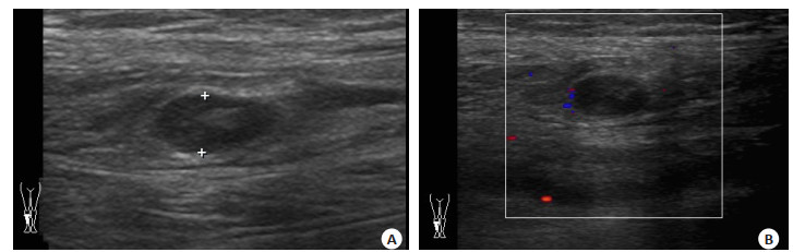
Objective To analyze the association of BI-RADS standardized ultrasound grade 4C lesion signs in the diagnosis of breast invasive carcinoma and the age distribution of this type of cancer, and explore the value of BI-RADS classification criteria in the diagnosis of non-specific type of breast invasive carcinoma. Methods We randomly collected 88 cases of breast cancer patients with ultrasound BI-RADS grade 4C from May 2020 to May 2023 as study data, and classified the patients' age and pathological examination results. The correlation between lesion ultrasound characteristics and pathological characteristics of patients with different age groups with different types of breast diseases were analyzed. Results The ultrasound examination and pathological tracking analysis showed that among 88 patients in BI-RADS 4C, 90% were patients younger than 40 years; more than 95% were 40 years and 5% were patients younger than 40 years. Simple invasive non-special carcinoma accounted for 64.77% of the total pathological examination results, while the remaining non-simple invasive non-special carcinoma accounted for 35.23. Conclusion Comprehensive data analysis the detection rate of ultrasound examination and BI-RADS grade in simple invasive non-special cancer is obviously high, which is conducive to the formulation of corresponding treatment measures, and highlights the detection rate and application of BI-RADS grade in simple breast invasive non-special cancer.
2023, 46(6): 1060-1064.
doi: 10.12122/j.issn.1674-4500.2023.06.17
Abstract:
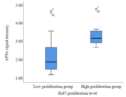
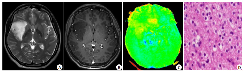
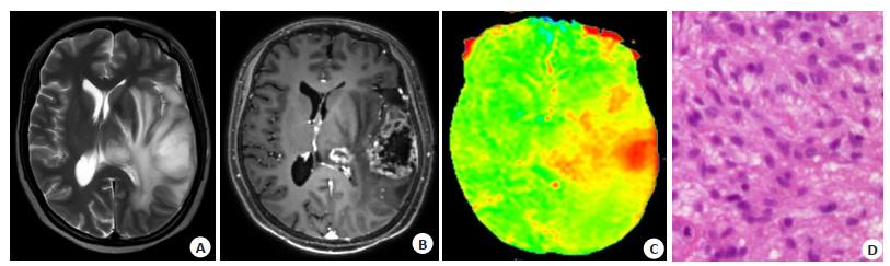
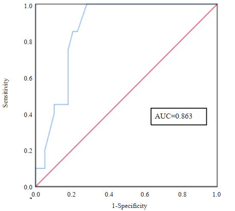
Objective To explore the correlation between amide proton transfer imaging (APTw) and the Ki67 proliferation index of glioma and analyze the accuracy of the predicting model. Methods 63 glioma patients from Beijing Tiantan Hospital were accepted APTw imaging within 2 weeks before surgery, and 59 glioma patients were finally included. The patients were divided into two groups according to immunohistochemistry results: low proliferation group (cell proliferation index < 20%, n=39) and high proliferation group (cell proliferation index 20%, n=20), and the mean APTw signal intensities of gliomas were measured. The correlaction between APTw signal intensity and Ki67 proliferation index was evaluated by Pearson test. The differences of mean APTw signal intensity between two proliferation groups were evaluated by Mann-Whitney U test. The predicting model by mean APTw signal intensity as an imaging biomarker was established, and then ROC curve was drawn to assess their diagnostic performance. Finally, the cut-off value of the APTw signal intensity for this model was obtained by Yuden index. Result There was a positivity correlaction between APTw signal intensity and Ki67 proliferation index (r=0.629, P < 0.001), and there was a significant difference between two proliferation groups (Z=4.539, P < 0.001). The AUC for predicting model was 0.863 (95% CI: 0.770-0.956), with sensitivity of 1, and specificity of 0.718. Youden index showed the cut-off value was 2.55. Conclusion APTw signal intensity can be served as an imaging biomarker for the predicting model of glioma Ki-67 proliferation level, which has a good diagnostic effectiveness and good sensitivity; When the mean APTw signal intensity is greater than 2.55, the glioma prefers high proliferation level, otherwise, the glioma prefers low proliferation level.
2023, 46(6): 1065-1069.
doi: 10.12122/j.issn.1674-4500.2023.06.18
Abstract:
Objective To explore the correlation between clinical diagnosis and prognosis in patients with acute pulmonary thrombus embolism (PTE) using multi-mode imaging. Methods Fifty-five patients with PTE confirmed in Beijing Hospital of Traditional Chinese Medicine from January 2018 to January 2022 were evaluated retrospectively. Echocardiography, twodimensional speckle tracking imaging, ultrasound, computer tomography pulmonary angiography (CTPA) and lab marker, including D-dimer, NT-pro brain natriuretic peptide (NT-proBNP) and C-reactive protein results were reviewed. 55 patients with PTE were ranked 5 classes according to CTPA results. Associations and correlations between CTPA classes of D-dimer, NT-proBNP, C-reactive protein, velocity of tricuspid regurgitation (VTR), pulmonary systolic pressure (PAPS) and longitudinal strain in right ventricular free wall (RLS) were analysed. Results NT-proBNP, VTR and PAPS showed a gradually increased trend with increased CTPA class and the absolute value of RLS showed a gradually decreased trend with increased CTPA class. The statistical difference among CTPA grading groups was significant (P < 0.05). Positive correlations were found between increased NT-proBNP, VTR, PAPS, decreased absolute value of RLS with higher CTPA class (r=0.25, 0.24, 0.32, 0.28, P < 0.05). Conclusion Increased NT-proBNP, VTR, PAPS and RLS are significantly associated with the severity of acute PTE patients, which has important clinical value for the diagnosis, treatment and prognosis evaluation of acute PTE patients.
2023, 46(6): 1070-1075.
doi: 10.12122/j.issn.1674-4500.2023.06.19
Abstract:
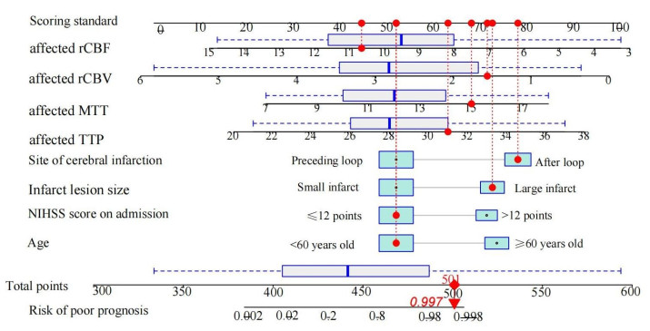
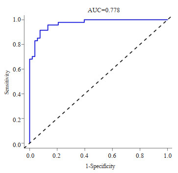
Objective To investigate the risk factors of poor prognosis in patients with ischemic cerebral infarction, and construct a nomogram model of poor prognosis in patients with ischemic cerebral infarction based on cerebral MRI perfusion imaging and clinical indicators. Methods A total of 100 patients with ischemic cerebral infarction who came to our hospital from July 2021 to January 2023 were selected as the study objects, and cerebral MRI perfusion imaging was performed on the patients. According to the prognosis, patients were divided into poor prognosis group (n=47) and good prognosis group (n=53). Logistic regression analysis was used to screen the risk factors of poor prognosis in patients with ischemic cerebral infarction. R4.2.0 software was used to establish and verify the poor prognosis of patients with ischemic cerebral infarction. Results Logistic regression analysis showed that age≥60 years old, NIHSS score>12 points at admission, major infarction, posterior circulation cerebral infarction, affected time to peak and affected mean transit time were risk factors for poor prognosis in patients with ischemic cerebral infarction, and affected cerebral blood volume and affected cerebral blood flow were protective factors for poor prognosis in patients with ischemic cerebral infarction (P < 0.05). The results of the constructed nomogram model for poor prognosis of patients with ischemic cerebral infarction showed that the actual value of the correction curve was in good agreement with the predicted value, the area under ROC curve of the model was 0.778 (95% CI: 0.735-0.821). When the decision curve showed that the threshold probability was 1%-100%, the net benefit value of the histogram predicting the poor prognosis of patients with ischemic cerebral infarction was higher. Conclusion The nomogram model of poor prognosis of patients with ischemic cerebral infarction based on cerebral MRI perfusion imaging and clinical indicators has high accuracy and clinical application value, and can be used to predict poor prognosis of patients with ischemic cerebral infarction.

2023, 46(6): 1076-1080.
doi: 10.12122/j.issn.1674-4500.2023.06.20
Abstract:
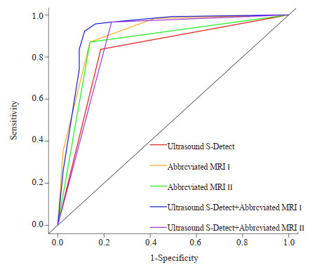
Objective To investigate the value of abbreviated MRI combined with ultrasound S-Detect model for benign and malignant differentiation of breast masses. Methods A total of 154 patients (159 lesions) who underwent breast ultrasound and MRI from March 2021 to January 2023 at Tangshan Maternal and Child Health Hospital affiliated to North China University of Science and Technology were selected as study subjects. Pathologic biopsy results were used as the gold standard. The abbreviated MRI Ⅰ represented the Breast Imaging Reporting and Data System (BI-RADS) classification model. For abbreviated MRI Ⅱ, BI-RADS classifications of 4A and below were deemed benign, while classifications of 4B and above were deemed malignant. The consistency between the results of different methods for identifying benign and malignant breast masses and pathology results was analyzed with the Kappa test. Logistic regression was used to construct models for identifying benign and malignant breast masses by abbreviated MRI Ⅰ and ultrasound S-Detect + abbreviated MRI Ⅰ. The value of different methods to identify benign and malignant breast masses was evaluated using the ROC curve and decision curve analysis. Results Pathologic findings showed 43 benign and 116 malignant breast masses. The accuracy of ultrasound S-Detect+abbreviated MRI Ⅰ in identifying benign and malignant breast masses was higher than that of ultrasound S-Detect alone (P < 0.05). It was also comparable to the accuracy of abbreviated MRI Ⅰ, abbreviated MRI Ⅱ, and ultrasound S-Detect + abbreviated MRI Ⅱ (P > 0.05). The Kappa value of ultrasound S-Detect+abbreviated MRI Ⅰ was higher than that of ultrasound S-Detect, abbreviated MRI Ⅰ, abbreviated MRI Ⅱ, and ultrasound S-Detect + abbreviated MRI Ⅱ. The area under the ROC curve for identifying benign and malignant breast masses with ultrasound S-Detect + abbreviated MRI Ⅰ was higher than that with ultrasound S-Detect, abbreviated MRI Ⅰ, abbreviated MRI Ⅱ and ultrasound S-Detect + abbreviated MRI Ⅱ (P < 0.05). Decision curve analysis results showed that within the full risk threshold, the net benefit of identifying breast masses with ultrasound S-Detect+abbreviated MRI Ⅰ was higher than that with ultrasound S-Detect and abbreviated MRI Ⅱ. In the vast majority of the risk threshold range, the net benefit of ultrasound S-Detect + abbreviated MRI Ⅰ in identifying benign and malignant breast masses was higher than that of abbreviated MRI Ⅰ and ultrasound S-Detect + abbreviated MRI Ⅱ. Conclusion The model constructed by abbreviated MRI combined with ultrasound S-Detect can help identify benign and malignant breast masses with a higher value than abbreviated MRI and ultrasound S-Detect alone.

2023, 46(6): 1081-1085.
doi: 10.12122/j.issn.1674-4500.2023.06.21
Abstract:
Objective To explore the clinical application of CT angiography (CTA) and magnetic resonance angiography (MRA) in the diagnosis of spinal cord vascular malformations. Methods This prospective study included 30 patients with spinal cord vascular malformation diagnosed and treated in our hospital from February 2020 to December 2022 as the experimental group. In addition, 30 patients without spinal vascular malformation who underwent vertebra MR Scan plus enhancement were selected as the control group. Contrast enhanced magnetic resonance angiography (CE-MRA) sequence and reconstructed spinal vascular CTA were performed on all enrolled subjects. The diagnostic consistency of CE-MRA sequence and reconstructed spinal vascular CTA was compared, and the combined diagnostic efficacy of CE-MRA sequence and reconstructed spinal vascular CTA was analyzed. Results There was no statistical significance in dural arteriovenous fistula, spinal arteriovenous malformation, and perimedullary arteriovenous fistula in CE-MRA and CTA examination (P > 0.05). In the analysis of fistula, there was no statistical significance in the display of supplying artery and fistula in CE-MRA and CTA examination (P > 0.05). CE-MRA had a good consistency by compare with the gold standard, with a kappa value of 0.758, and CTA had a good consistency by compare with the gold standard, with a kappa value of 0.881. The sensitivity of CE-MRA and CTA combined diagnosis were significantly higher than that of the single detection. ROC curve analysis showed that the area under the curve of CE-MRA and CTA combined diagnosis was significantly higher than that of single detection(P < 0.001). Conclusion In the diagnosis of spinal cord vascular malformations, the combined diagnosis of CTA and CE-MRA can significantly improve the diagnostic efficiency of patients, and can be used as an important basis for clinical diagnosis.
2023, 46(6): 1086-1091.
doi: 10.12122/j.issn.1674-4500.2023.06.22
Abstract:
Objective To investigate the clinical value of dynamic contrast-enhanced MR combined with high resolution MR in lymph node metastasis of rectal adenocarcinoma. Methods Clinical data of 262 patients with pathologically confirmed rectal cancer admitted to the First People's Hospital Affiliated to Shanghai Jiao Tong University from January 2018 to December 2022 were collected. All patients underwent high resolution MR and contrast-enhanced MR in the hospital. According to postoperative pathology, the patients were divided into two groups: no lymph node metastasis group (n=124) and lymph node metastasis group (n=72). The differences in clinical features, quantitative parameters of dynamic contrast-enhanced MR and high resolution MR between the two groups were analyzed, and the factors predicting lymph node metastasis of rectal cancer were screened by Logistic regression analysis. ROC curve was used to calculate the efficacy of different parameters in predicting lymph node metastasis. Results Univariate Logistic regression analysis showed that the risk factors for lymph node metastasis of rectal adenocarcinoma were age < 59.5 years old (OR: 0.329), mrN stage N1-N2 (OR: 6.857)、abnormal carcinoembryonic antigen (OR: 2.742), mrT stage T3 (OR: 2.959), mrMRF positive (OR: 5.577), Quantitative parameter Ktrans of dynamic contrast-enhanced MR < 0.298 min-1 (OR: 0.210) and mr EMVI positive (OR: 2.261). Multivariate Logistic regression analysis showed that mrN stage positive and Ktrans were independent predictors of lymph node metastasis in colorectal adenocarcinoma. The AUC of mrN stage + Ktrans (0.788) in predicting lymph node metastasis of rectal adenocarcinoma was higher than single mrN stage (0.713) and Ktrans (0.650). Conclusion Quantitative parameter Ktrans of dynamic contrast-enhanced MR combined with mrN stage can improve the prediction ability of patients with rectal adenocarcinoma lymph node metastasis.
2023, 46(6): 1092-1097.
doi: 10.12122/j.issn.1674-4500.2023.06.23
Abstract:
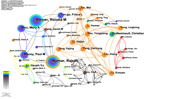
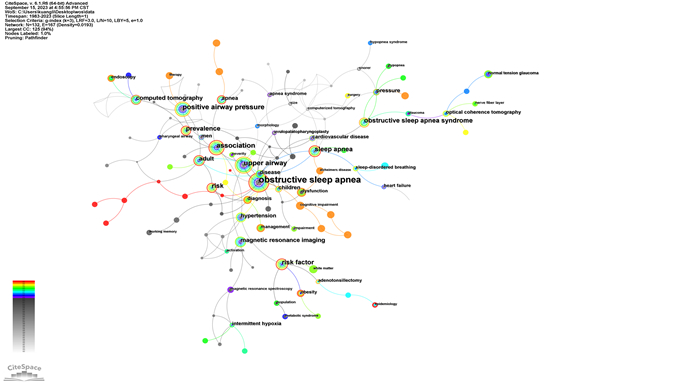
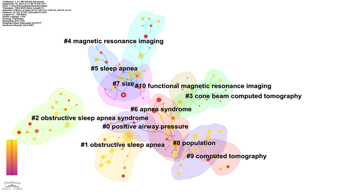
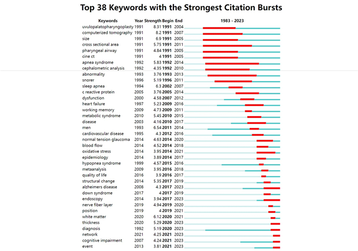
Objective To analyze the current status and hot issues in the field of Obstructive sleep apnea hypopnea syndrome (OSAHS) imaging research. Methods The literature on imaging studies of OSAHS in the Web of Science core database from the establishment of the database to September 2023 was retrieved, and CiteSpace 6.1.R6 software was used for visual analysis. Results A total of 1016 articles were retrieved, and the number of publications showed a fluctuating upward trend. The research countries/regions were concentrated in the United States, China and Turkey. The main journals were Sleep, Am J Resp Crit Care and Chest. 210 research institutions, represented by Univ Calif Los Angeles, Stanford Univ, Capital Med Univ, etc. There were 331 researchers, represented by Kumar R, Harper RM, Macey PM, Woo MA, Yan-go FL, etc. Keyword analysis suggested that the current research hotspots were related research on OSAHS imaging diagnostic techniques, OSAHS multidisease coexistence, and the pathogenesis of OSAHS comorbidity. Conclusion At present, OSAHS imaging research in China has gradually become a hot spot, but the cooperation between clinical research institutions needs to be strengthened. The new technology of OSAHS imaging diagnosis needs more research and verification with larger samples and more standardized methods. The research on the reverse relationship between OSAHS and comorbidity, the study of different pathogenic mechanisms, and the combination of neuroimaging markers and negative cognitive results need to be strengthened.
2023, 46(6): 1098-1101.
doi: 10.12122/j.issn.1674-4500.2023.06.24
Abstract:
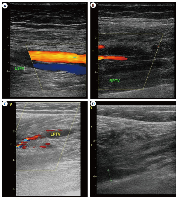
Objective To analyze the clinical application of vascular ultrasound in the diagnosis of lower limb venous thrombosis during perioperative period of joint replacement surgery. Methods A retrospective study was conducted on 868 patients who underwent initial unilateral hip or knee arthroplasty from January 2021 to June 2023 as the observation group. The group under observation was contrasted with 868 healthy volunteers who had hospital physical examinations within the same period, forming the control group. The observation group was subject to multimodal thrombus prevention measures including anticoagulants, calf venous pressure pump, strengthened ankle pump, early mobilization, and reduction of perioperative dehydration. The observation group underwent color doppler ultrasound examination of bilateral lower limb venous vessels pre- surgery and 3- 5 d post- surgery. The detection accuracy for lower limb venous thrombosis and the differences in vein diameter of the superficial femoral vein, medial sural vein, and posterior tibial vein between both groups were analyzed. Results 152 patients in the observation group were reported to have deep vein thrombosis (DVT) after surgery with an incidence rate of 17.5%. All cases were asymptomatic DVTs. The breakdown was as follows: 138 cases were on the side of the surgery, 14 cases on both sides, 110 cases of peripheral venous embolization, 22 cases of central venous embolization and 20 cases of mixed venous embolization. In the control group, 26 cases of peripheral lower limb thrombosis were found. Comparatively, the diameters of the superficial femoral vein, medial sural vein, and posterior tibial vein in the observation group were significantly larger than those in the control group (P < 0.05). Conclusion Vascular ultrasound examination can accurately diagnose lower limb venous thrombosis in patients post joint replacement surgery. It can display various imaging characteristics, which is beneficial for disease status assessment and provides clinical evidence for early treatment.
2023, 46(6): 1102-1106.
doi: 10.12122/j.issn.1674-4500.2023.06.25
Abstract:
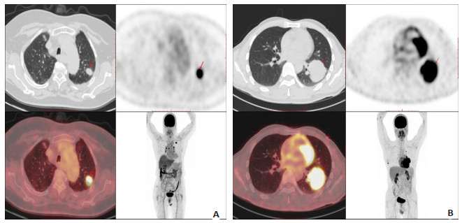
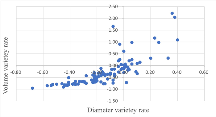
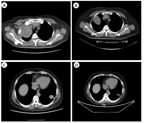
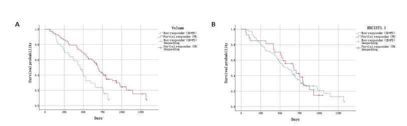
Objective To analyze whether tumor volume and tumor maximum diameter [based on the criteria for evaluating the efficacy of solid tumors (RECIST) version 1.1] are correlated with each other in evaluating patients with advanced non-small cell lung cancer (NSCLC) after non-surgical treatment and to assess the value of the application of the changes in tumor volume of advanced NSCLC in the evaluation of efficacy. Methods We retrospectively analyzed the CT imaging data of 108 patients with pathologically confirmed NSCLC by puncture biopsy and followed them up, measured the volume of the lesions before and after treatment using volumetric software, recorded the volume measurement values with the maximum diameter of the lesions, and then evaluated the therapeutic efficacy by the volumetric method and RECIST version 1.1, respectively. The complete response and partial response were categorized into the partial responders, and stable disease and progressive disease were categorized into the non-responders according to different criteria. Spearman correlation coefficients were used to assess the correlation between the maximum diameter of RECIST and the rate of change in volume before and after treatment, and Kaplan-Meier curves were used to evaluate the association between the survival of patients in different evaluation criteria. Results The rate of change of volume versus diameter relative to baseline measurements was calculated by baseline and first follow-up scans, and the assessment showed a moderate correlation (r=0.881, P < 0.01). The difference between the Kaplan-Meier curves of the responders and non-responders as assessed by the volume criterion at first follow up was statistically significant (P=0.002), and the difference between the two groups was not statistically significant when assessed according to the RECIST criterion (P=0.828). Conclusion Volumetric measurements have good application value for the assessment of the efficacy of NSCLC after treatment.
2023, 46(6): 1107-1111.
doi: 10.12122/j.issn.1674-4500.2023.06.26
Abstract:
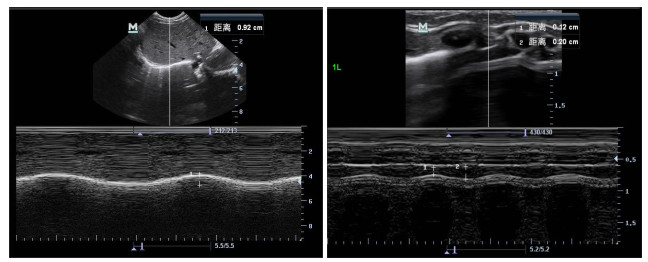
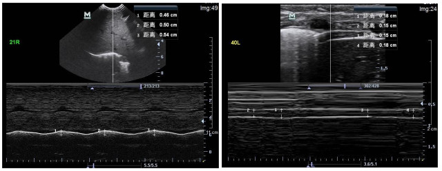
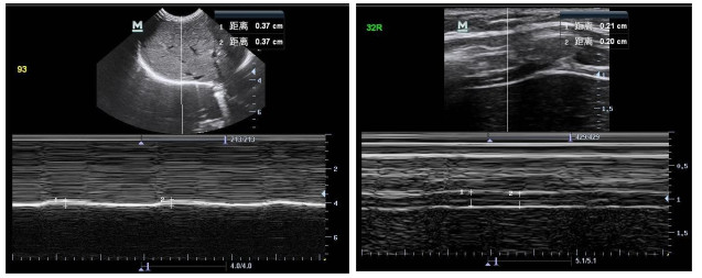
Objective To investigate the application of ultrasound to quantify the diaphragmatic motor function of neonates and infants and the change of diaphragmatic motor in children with broncho-pulmonary dysplasia (BPD), and to evaluate the predictive value of diaphragmatic ultrasound for the results of mechanical ventilation weaning of BPD children. Methods A total of 23 children with mechanical ventilation of BPD in NICU, PICU and SNICU in Guangzhou Women and Children's Medical Center were included from August 2022 to August 2023. The children were divided into a successful group (n=18) and a failed group (n=5) according to whether they could breathe autonomically after clinical weaning. The basic clinical characteristics of the children were recorded. Meanwhile, 23 newborns and infants in the same period were selected as the normal control group. Diaphragmatic motion (DM), diaphragmatic thickness at the end of inspiration (DTinsp), diaphragmatic thickness at the end of expiration (DTexp) and diaphragmatic thickening fraction (DTF) were measured by ultrasonography in all groups. The difference of diaphragmatic muscle movement between left and right sides in different groups was compared. Results The levels of DM in BPD group were significantly lower than those in control group before and after weaning (P < 0.05). DTinsp, DTexp and DTF had no statistical significance compared with the control group (P > 0.05). The levels of DM, DTinsp and DTF in the failed group were significantly lower than those in the successful group (P < 0.05), but there was no significant difference in the DTexp group (P > 0.05). Retrospective comparison before weaning: DM and DTF in the failed BPD group were lower than those in the successful BPD group, and the difference was statistically significant (P < 0.05), there was no significant difference in DTexp and DTinsp between the two groups (P > 0.05). Conclusion The changes of diaphragmatic motor function in children with BPD undergoing mechanical ventilation can be evaluated by bedside ultrasound, which has a good value in predicting the outcome of weaning from mechanical ventilation.
2023, 46(6): 1112-1117.
doi: 10.12122/j.issn.1674-4500.2023.06.27
Abstract:
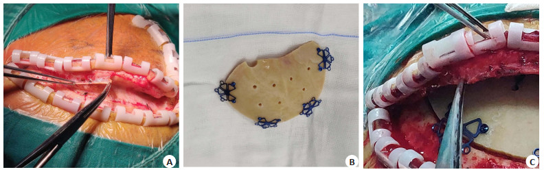
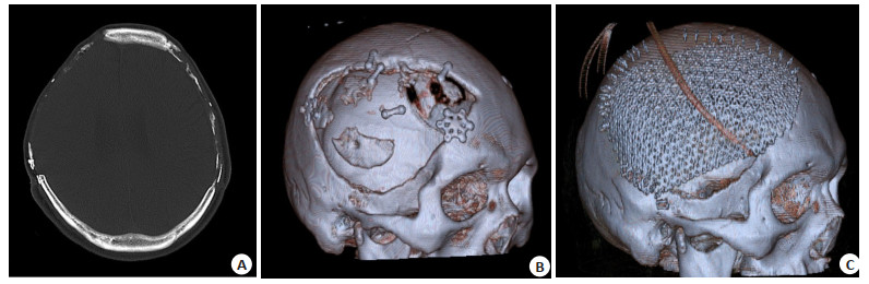
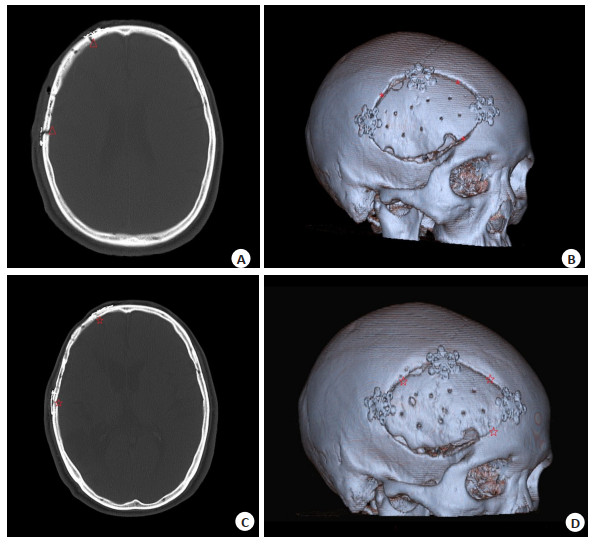
Objective To observe the clinical efficacy and safety of autogenous skull preserved by bone tissue bank technology in cranioplasty. Methods Hospitalized patients with supratentorial skull defect repair surgery in the Department of Neurosurgery, Shunde Hospital of Southern Medical University from September 2019 to July 2022 were enrolled. According to different repair materials, the patients were divided into the autologous skull repair group (n=30, autologous skull repair surgery preserved by bone tissue bank) and the 3D titanium mesh group (n=30, skull repair surgery with 3D digital shaped titanium mesh). The general data of the two groups was comparable (P > 0.05). Indicators of complications directly related to surgery including infection rate within 1 month after surgery, incidence of epidural effusion/hematoma, incidence of poor healing of surgical incision, epilepsy incidence and incidence of intracerebral hemorrhage were compared between the two groups and bone absorptivity based on skull CT images was measured in the autologous skull repair group. Results Both groups were followed up for at least 6 months after surgery. There was no significant difference in the infection rate within 1 month after surgery (0 case vs 0 case), incidence of epidural effusion/hematoma (3 cases vs 7 cases), incidence of poor healing of surgical incision (0 case vs 0 case), epilepsy incidence (0 case vs 0 case) and incidence of intracerebral hemorrhage (0 case vs 1 case) between the autologous skull group and 3D titanium mesh group (P > 0.05). Bone resorption occurred in 4 patients (13.3%) in the autogenous skull group 6 months after operation, and one of the patients with severe bone resorption required re-repair with artificial materials. There was no significant difference in the total complication rate between the autologous skull group and 3D titanium mesh group [7 cases (23%) vs 8 cases (35%), P > 0.05]. Conclusion The technique of preserving autogenous skull through bone tissue bank is one of the safe and effective methods in cranioplasty while remaining a risk of bone resorption, it can be an alternative depending on the specific situation of patients clinically.
2023, 46(6): 1118-1121.
doi: 10.12122/j.issn.1674-4500.2023.06.28
Abstract:
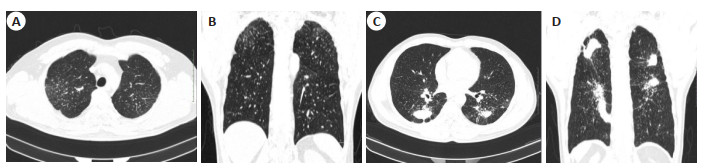
Objective To explore the diagnostic value of CT in the staging of silicosis. Methods Eighty silicosis patients from our hospital from January 2022 to January 2023 were selected, and all patients underwent CT examination. The diagnostic results of CT for silicosis staging and the consistency of clinical diagnostic results were compared, and the diagnostic efficacy of CT for silicosis staging was calculated. Results The CT sign analysis showed that among 80 silicosis patients, 20 patients had nodules in the upper, middle, and lower regions of both lungs, 36 patients had nodules in 4 lung regions, and 24 patients had nodules in 2 lung regions; 39 patients had no masses, 13 patients had masses ranging from 1 to 3cm, and 28 patients had masses greater than 3cm; 54 patients had increased pulmonary nodule density; 80 patients had different degrees of Lymphadenopathy, 57 patients had lymph nodes in hilar region, 23 patients had in mediastinum; 60 patients had pleural thickening, involving interlobular pleura and visceral pleura. The clinical diagnosis results showed that there were 24 cases in stage Ⅰ, 36 cases in stage Ⅱ, and 20 cases in stage Ⅲ among 80 silicosis patients; The CT examination results showed that there were 26 cases in Phase Ⅰ, 35 cases in Phase Ⅱ, and 19 cases in Phase Ⅲ. The Kappa value was 0.884, 0.823, 0.898, respectively, indicating good consistency between the two. The overall accuracy of CT in staging diagnosis of silicosis was 91.25%. The accuracy of CT in staging diagnosis of stage Ⅰ, stage Ⅱ and stage Ⅲ was 95.00%, 91.25%, 96.25%, respectively. The sensitivity was 95.83%, 88.89%, 90.00%, respectively. The specificity was 94.64%, 93.18%, 98.33%, respectively. The positive predictive value was 88.46%, 91.43%, 94.74%, respectively, and the negative predictive values was 98.15%, 91.11%, 96.72%, respectively. Conclusion CT plays a positive role in the diagnosis of silicosis patients, can improve the diagnostic accuracy of silicosis staging, and has good diagnostic efficacy.
2023, 46(6): 1127-1132.
doi: 10.12122/j.issn.1674-4500.2023.06.30
Abstract:
Cardiovascular disease is one of the leading causes of death and disease burden worldwide. Accurate evaluation of myocardial viability is helpful for clinical decision- making and has important prognostic value for patients with cardiovascular disease. Positron- emission tomography (PET) has good image quality and high diagnostic accuracy with known or suspected coronary artery disease. The value of classical PET myocardial perfusion and metabolic imaging in evaluating myocardial viability has been recognized. At present, the image analysis of PET myocardial viability assessment mainly relies on visual assessment or semi-quantitative scoring. With the promotion and application of automatic analysis by various software, the inclusion of quantitative indicators provides more valuable reference information for clinical practice. Continuous advances in software and technical equipment have also facilitated better use of PET in assessing myocardial viability. The rise of artificial intelligence technology and the exploration and application of integrated PET/MR provide new means for PET myocardial viability imaging and image interpretation, but the opportunities of new technology are accompanied by challenges as well. This review briefly describes the common software, image display, different image analysis methods and progress in PET myocardial viability assessment.
Cardiovascular disease is one of the leading causes of death and disease burden worldwide. Accurate evaluation of myocardial viability is helpful for clinical decision- making and has important prognostic value for patients with cardiovascular disease. Positron- emission tomography (PET) has good image quality and high diagnostic accuracy with known or suspected coronary artery disease. The value of classical PET myocardial perfusion and metabolic imaging in evaluating myocardial viability has been recognized. At present, the image analysis of PET myocardial viability assessment mainly relies on visual assessment or semi-quantitative scoring. With the promotion and application of automatic analysis by various software, the inclusion of quantitative indicators provides more valuable reference information for clinical practice. Continuous advances in software and technical equipment have also facilitated better use of PET in assessing myocardial viability. The rise of artificial intelligence technology and the exploration and application of integrated PET/MR provide new means for PET myocardial viability imaging and image interpretation, but the opportunities of new technology are accompanied by challenges as well. This review briefly describes the common software, image display, different image analysis methods and progress in PET myocardial viability assessment.
2023, 46(6): 1133-1137.
doi: 10.12122/j.issn.1674-4500.2023.06.31
Abstract:
Breast cancer is the leading malignant tumor in women worldwide. Precision medicine and improving prognosis are crucial. Breast cancer is a tumor with strong heterogeneity. Preoperative overall assessment of prognostic factors is very important for clinical treatment options and monitoring efficacy. In recent years, new ultrasound technologies such as shear wave elastography, contrast-enhanced ultrasound, three- dimensional ultrasound, ultrasound radiomics, and artificial intelligence breast ultrasound have made satisfactory progress in non-invasive assessment of breast cancer prognosis. This article reviews the prognostic factors of breast cancer and its clinical significance, research progress on correlation between characteristics and prognostic factors of breast cancer diagnosed by new ultrasound techniques in recent 5 years.
Breast cancer is the leading malignant tumor in women worldwide. Precision medicine and improving prognosis are crucial. Breast cancer is a tumor with strong heterogeneity. Preoperative overall assessment of prognostic factors is very important for clinical treatment options and monitoring efficacy. In recent years, new ultrasound technologies such as shear wave elastography, contrast-enhanced ultrasound, three- dimensional ultrasound, ultrasound radiomics, and artificial intelligence breast ultrasound have made satisfactory progress in non-invasive assessment of breast cancer prognosis. This article reviews the prognostic factors of breast cancer and its clinical significance, research progress on correlation between characteristics and prognostic factors of breast cancer diagnosed by new ultrasound techniques in recent 5 years.
2023, 46(6): 1138-1142.
doi: 10.12122/j.issn.1674-4500.2023.06.32
Abstract:
Molecular imaging is a non-invasive technique for visualization and quantitative analysis of cells or subcells in vivo. At present, molecular imaging is mainly used for early diagnosis, staging, prognosis and personalized treatment of diseases. Ophthalmic tumors are a kind of important ophthalmic diseases which can cause death, blindness and disability. Based on the disease classification, clinical manifestations and treatment status of primary ocular malignant tumors, this paper reviews the research progress of molecular imaging technology in the diagnosis of ophthalmic tumors.
Molecular imaging is a non-invasive technique for visualization and quantitative analysis of cells or subcells in vivo. At present, molecular imaging is mainly used for early diagnosis, staging, prognosis and personalized treatment of diseases. Ophthalmic tumors are a kind of important ophthalmic diseases which can cause death, blindness and disability. Based on the disease classification, clinical manifestations and treatment status of primary ocular malignant tumors, this paper reviews the research progress of molecular imaging technology in the diagnosis of ophthalmic tumors.
2023, 46(6): 1143-1148.
doi: 10.12122/j.issn.1674-4500.2023.06.33
Abstract:
Magnetic resonance imaging technology has unique advantages in displaying brain tissue structure and identifying intracranial lesions. With the continuous development of imaging technology, many new functional magnetic resonance imaging technologies have emerged. Diffusion tensor imaging is a new sequence developed on the basis of diffusion weighted imaging sequence. Based on the intrinsic directionality of water molecules diffusion in brain tissue, diffusion tensor imaging can fully display the shape of nerve fiber bundles, and is currently the only non-invasive imaging method that can evaluate the microstructure of nerve fiber bundles in vivo. Neural cells are permanent cells that do not have the ability to regenerate. Common sequelae after brain tissue lesions (such as central hemiplegia, consciousness disorders, sensory disorders) are often caused by ischemic damage to nerve cells at the lesion site and interruption of intracranial nerve fiber transmission. Early assessment of the degree of nerve damage in the lesion area through diffusion tensor imaging is beneficial for clinical determination of more accurate diagnosis and treatment plans, which can lead to early intervention and improved prognosis. This article mainly focuses on diseases that can cause neurodegeneration (such as stroke, brain tumors, degenerative diseases of the nervous system, epilepsy, and developmental disorders of the brain), explore the clinical application value and progress of diffusion tensor imaging in assisting early diagnosis, assisting clinical treatment, and predicting long- term prognosis of patients.
Magnetic resonance imaging technology has unique advantages in displaying brain tissue structure and identifying intracranial lesions. With the continuous development of imaging technology, many new functional magnetic resonance imaging technologies have emerged. Diffusion tensor imaging is a new sequence developed on the basis of diffusion weighted imaging sequence. Based on the intrinsic directionality of water molecules diffusion in brain tissue, diffusion tensor imaging can fully display the shape of nerve fiber bundles, and is currently the only non-invasive imaging method that can evaluate the microstructure of nerve fiber bundles in vivo. Neural cells are permanent cells that do not have the ability to regenerate. Common sequelae after brain tissue lesions (such as central hemiplegia, consciousness disorders, sensory disorders) are often caused by ischemic damage to nerve cells at the lesion site and interruption of intracranial nerve fiber transmission. Early assessment of the degree of nerve damage in the lesion area through diffusion tensor imaging is beneficial for clinical determination of more accurate diagnosis and treatment plans, which can lead to early intervention and improved prognosis. This article mainly focuses on diseases that can cause neurodegeneration (such as stroke, brain tumors, degenerative diseases of the nervous system, epilepsy, and developmental disorders of the brain), explore the clinical application value and progress of diffusion tensor imaging in assisting early diagnosis, assisting clinical treatment, and predicting long- term prognosis of patients.