

 Find Duplicates
 Check Document
 Submission(new)
 Experts Office
 Editorial Office
2020 Vol. 43, No. 3
column
Display Method:
2020, 43(3): 365-368.
doi: 10.12122/j.issn.1674-4500.2020.03.01
Abstract:
Lung cancer is the malignant tumor with the highest mortality rate. Early diagnosis of lung nodules is the key to reducing the mortality of lung cancer. Artificial intelligence technology based on deep learning can continuously improve the accuracy of lung nodule detection and diagnosis through self-learning, which is an important means to achieve computer-aided diagnosis. This article first briefly introduces the concepts of artificial intelligence, machine learning, deep learning, and the relationship between the three. The paper describes four common deep learning models: convolutional neural network, massive-training artificial neural network, auto-encoder, and deep belief network. The convolutional neural network is the most commonly used deep learning model, mainly including two dimensional convolutional neural network, three dimensional convolutional neural network and multi-stream multi-scale convolutional neural network, of which multi-stream multi-scale convolutional neural network it is more conducive to the classification of lung nodules. The massive-training artificial neural network has advantages in limited lung nodule training samples. The auto-encoder can detect lung nodules in a lower dimensional space.The deep belief network is a generation mode. Combining with extreme learning machine could improve the diagnosis rate of pulmonary nodules. Finally, it analyzes the current problems of artificial intelligence: too few labeled images; insufficient interpretability and controllability. There are ethical and legal issues. In short, artificial intelligence based on deep learning has changed not only imaging, but also all other medical fields, and has broad application prospects.
Lung cancer is the malignant tumor with the highest mortality rate. Early diagnosis of lung nodules is the key to reducing the mortality of lung cancer. Artificial intelligence technology based on deep learning can continuously improve the accuracy of lung nodule detection and diagnosis through self-learning, which is an important means to achieve computer-aided diagnosis. This article first briefly introduces the concepts of artificial intelligence, machine learning, deep learning, and the relationship between the three. The paper describes four common deep learning models: convolutional neural network, massive-training artificial neural network, auto-encoder, and deep belief network. The convolutional neural network is the most commonly used deep learning model, mainly including two dimensional convolutional neural network, three dimensional convolutional neural network and multi-stream multi-scale convolutional neural network, of which multi-stream multi-scale convolutional neural network it is more conducive to the classification of lung nodules. The massive-training artificial neural network has advantages in limited lung nodule training samples. The auto-encoder can detect lung nodules in a lower dimensional space.The deep belief network is a generation mode. Combining with extreme learning machine could improve the diagnosis rate of pulmonary nodules. Finally, it analyzes the current problems of artificial intelligence: too few labeled images; insufficient interpretability and controllability. There are ethical and legal issues. In short, artificial intelligence based on deep learning has changed not only imaging, but also all other medical fields, and has broad application prospects.
2020, 43(3): 369-374.
doi: 10.12122/j.issn.1674-4500.2020.03.02
Abstract:
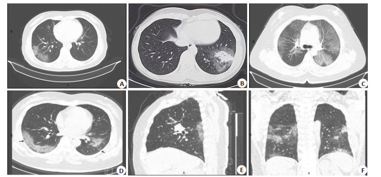
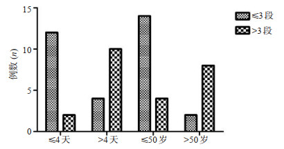
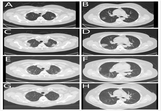
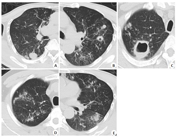
ObjectiveTo explore the typical and atypical HRCT manifestations of novel coronavirus and its value in clinical diagnosis and treatment. MethodsThe chest HRCT manifestations of 28 COVID-19 patients diagnosed by clinical and nucleic acid testing were collected and analyzed. There were 16 males and 12 females, with age of 25-71 years(46±14.9 years). The distribution, range, shape and density of the lesion, the presence of lymph nodes in the mediastinum and hilum, and the presence of abnormalities in the thorax and pleura were analyzed. According to the 4-day interval between the first chest HRCT examination at the age of 50 and after fever, the age and time interval were divided into groups. The number of lung segments involved by COVID-19 in different groups was compared. ResultsTypical HRCT manifestations of COVID-19: Subpleural or multicentric distribution in 20 cases, single or multiple patchy or segmental ground glass density shadow (GGO) in 11 cases. GGO was associated with septal thickening in 14 cases, of which 5 showed typical "paving signs". Eight cases of solid change with GGO. Consolidation was observed in 5 cases with internal air bronchus. Atypical manifestations: 5 cases presented diffuse hyaline nodules in the center of lobule. In 5 cases, there were meshes and honeycomb fiber strip shadows. In 3 cases, the distribution was segmental around the bronchus. Pleural and mediastinal manifestations: pleural thickening in 1 case. There was no pleural effusion and lymph node enlargement in this group. Patients who underwent HRCT for the first time after fever at a time interval of more than 4 days involved more than 3 pulmonary segments (P=0.002). Patients over 50 years of age with more than 3 lung segments were more likely to be involved than patients under 50 years of age (P=0.003) ConclusionsThe chest HRCT manifestations of COVID-19 have certain characteristics, which not only become the main means of early screening and diagnosis of COVID-19, but also control and early treatment of highly suspected patients.It can evaluate the clinical course, severity, efficacy and prognosis of patients, so as to control the epidemic situation and improve the cure rate.

2020, 43(3): 375-380.
doi: 10.12122/j.issn.1674-4500.2020.03.03
Abstract:
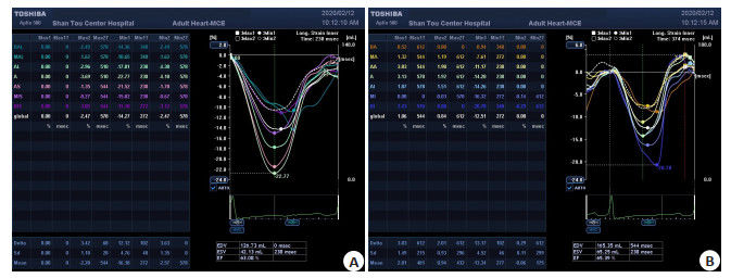
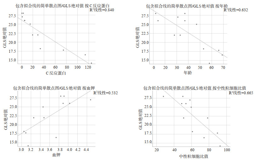
ObjectiveTo analyze the clinical manifestations of new coronavirus pneumonia (COVID-19) combined with early heart damage, and explore indicators that can identify COVID-19 heart damage early. MethodsThe clinical manifestations, laboratory examinations, and cardiac color Doppler ultrasound of 12 COVID-19 patients admitted to our hospital from January 20, 2020 to February 20, 2020 were collected. Among the patients, left ventricular ejection fraction (LVEF) and general left ventricular longitudinal strain (GLS) were collected by cardiac color Doppler ultrasound. We used the data above to analyze the occurrence of COVID-19 early heart damage. The sinus tachycardia, new ECG changes and GLS were compared as possible identification indicator of COVID-19 combined early heart damage performance. The risk factor of COVID-19 early heart damage was analyzed. ResultsThe 12 COVID-19 patients included 2 severe cases, 8 normal cases, and 2 mild cases. All patients had no significant abnormalities in troponin Ⅰ and left ventricular ejection fraction. However, abnormal rates of sinus tachycardia, new changes in ECG and GLS were all 33.3%. The abnormal rates of CKMB and BNP were both 8.3%. The absolute value of GLS, the possible index of COVID-19 with early heart damage, were negatively correlated with age, neutrophil ratio, and C-reactive protein (r=-0.795, -0.816, -0.917, P < 0.05). They were positively correlated with hypokalemia (r= 0.73, P < 0.05). ConclusionsSinus tachycardia, new ECG changes and GLS have a certain abnormal proportion in COVID-19 patients, which may be used as indicators of early heart damage identification. In COVID-19 patients with normal aTNI and LVEF, observing whether they appear sinus tachycardia, new abnormal ECG changes, and abnormal GLS will help to identify COVID-19 with early heart damage.
2020, 43(3): 381-386.
doi: 10.12122/j.issn.1674-4500.2020.03.04
Abstract:
ObjectiveTo analyze the epidemiology and clinical characteristics of patients with COVID-19, and provide reference for prevention and control strategies. MethodsWe selected 41 patients with COVID-19 of Linyi, including 9 patients with mild symptom, 25 patients with general symptom, 7 patients with severe and critical symptom. The epidemiological and clinical characteristics were analyzed. ResultsThe median age was 41 years old (31.6-53.6 years old), the male to female ratio 1.56:1, median BMI 25.1 kg/m2(22.4-27.6 kg/m2). The severe and critical patients were older, more obese, and had more underlying diseases. Fifteen cases(36.6%) were clearly related to Hubei, 33 clustered cases (80.5%), the median incubation period 6.5 d (4.0-11.5 d).The main first symptoms of the patients included fever (65.9%), dry cough (78.0%), the proportion of fever, sputum, and headache. The dizziness in severe and critical patients was significantly higher than that in mild group. For severe and critical patients, PLT was significantly reduced. AST, Cr, Myo, and cTnI were significantly increased. CT images showed multiple patchy shadows or ground glass shadows on one or both sides. The number of antiviral drugs, antibiotics, glucocorticoids, gamma globulin, thymus fascin and oxygen therapy were higher in severe and critical patients. ConclusionsPatients with multiple underlying diseases, advanced age, and obesity are more likely to develop into severe or critical. It causes abnormalities in multiple systems or organs.
2020, 43(3): 387-393.
doi: 10.12122/j.issn.1674-4500.2020.03.05
Abstract:
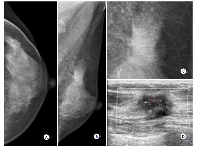
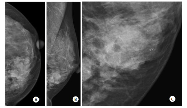
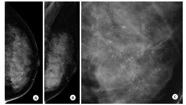
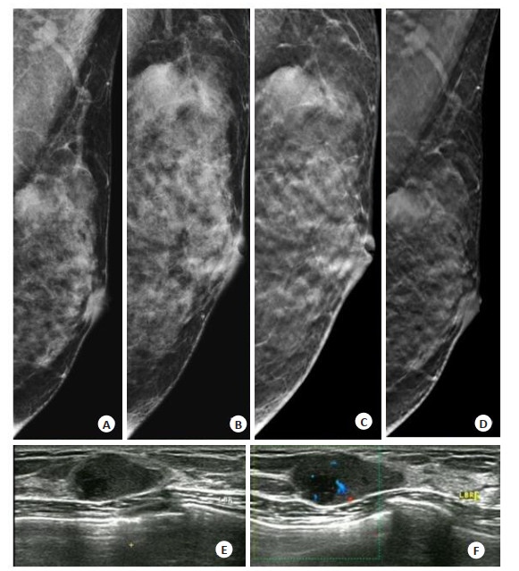
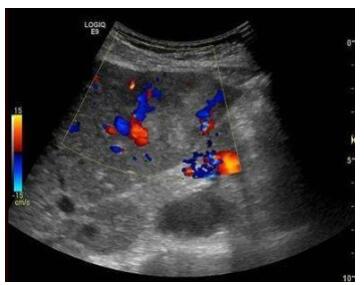
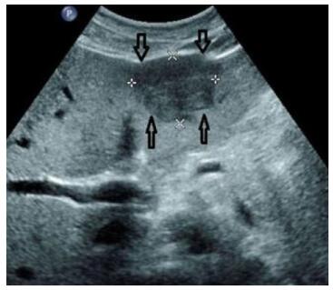
ObjectiveTo investigate the clinicopathological features and the main points of X-ray and ultrasound diagnosis of primary young breast cancer with different molecular types. MethodsA total of 140 cases of primary young breast cancer confirmed by pathology were collected. The clinicopathological data, X-ray and ultrasound findings of each subtype were analyzed retrospectively. ResultsThe molecular typing order of young breast cancer were Luminal B, HER2 overexpression, triple negative and Luminal A type. The mammography features was significantly different between different molecular phenotype in young breast cancer. Luminal A showed irregular mass with calcification and marginal burr and luminal B showed irregular mass with calcification and blurred margin. The overexpression of HER2 showed simple calcification, fine pleomorphism or/and thin line, segment-like distribution, and triple negative showed simple irregular mass with blurred edge. Different subtypes of ultrasound showed irregular solid hypoechoic masses with calcification and blurred edges. There were differences in echo properties and blood supply. Although the pathological types of different subtypes of young breast cancer were significant, most of them were IDC grade 2 and Luminal B type accounted for the highest proportion Luminal B type was the most common subtype of young breast cancer with lymph node metastasis and distant metastasis, but the difference was not statistically significant. ConclusionsThe main ultrasonographic signs of this group of cases are not characteristic, mixed echoes are more common in the triple negative type, and the blood supply of each molecular subtype is also different. Therefore, a preliminary molecular classification can be made according to the comprehensive manifestations of X-ray and ultrasound before operation.
2020, 43(3): 394-398.
doi: 10.12122/j.issn.1674-4500.2020.03.06
Abstract:
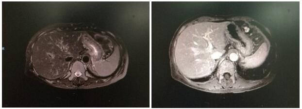
Objective To explore the relationship between the peak time, peak acceleration time, enhancement rate and the degree of histological differentiation and malignant behavior of cancer cells in PLC patients. Methods From February 2015 to February 2019, 60 patients with primary liver cancer were selected. According to the degree of tissue differentiation, there were 21 patients with low differentiation, 19 patients with medium differentiation, 20 patients with high differentiation. All patients underwent pathological examination, 35 patients in stage I-II, 25 patients in stage III-IV and 32 patients with lymph metastasis. To analyze the difference among the different pathological state, the degree of tissue differentiation and the condition of lymph node metastasis in the beginning time, peak time, peak enhancement intensity, enhancement rate and 50% tilt rate were compared. Results The differences of the starting time, peak time, peak enhancement intensity, enhancement rate and 50% tilt rate between the low differentiation group, the high differentiation group and the middle differentiation group were significant. The peak time, peak enhancement intensity and 50% tilt rate of the patients were divided into low differentiation group, middle differentiation group and high differentiation group. The time and rate of enhancement in the stage Ⅲ-Ⅳ group were significantly higher than those in the stage Ⅰ-Ⅱ group. The peak time, peak intensity and 50% tilt rate in the stage Ⅲ-Ⅳ group were significantly lower than those in the stage Ⅰ-Ⅱ group. The time and rate of enhancement in the stage Ⅲ-Ⅳ group were significantly higher than those in the stage Ⅰ-Ⅱ group. The peak time, peak intensity and 50% tilt rate in the stage Ⅲ-Ⅳ group were significantly lower than those in the stage Ⅰ-Ⅱ group. In the group without lymph node metastasis, the lymph node metastasis, pathological state and the degree of tissue differentiation were positively correlated with the beginning time and the rate of enhancement respectively. The lymph node metastasis, pathological state and the degree of tissue differentiation were negatively correlated with the peak time, the peak intensity of enhancement and the 50% tilt rate respectively. Conclusions The peak time, peak acceleration time and enhancement rate measured by contrast-enhanced ultrasound are significantly related to the degree of histological differentiation and malignant behavior of cancer cells in patients with primary liver cancer.
2020, 43(3): 399-403.
doi: 10.12122/j.issn.1674-4500.2020.03.07
Abstract:
Magnetic resonance diffusion kurtosis imaging (DKI) is an extension of diffusion tensor imaging (DTI) technology. It can quantify the non-Gaussian diffusion characteristics of water molecules in tissues, and provide more real and accurate tissue microstructure information than DWI and DTI. DKI is mainly used in clinical diagnosis and treatment of central nervous system diseases. Early and accurate diagnosis of these diseases and timely formulation of a reasonable treatment plan can improve the prognosis of patients. Therefore, as a supplement and extension of DWI and DTI technology, DKI technology can achieve complementary advantages through quantitative analysis of the non-normal diffusion characteristics of water molecules, which is of great significance for the early diagnosis and prognosis evaluation of central nervous system diseases. In recent years, DKI has more widely used in clinic, but the optimal selection of b value and how to shorten the scanning time need to be further studied. Combined with the current research, this paper introduces the basic principle and related parameters of DKI, summarizes the application of DKI in the early diagnosis of mild brain injury and acute cerebral infarction, the monitoring and evaluation of brain degenerative diseases, and the preoperative grading of brain tumors.
Magnetic resonance diffusion kurtosis imaging (DKI) is an extension of diffusion tensor imaging (DTI) technology. It can quantify the non-Gaussian diffusion characteristics of water molecules in tissues, and provide more real and accurate tissue microstructure information than DWI and DTI. DKI is mainly used in clinical diagnosis and treatment of central nervous system diseases. Early and accurate diagnosis of these diseases and timely formulation of a reasonable treatment plan can improve the prognosis of patients. Therefore, as a supplement and extension of DWI and DTI technology, DKI technology can achieve complementary advantages through quantitative analysis of the non-normal diffusion characteristics of water molecules, which is of great significance for the early diagnosis and prognosis evaluation of central nervous system diseases. In recent years, DKI has more widely used in clinic, but the optimal selection of b value and how to shorten the scanning time need to be further studied. Combined with the current research, this paper introduces the basic principle and related parameters of DKI, summarizes the application of DKI in the early diagnosis of mild brain injury and acute cerebral infarction, the monitoring and evaluation of brain degenerative diseases, and the preoperative grading of brain tumors.
2020, 43(3): 404-409.
doi: 10.12122/j.issn.1674-4500.2020.03.08
Abstract:
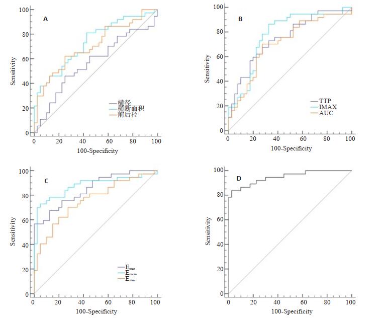
Objective To explore the application of multimodal ultrasound imaging technology (mUS) in the diagnosis of diabetic peripheral neuropathy (DPN). Methods From January 2019 to October 2019, 38 patients with T2DM and DPN (group A), 40 simple T2DM patients (group B) and 36 healthy people (group C) in our hospital were selected. They were given conventional two-dimensional ultrasound, ultrasound contrast and shear wave elastography (SWE) and neuroelectrophysiological examination. The results in different groups were compared. Receiver operating characteristic curve (ROC) was drawn and the area under the curve (AUC) was calculated to analyze the diagnostic value of each parameter on DPN. Results The incubation period in group A was significantly higher than that in groups B and C (P < 0.05). The MCV and amplitude were significantly lower than those in groups B and C (P < 0.05). The differences of the indicators between group B and group C were not significant(P > 0.05). The anterior-posterior diameter, transverse diameter and cross-sectional area of tibial nerve in group A were higher than those in groups B and C. The anterior-posterior diameter, transverse diameter and cross-sectional area of tibial nerve in group B were higher than those in group C (P < 0.05). Time to peak (TTP) in group A was higher than that in groups B and C, while the peak intensity (Imax) and AUC were lower than those in groups B and C. The TTP in group B was higher than that in group C, while the Imax and AUC were lower than those in group C(P < 0.05). The maximum (Emax), minimum (Emin) and mean value (Emean) of elasticity of tibial nerve in group A were higher than those in group B and C. The Emax, Emin and Emean of tibial nerve in group B were higher than those in group C (P < 0.05). The cross-sectional area, Imax and Emean had the highest diagnostic efficacy in diagnosing DPN in conventional two-dimensional ultrasound, ultrasound contrast and SWE, with AUC values of 0.733, 0.775 and 0.876. The AUC of combination of the three was 0.948. The sensitivity and specificity were up to 83.78% and 97.50%. Conclusions mUS technology can synthesize the data information of conventional two-dimensional ultrasound, ultrasound contrast and SWE examination models.It can provide reference basis for evaluating the diagnosis of DPN, such as peripheral nerve morphological characteristics, microcirculation blood perfusion and elasticity index.
2020, 43(3): 410-414.
doi: 10.12122/j.issn.1674-4500.2020.03.09
Abstract:
Objective To improve the preoperative diagnosis rate of Primary duodenal stromal tumor (PDST) by analyzing the MSCT enhancement of PDST with different pathological risks. Methods The clinical and imaging data of 20 cases confirmed by surgery pathology of primary duodenal stromal tumor were retrospectivly analyzed. The tumor location, shape, size, density, boundary, enhanced scan, arterial and venous phase and delay characteristic were observed. According to the pathological grading into low-risk and high-risk group, the signs of rank and inspection were analyzed. Results Three of the 20 cases with PDST were located in the bulb, 11 cases were descending, and 6 cases were horizontal.The lesions were solid-cystic in 8 cases and solid in 12 cases.The lesions grew into the lumen in 1 case, inside and outside the lumen in 8 cases, and outside the lumen in 11 cases.There were 3 cases of very low risk, 9 cases of low risk, 0 cases of moderate risk, and 8 cases of high risk. At the enhancement stage, the enhancement was obviously uniform or non-uniform, 13 cases presented vascular enhancement, the bigger the lesion and the more non-uniform the enhancement. Eight were in the high-risk group with an average size of 5.06 ± 1.64 cm. 12 were in the low-risk group with an average size of 2.83 ± 1.79 cm. The size and venous difference of the low-risk group were significantly differrent from the high-risk group (P < 0.05). Conclusions MSCT enhancement and multilevel reconstruction are conducive to the diagnosis of PDST. Tumor size and venous phase difference can effectively evaluate the primary PDST risk grading. The clinical characteristics and CT enhancement of the low-risk group and high-risk group of interstitial tumors are different.
2020, 43(3): 415-421.
doi: 10.12122/j.issn.1674-4500.2020.03.10
Abstract:
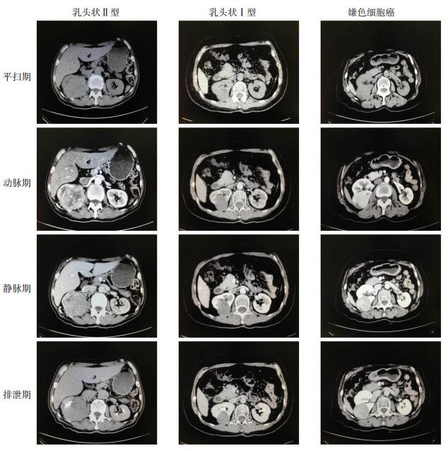
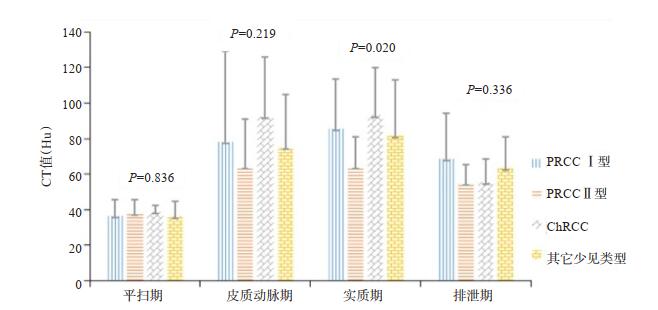
Objective To investigate the value of multislice computed tomography enhanced scan in the differential diagnosis of rare subtypes of renal cell carcinoma. Methods We retrospectively collected the data of rare renal cell carcinoma (RCCs), diagnosed by pathology in our hospital, including 15 cases of papillary carcinoma (PRCC) type Ⅰ, 16 cases of PRCC type Ⅱ, 16 cases of chromocytic cell carcinoma (ChRCC), and 13 cases of other rare RCCs. All the patients with RCC underwent examination of enhanced CT, using GE LightSpeed VCT. The CT imaging characteristics of each subtype and the ratio of CT value of each phase to that of abdominal aorta were compared and analyzed. Results There were significant differences among four groups on whether the margin was smooth and whether there was lymph node enlargement in the hilum and retroperitoneum. The margin smooth rate of other rare renal cell carcinoma was only 53.8%, which was significantly lower than that of PRCC type Ⅰ (86.7%), PRCC type Ⅱ (75.0%) and CHRCC (100.0%) (P < 0.05). The occurrence rates of hilar and retroperitoneal lymph node enlargement in PRCC type Ⅱ and other rare types were 37.5% and 53.8%, respectively, which were significantly higher than those in PRCC type Ⅰ (13.3%) and CHRCC type (12.5%) (P < 0.05).The difference of CT value of solid tumors among the four groups was significant (P < 0.05). The tumor CT value of enhanced three phases in PRCC type Ⅱ was lower than that in PRCC type Ⅰ. The CT value in ChRCC tumors was obviously lower in excretion phase, but significantly higher in the rest three phases than those of the other three groups. The ratio of PRCC type Ⅱ in parenchymal phase was significantly different from those in ChRCC and other rare types (P < 0.05). Conclusions Enhanced CT can be used for the initial identification of different RCCs subtypes. It is helpful in the preoperative diagnosis of RCCs.
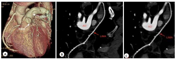
2020, 43(3): 422-427.
doi: 10.12122/j.issn.1674-4500.2020.03.11
Abstract:
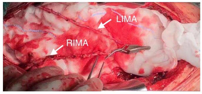
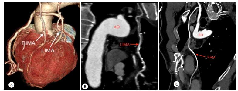
Objective To explore the short-term (3 months) results of bilateral internal mammary artery (BIMA) grafting in elderly diabetic patients (60-75 years old). Methods From December 2015 to August 2017, 64 elderly patients who underwent BIMA grafting in our department were enrolled. They were divided into two groups: diabetic group and non-diabetic group. The diabetic group had 36 cases, including 25 males, 11 females, with an average age of 62.83±2.60 years old. Non-diabetic group had 28 cases, including 23 males and 5 females, with an average age of 62.29±1.76 years old. The clinical records, intra-operative data (aortic clamp time, cardiopulmonary bypass time, operation time, etc.), short-term complications, and imaging follow-up data of these patients were retrospectively analyzed. Results The incidence rate of left main + triple-vessel disease and preoperative HbA1c level of diabetic group were higher than those of non-diabetic group (P=0.025, 0.001). The differences of the internal mammary artery harvest technique, operation time, aortic clamp time, cardiopulmonary bypass time as well as the flow and pulsatility index of internal mammary artery grafts between two groups were not significant (P > 0.05). The difference of sternal wound complications and other common complications between these groups was not significant (P > 0.05). Four patients suffered sternal wound complications. The preoperative HbA1c level of them was significantly higher than the other's (P < 0.001). The difference of grafts occlusion between two groups was not significant (P > 0.05), according to the results of coronary CTA three months after operation. Conclusions Compared with the elderly patients without diabetes mellitus, the coronary artery lesions in the elderly (60-75 years old) diabetic patients are more serious. However, the difficulty of BIMA grafting for them are not increased. Satisfactory short-term results could be achieved on the basis of appropriate treatment. Coronary CTA as a follow-up method for these patients achieves satisfactory results.
2020, 43(3): 428-433.
doi: 10.12122/j.issn.1674-4500.2020.03.12
Abstract:
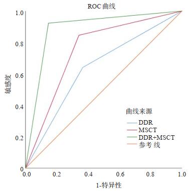
Objective To explore the diagnostic value of multi-slice spiral CT (MSCT) and direct digital radiography (DDR) in active pulmonary tuberculosis (ATB) and non-active pulmonary tuberculosis (non-ATB). Methods From February 2018 to October 2019, 80 cases of tuberculosis in our hospital were admitted, including 39 cases of ATB and 41 cases of non-ATB. All patients were examined by MSCT and DDR. The DDR and MSCT features of ATB and non-ATB patients were observed. The diagnostic value of MSCT, DDR and DDR+MSCT were analyzed by receiver operator characteristics curve(ROC)based on the pathological results. Results The detection rate of pulmonary tuberculosis by DDR was 65.00%. The detection rate of patchy, indistinct edge and unevenly dense shadow in ATB group were higher than that in non-ATB group (P < 0.05). Pulmonary tuberculosis detection rate was 76.25% by MSCT. The MSCT signs in group ATB were to give priority to ground glass density shadow, nodules, tree bud sign, thickening of bronchial wall, fuzzy consolidation at the edge, mainly cavity, while the MSCT signs in group non-ATB were high-density nodules, stripe shadow and lung structure distortion. According to the pathological results, the sensitivity and specificity of DDR, MSCT and DDR+MSCT in diagnosing ATB were 64.10%, 84.62%, 92.31%;65.85%, 68.29%, 87.80%, respectively, and the AUC was 0.638, 0.752, 0.888, respectively. Conclusions MSCT and DDR have certain value in diagnosing pulmonary tuberculosis. DDR+MSCT can improve the detection rate of pulmonary tuberculosis and the value of ATB differential diagnosis.
2020, 43(3): 434-438.
doi: 10.12122/j.issn.1674-4500.2020.03.13
Abstract:
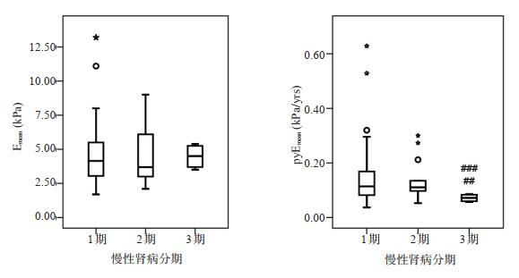
Objective To investigate the value of RT-SWE technology in early damage of chronic kidney disease. Methods Emean modulus of renal cortex in patients with chronic kidney disease (stage 1-3) was detected by RT-SWE technique, which was compared with 24-hour urinary protein quantification, serum creatinine value and interstitial fibrosis degree in with renal biopsy. Age factor was introduced to adjust the Emean value (pyEmean). The differences of pyEmean value in patients with early chronic kidney disease at all stages were compared. Results The difference in Emean value between the groups of patients with chronic kidney disease stage 1-3 was not significant (P > 0.05).The difference in pyEmean value obtained by age correction between stage 1 and stage 2 chronic kidney disease patients was not significant (P > 0.05). The difference between stage 1, 2 and 3 was significant (P < 0.05). Conclusions pyEmean excludes the influence of age on renal hardness. It has important potential application value in the evaluation of renal fibrosis in early chronic kidney disease patients. RT-SWE technology can be used in the evaluation of early renal damage in chronic kidney disease.

2020, 43(3): 439-443.
doi: 10.12122/j.issn.1674-4500.2020.03.14
Abstract:
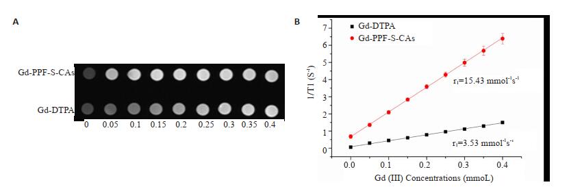
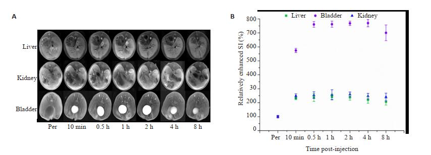
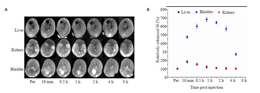
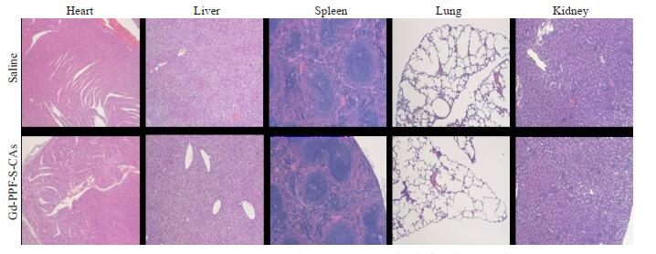
Objective To explore the imaging of a novel gadolinium-containing magnetic resonance contrast agent (Gd-PPF-S-CAS) in normal organs in mice. Methods Firstly, the relaxation performance of GD-PPF-S-CAS and DTPA-GD in vitro was compared. Secondly, 10 normal Balb/C mice were randomly divided into two groups. One group was injected with Gd-PPF-S-CAs, and the other was injected with clinical DTPA-Gd. The liver, kidney and bladder of the mice were scanned respectively. The Gd-PPF-S-CAs group was compared with clinical DTPA-Gd in terms of in vivo enhancement effect, enhancement amplitude and duration of enhancement. Results The relaxation rate of Gd-PPF-S-CAs in the experimental group was 15.43 mmol-1s-1, which was five times that of the clinical DTPA-Gd relaxation rate of 3.53 mmol-1s-1. By analyzing the enhancement effect, amplitude and duration of liver, kidney and bladder in mice, GD-PPF-S-CAS had more obvious enhancement effect, higher enhancement amplitude and longer duration of enhancement than DTPA-Gd in mice liver, kidney and bladder. Conclusions Gd-PPF-S-CAs have a high relaxation effect in vitro. Normal organs in mice have obvious enhancement effects and long-lasting enhancement duration, which can effectively solve the shortcomings of small molecular DTPA-Gd enhancement, low tissue contrast and short imaging window time. At the same time, Gd-PPF-S-CAs have the characteristics of enzymatic degradation and can be metabolized in the body quickly, which effectively solves the potential toxicity of Gd3++ contrast agent and has good clinical application prospects.
2020, 43(3): 444-448.
doi: 10.12122/j.issn.1674-4500.2020.03.15
Abstract:
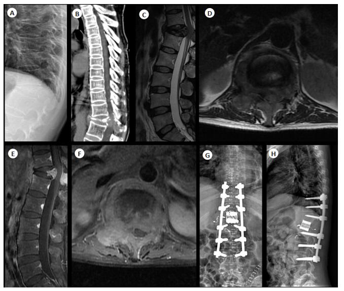
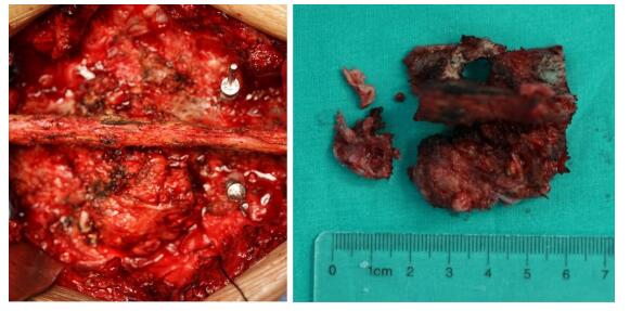
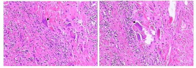
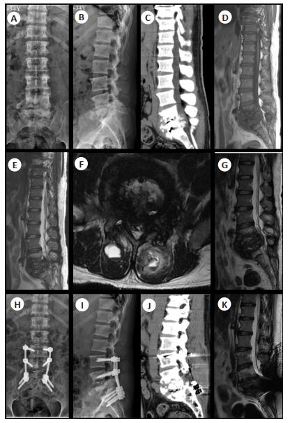
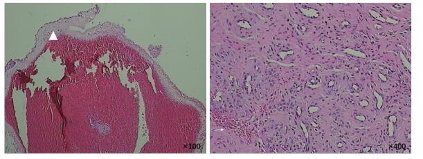
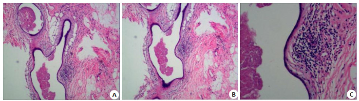
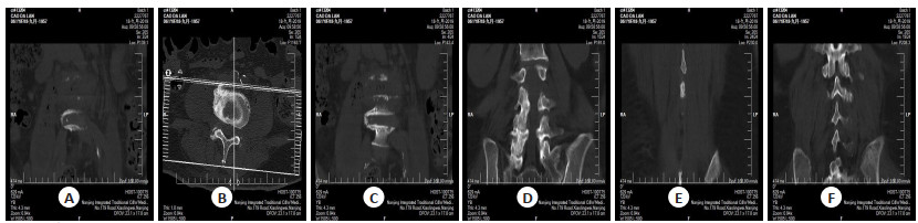
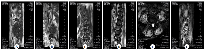
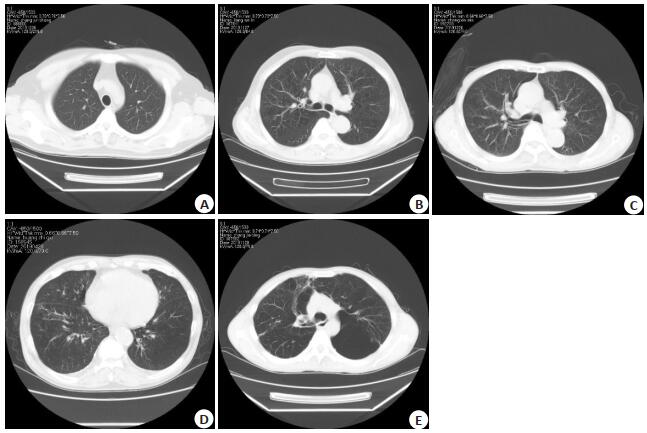
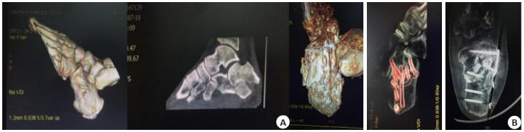
Objective To analyze the diagnosis and treatment of syphilitic myelitis. Methods The clinical features, imaging data, pathological manifestations and treatment of 2 patients with syphilitic myelitis diagnosed by operation and pathology in our department were reported. The clinical data were analyzed and summarized combined with the review of related literature. Results Case 1: a 47-year-old female patient with lumbosacral pain for more than 2 years. The serology showed syphilis (+) and posterior thoracic puncture biopsy was performed. T12 tumor was diagnosed, and posterior debridement of T12 vertebrae was performed. Autogenous iliac bone graft fusion and internal fixation was performed. The pathological findings were consistent with the changes of syphilis. Case 2: a 34-year-old male patient was diagnosed with tertiary syphilis for more than 1 year and low back pain with lameness for more than 2 months. Lumbar puncture biopsy showed that L5/S1 syphilis spondylitis complicated with spinal tuberculosis. Posterior spinal canal decompression, focus debridement, discectomy, interbody fusion and internal fixation were performed. Two patients were treated with benzylpenicillin anti-syphilis before and after operation. The postoperative symptoms were significantly improved, the reexamination index decreased, and the imaging data indicated that the focus was reduced. Conclusions Syphilitic spondylitis is extremely rare in clinic. After systemic antisyphilis treatment and surgical treatment, the focus is reduced and the symptoms are improved, which has a good prognosis and curative effect.
2020, 43(3): 449-452.
doi: 10.12122/j.issn.1674-4500.2020.03.16
Abstract:
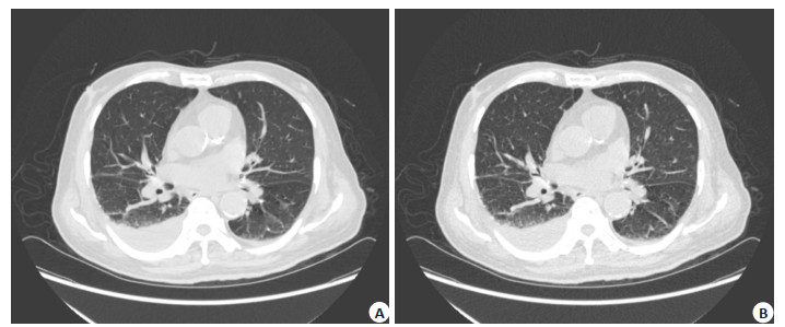
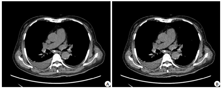
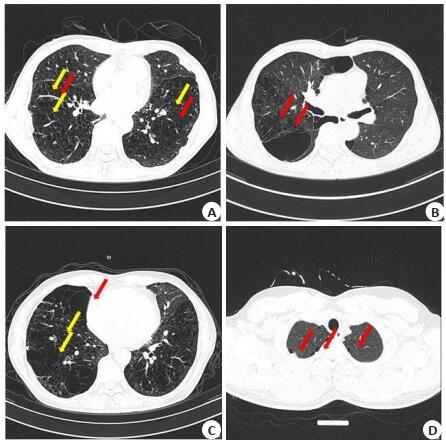
ObjectiveTo investigate the effect of multi-model iterative reconstruction technique on imaging quality of low-dose GE revolution CT in early lung cancer. MethodsA total of 240 patients with early lung cancer who visited Beijing Shijitan Hospital from January 2018 to June 2019 were collected. All patients underwent low-dose GE revolution CT imaging. The images were reconstructed by multi-model iterative reconstruction technique. The image quality of lung window, mediastinal window and image quality parameters before and after image reconstruction were compared. ResultsCompared with low-dose GE revolution CT images, the image quality of lung window and mediastinal window reconstructed by multi-model iterative reconstruction technique was improved with reduced image noise (8.83±1.95 vs 9.21±2.17 Hu) and increased signal-to-noise ratio (SNR, 7.21±1.30 vs 6.89±1.22) (P < 0.05). The difference of the CT value was not significant(65.01±7.94 vs 65.38±8.26 HU, P > 0.05). ConclusionMulti-model iterative reconstruction technique can improve the imaging quality of low-dose GE revolution CT for early lung cancer. It has important clinical value for screening early lung cancer.
2020, 43(3): 453-456.
doi: 10.12122/j.issn.1674-4500.2020.03.17
Abstract:
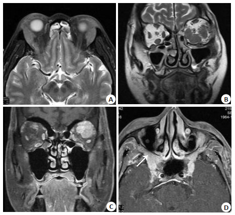
ObjectiveTo evaluate MRI findings of IgG4-related ophthalmic disease. MethodsWe included 18 patients with histopathologically proven IgG4-ROD. MR images were retrospectively evaluated for location, laterality, shape, margin, T1 and T2 signal intensity on precontrast MRI, internal architecture, ocular adnexal lesion enhancement patterns. ResultsThe lesions involved the lacrimal gland (n=6), focal mass (n=7), extraocular muscles (n=1) and multiple areas (n=4). All lacrimal gland lesions presented as diffuse enlargements. 4 patients had lesions involving multiple areas which extended along the trigeminal nerve, accompanied by expansion of neural foramina along their courses, with no signs of bone destruction. Infraorbital nerve enlargement was present in 8 cases. All ocular adnexal lesions showed isointensity on T1- and hypointensity on T2-weighted images, homogenous enhancement patterns and bone remodelling without destruction. TIC exhibited a rapidly enhancing and slow washout pattern in 14 patients. ConclusionIgG4-ROD can involve the lacrimal gland, extraocular muscles, medial canthus, extraconal space, and infraorbital nerves, and pterygopalatine fossa. Recognition of the typical radiological features of IgG4-ROD may help in the diagnosis of this benign clinical entity.
2020, 43(3): 457-461.
doi: 10.12122/j.issn.1674-4500.2020.03.18
Abstract:
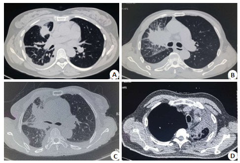
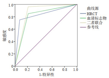
ObjectiveTo explore the diagnostic value of high-resolution CT (HRCT) combined with detection of serum tumor markers on early lung cancer. MethodsFrom March 2016 to November 2019, 80 patients with early lung cancer and 80 patients with benign lung lesions in the hospital were selected. HRCT examination and detection of lung cancer serum markers [carcinoembryonic antigen (CEA), carbohydrate antigen CA 125, neuron-specific enolase (NSE), cytokeratin 19 fragment (CYFRA21-1)] were performed on all patients. The HRCT imaging results and serum markers levels were compared between the two groups. The pathological results were taken as the gold standards to analyze the diagnostic efficacy of HRCT, serum markers and the combination of the two. ResultsThe differences of the imaging features of lobulation sign, spicule sign, interfaces and cystic lucency shadow between lung cancer group and benign group were significant (P < 0.05). The levels of serum CEA, CA125, NSE and CYFRA21-1 in lung cancer group were significantly higher than those in benign group (P < 0.05). With the pathological results as the gold standards, the sensitivity, specificity and accuracy of HRCT in the diagnosis of lung cancer were75.00%, 92.50% and 83.75%, the above indexes of serum markers were 85.00%, 78.75% and 81.75% respectively, and above indexes of the combination of the two were 96.25%, 77.50% and 86.87%. The values of area under the receiver operating characteristic curve (ROC curve) of HRCT, serum tumor markers and combined detection of early lung cancer were 0.838, 0.819 and 0.875(P < 0.001). ConclusionHRCT and lung cancer serum markers have certain diagnostic value on early lung cancer. The combined examination of the two can further improve the diagnostic sensitivity and accuracy.
2020, 43(3): 462-465.
doi: 10.12122/j.issn.1674-4500.2020.03.19
Abstract:
Radiomics has made many achievements in the diagnosis, differentiation and prognosis of oncology. With the continuous development on oncology, it has made gratifying progress in the field of clinical non-neoplastic disease with its advantages of high throughput and big data. The past reports analyzed measurements of brain volume, attention deficit hyperactivity disorder, Schizophrenia, differentiation of hypertrophic cardiomyopathy and hypertensive heart Disease, the characteristics of acute coronary syndrome and atheromatous plaque, and the diagnosis of diseases such as cirrhosis. The results showed that compared with the conventional imaging methods, radiomics showed more accurate diagnostic advantages. Although relevant research methods are different, several studies have shown that ICC and AUC values can reach around 0.9 or even close to 1. The above research is summarized in detail as follows.
Radiomics has made many achievements in the diagnosis, differentiation and prognosis of oncology. With the continuous development on oncology, it has made gratifying progress in the field of clinical non-neoplastic disease with its advantages of high throughput and big data. The past reports analyzed measurements of brain volume, attention deficit hyperactivity disorder, Schizophrenia, differentiation of hypertrophic cardiomyopathy and hypertensive heart Disease, the characteristics of acute coronary syndrome and atheromatous plaque, and the diagnosis of diseases such as cirrhosis. The results showed that compared with the conventional imaging methods, radiomics showed more accurate diagnostic advantages. Although relevant research methods are different, several studies have shown that ICC and AUC values can reach around 0.9 or even close to 1. The above research is summarized in detail as follows.
2020, 43(3): 466-470.
doi: 10.12122/j.issn.1674-4500.2020.03.20
Abstract:
ObjectiveTo explore CT imaging features of superior mesenteric artery (SMA) calcification in elderly patients, and further analyze its clinical features. MethodsA total of 186 patients who had completed SMA CT examination in the hospital between June 2016 and April 2020 were selected as the research subjects, and their general clinical data were retrospectively analyzed. According to the presence or absence of ischemic bowel disease (IBD), the patients were divided into IBD group (n=26) and non-IBD group (n=160). The detection rate of SMA calcification, location, morphology, number and stenosis of calcified plaques were compared between the two groups. CT imaging features of SMA calcification and its correlation with IBD were discussed. ResultsThe number of SMA calcified plaques in IBD group was significantly larger than that in non-IBD group (P < 0.05). SMA calcified plaques in IBD group were mainly located in the distal segment of SMA, followed by the proximal segment, while those in non-IBD group were mainly located in proximal and middle segments of SMA. Differences in location distribution were significant (P < 0.05). Vascular angles of SMA plaques in IBD group were significantly smaller than those in non-IBD group (P < 0.05), and difference between the two groups in opening stenosis was also statistically significant (P < 0.05). The degree of SMA plaque calcification was positively correlated with the degree of opening stenosis in IBD group (P < 0.05). ConclusionSMA calcified lesions in elderly patients are mostly located in distal segment, and opening stenosis ≥25% may indicate the presence of IBD.
2020, 43(3): 471-475.
doi: 10.12122/j.issn.1674-4500.2020.03.21
Abstract:
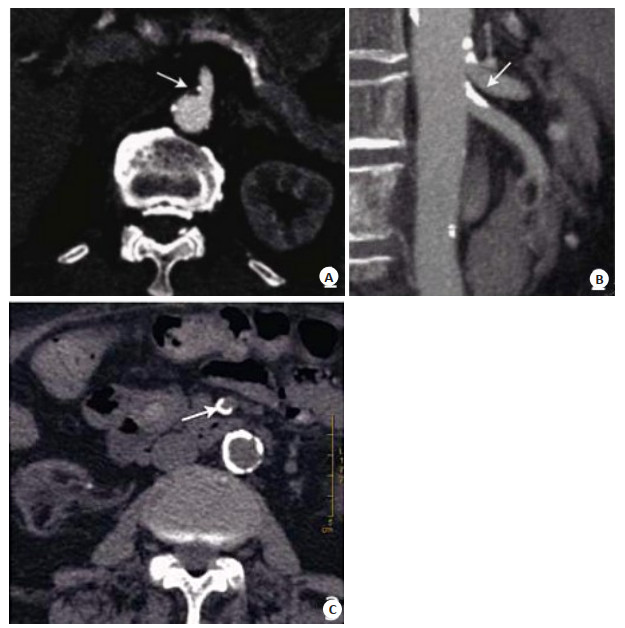
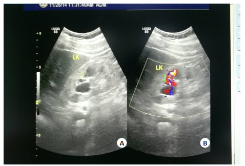
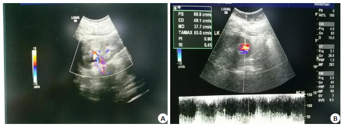
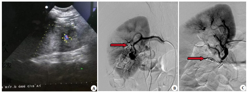
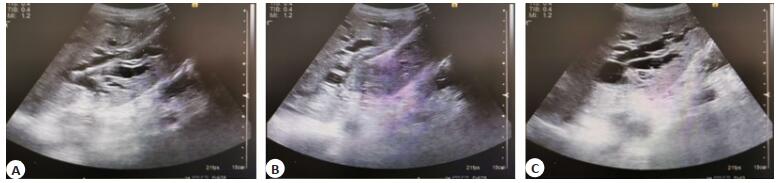
ObjectiveTo investigate the clinical value of bedside color doppler ultrasound in the diagnosis of renal pseudoaneurysm hemorrhage after percutaneous nephrolithotripsy (PCNL). MethodsNine patients with renal hemorrhage after PCNL treated by interventional embolization in Southern Hospital from November 2017 to November 2019 were enrolled. The bedside color doppler ultrasonography, clinical data and digital subtraction angiography were analyzed retrospectively. ResultsAmong 9 patients, 5 cases were finally diagnosed with arterial injury, 4 cases with pseudoaneurysm and no cases of arteriovenous fistula. All participants were examined by bedside color doppler ultrasound before arterial embolization. Four cases of renal pseudoaneurysm were initially diagnosed by bedside color Doppler Ultrasound and finally confirmed by renal arteriography. ConclusionBedside color doppler ultrasound. It has great superiority and clinical value in the diagnosis of renal pseudoaneurysm.
2020, 43(3): 476-480.
doi: 10.12122/j.issn.1674-4500.2020.03.22
Abstract:
ObjectiveTo investigate the clinical effect of ultrasound-based EnCor minimally invasive surgery on patients with plasma cell mastitis (PCM). MethodsA total of 120 PCM patients in our hospital from March 2017 to February 2019 were enrolled. They were divided into two groups according to the random number table methods, with 60 cases in each group. The open group received the open surgery, while the minimally invasive group received the EnCor minimally invasive surgery. The perioperative indexes, mass clearance rate, postoperative pain, complications, recurrence rate and satisfaction degree were compared between the two groups. ResultsThe operation time, wound healing time, scar length and intraoperative blood loss in the minimally invasive group were significantly lower than those in the open group (P < 0.05). The mass clearance rate in the minimally invasive group was significantly higher than that in the open group (96.00% vs 81.75%, P < 0.05).The difference in postoperative pain grading between the two groups was significant (P < 0.05). The incidence rates of postoperative breast deformity, total complications and postoperative recurrence rate in the minimally invasive group were significantly lower than those in the open group (0.00% vs 3.33%, 3.33% vs 23.33%, 6.67% vs 40.00%, P < 0.05). Satisfaction rate of the minimally invasive group was significantly higher than that of the open group (93.33% vs 70.33%, P < 0.05). ConclusionUltrasound-based EnCor minimally invasive surgery can effectively ameliorate the improve perioperative indicators, remove lesions, relieve pain, reduce postoperative complications and recurrence rate for PCM patients, which also obtains high satisfaction degree.
2020, 43(3): 481-484.
doi: 10.12122/j.issn.1674-4500.2020.03.23
Abstract:
ObjectiveTo investigate the characteristics and manifestations of CT and MRI in the diagnosis and differentiation of spinal tuberculosis. MethodsEighty patients with spinal tuberculosis admitted to our hospital from January 2014 to January 2019 were selected. The patients received the CT and MRI examinations. All images were read by 3 experienced radiologists. Pathological and anatomical results were taken as criteria. The positive status, invasion degree and pathological stage of spinal tuberculosis were compared between groups. ResultsThe positive rates of lesion sites presented by CT, MRI and CT combined with MRI were significant different (P < 0.05), which was highest by CT combined with MRI, followed by MRI and CT (P < 0.05). The positive rates of CT, MRI, CT combined with MRI in the diagnosis of bone, disc destruction, paravertebral mass or abscess, epidural or spinal canal invasion, spinal meningeal enhancement and calcification had significant difference (P < 0.05). The CT, MRI, CT combined with MRI had no significant difference in the diagnosis of subcortical diffusion (P > 0.05). The positive rates of bone, disc destruction, paravertebral mass or abscess, and spinal membrane enhancement were the highest by CT combined with MRI (P < 0.05). The positive rate of epidural or spinal canal invasion and spinal membrane enhancement was the lowest presented by CT, and those of MRI and CT combined MRI were the same. The positive rates of CT, MRI and CT combined with MRI in the diagnosis of exudation, proliferation and caseous degeneration were significantly different (P < 0.05), which was highest by CT combined with MRI, followed by MRI and CT (P < 0.05). ConclusionThe diagnostic value and characteristics of CT and MRI of spinal tuberculosis are different. The combined diagnosis can improve the accuracy of spinal tuberculosis, and reduce the missed diagnosis and misdiagnosis.
2020, 43(3): 485-489.
doi: 10.12122/j.issn.1674-4500.2020.03.24
Abstract:
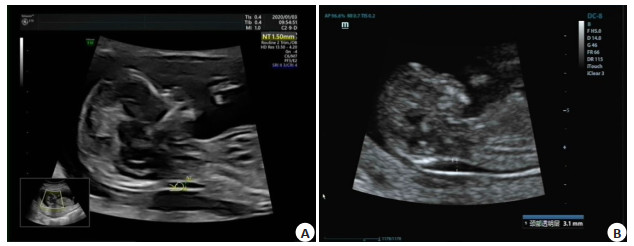
ObjectiveTo explore the relationship between the thickness of different cervical transparent layers (NT) and the outcome of fetal poor perinatal outcomes. MethodsThe clinical data of 496 pregnant women who were delivered in our hospital from March 2017 to March 2019 were retrospectively analyzed. Niety-eight pregnant women with abnormal fetal NT value were divided into 56 cases of pure fetal NT value thickening group and 42 cases of poor pregnancy outcome group according to pregnancy outcome. The NT value, age, serum Freeβ-hCG abnormal rate and NIPT chromosome abnormal rate were compared between the two groups. The value of abnormal thickening was divided into 4 groups of 2.50-3.00 mm group (group Ⅰ), 3.01-4.00 mm group (group Ⅱ), 4.01-5.00 mm group (group Ⅲ) and 5.00 mm group (group Ⅳ). The rates were compared. Logistic regression analysis was used to test the related factors of fetal perinatal outcomes and chromosomal abnormalities. ResultsA total of 56 patients with purely thickened NT values and 42 patients with poor pregnancy outcomes were found. The incidence of poor pregnancy outcomes was 42.86%. The NT value and age of the patients with poor pregnancy outcomes were significantly larger than those of the purely thickened NT patients. The abnormal rate of serum human chorionic gonadotropin free β-subunit (Freeβ-hCG) and the abnormal rate of NIPT chromosomes were significantly higher in the group than in the NT thickening group. The proportion of normal fetus in group Ⅰ was significantly higher than that in group Ⅱ, group Ⅲ and group Ⅳ. The incidence of fetal pregnancy in group Ⅲ was 100.00%, and the incidence of chromosomal abnormality was 71.43%, which was significantly higher than group Ⅰ and Ⅱ. The incidence of chromosomal abnormalities in the incidence of fetal pregnancy in group and group Ⅳ was significantly different (P < 0.05). ConclusionFetal NT value can be used as a predictor of fetal perinatal pregnancy outcome. The increase of fetal NT value will increase the risk of fetal adverse pregnancy outcome. The highest incidence of fetal chromosomal abnormalities between NT and 4.10-5.00 mm.
2020, 43(3): 490-494.
doi: 10.12122/j.issn.1674-4500.2020.03.25
Abstract:
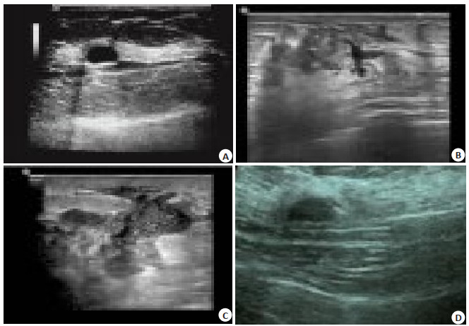
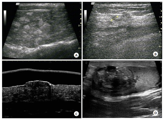
ObjectiveTo explore the clinical value of high frequency ultrasound in the diagnosis of superficial soft tissue and small organ diseases. MethodsWe retrospectively analyzed the clinical data of 78 patients with superficial soft tissue and small organ diseases admitted to the hospital from May 2014 to May 2017. All patients were diagnosed by high-frequency ultrasound and the diagnostic results were compared with the final pathological diagnostic results. The diagnostic accuracy of high-frequency ultrasound and the diagnostic accuracy rates for different diseases were calculated. The images were analyzed, and the excellent rate of images were calculated. ResultsAll patients were diagnosed by histopathological examination after admission. Among them, there were 15 cases of thyroid diseases, 29 cases of breast diseases, 18 cases of male external genital diseases and 16 cases of superficial soft tissue lesions. The detection rates of thyroid diseases, breast diseases, male external genital diseases and superficial soft tissue lesions by high-frequency ultrasound were 93.33%, 93.10%, 94.44% and 87.50%, respectively, without significant difference in the detection rates of the four superficial soft tissue or small organ diseases by high-frequency ultrasound (P < 0.05). The image quality of high-frequency ultrasound was evaluated excellent in 71 cases, good in 5 cases and poor in 2 cases. The excellent and good rate of images was 97.44%. ConclusionHigh-frequency ultrasound has high accuracy in the diagnosis of various superficial soft tissue and small organ diseases. The excellent and good rate of images is high. It is easy to operate and non-invasive, with high safety.
2020, 43(3): 495-499.
doi: 10.12122/j.issn.1674-4500.2020.03.26
Abstract:
ObjectiveTo explore the guiding and clinical effects of ultrasound combined with X-ray on percutaneous transhepatic cholangial drainage (PTCD) in patients with primary hepatolithiasis (PHL). MethodsA total of 114 patients with PHL in our hospital from June 2017 to June 2019 were selected. They were divided into combined group and X-ray group according to the random number table method, with 57 cases in each group. Combined group was given PTCD treatment under B-ultrasound combined with X-ray guidance, and X-ray group was given PTCD only under C-shaped arm X-ray perspective.The perioperative conditions, treatment effects, liver function, recurrence and complications were compared between the two groups. ResultsThe operative time, puncture frequency, intraoperative blood loss, recovery time of gastrointestinal peristalsis and hospital stay in combined group were significantly less than those in X-ray group (P < 0.05). The success rates of surgery in combined group and X-ray group were 98.15% and 90.38% respectively (P>0.05). The residual stone rates were 5.56% and 19.23% (P < 0.05). On the 3rd day after operation, the levels of serum TBIL, ALT and amylase were significantly increased in the two groups (P < 0.05). The levels of serum TBIL, ALT and amylase in combined group were lower than those in X-ray group (P < 0.05). The recurrence rates at 3 years after operation were 12.96 % and 30.77% (P < 0.05).The incidence rates of complications in two groups were 14.81% and 32.69%, respectively (P < 0.05). ConclusionPTCD treatment guided by B-ultrasound combined with X-ray for PHL patients can improve the accuracy of localization and puncture, enhance the stone removal effects and reduce the structural and functional damage of liver tissue. It can relieve the body inflammatory response and reduce occurrence of postoperative complications.
2020, 43(3): 500-503.
doi: 10.12122/j.issn.1674-4500.2020.03.27
Abstract:
ObjectiveTo explore the correlation of high flow nasal oxygen (HFNO) therapy and pulmonary CT imaging classification in patients with chronic obstructive pulmonary disease (COPD). MethodsA total of 100 patients with COPD disease worsen were selected as the research object. ALL patients were given lung CT examination, assessment of each patient's lung characteristics (including lobular centricity emphysema, whole panlobular emphysema, next to the interval emphysema and interstitial emphysema). the severity of patients with emphysema Goddard were conducted. All patients received HFNO treatment, and the efficacy was evaluated according to the results of blood gas analysis before and after treatment. The correlation of HFNO treatment and the efficacy of COPD patients with different lung CT image classification was analyzed. ResultsAmong the 100 patients with COPD, 32 were central lobular emphysema, 21 were total lobular emphysema, 20 were septal emphysema and 27 were interstitial emphysema. The Goddard score for the severity of emphysema: 0 score with 0 cases, 1 score with 23 cases, 2 score with 32 cases, 3 score with 22 cases, 4 score with 12 cases. The effective rates of high flow nasal oxygen treatment in patients with various types of emphysema were 75.00%, 66.67%, 85.00% and 25.93%, respectively, with a significant difference of effective rate between the groups (χ2=21.894, P < 0.001). There was no significant difference of the effective rate among patients with central lobular emphysema, total lobular emphysema and parietal emphysema (χ2=1.857, P=0.395). The effective rates of high- flow nasal oxygen treatment in patients with different Goddard scores of emphysema severity were 95.65%, 90.63%, 30.30% and 8.33%, respectively (χ2=50.927, P < 0.001). There was no significant difference in the response rates of patients with Goddard scores of 1 and 2 (χ2=0.501, P=0.632). The difference of the response rates between patients with Goddard scores of 3 and 4 was not significant (χ2=2.620, P=0.141). ConclusionHFNO treatment is suitable for the COPD patients in those central lobular emphysema, total lobular emphysema, parietal emphysema, and the patients with the Goddard score of 1 and 2. The curative effect is satisfactory within 1 hour. The curative effect is worse for the patients with the interstitial emphysema and the Goddard score of more than 3.
2020, 43(3): 504-507.
doi: 10.12122/j.issn.1674-4500.2020.03.28
Abstract:
ObjectiveTo observe the application value of 16-slice spiral CT in clinical diagnosis and postoperative evaluation of comminuted calcaneal fractures. MethodsWe selected 154 patients with suspected comminuted calcaneal fractures who were admitted to the hospital between March 1st, 2019 and March 1st, 2020 and confirmed by surgical findings. All patients completed X-ray film, 16-slice spiral CT and 64-slice spiral CT examinations before surgery. The diagnostic accuracy rates of the 3 methods were compared. After surgery, 16-sice spiral CT examination was performed. The calcaneal Bohler angle and Gissane angle were compared before and after surgery. ResultsA total of 154 cases (174 feet) with comminuted calcaneal fractures were found by surgery. The diagnostic accuracy rates of 16-slice spiral CT and 64-slice spiral CT (91.38% and 92.53%) were significantly higher than that of X-ray film (82.18%, P < 0.05). The difference of diagnostic accuracy between 16-slice spiral CT and 64-slice spiral CT was not significant (P>0.05). The calcaneal Bohler angle and Gissane angle in patients with comminuted calcaneal fracture were significantly increased after surgery (P < 0.05). ConclusionCompared with X-ray film, 16-slice spiral CT can effectively improve the clinical diagnostic accuracy of comminuted calcaneal fractures, and provide a reliable basis for postoperative fracture recovery evaluation.
2020, 43(3): 508-511.
doi: 10.12122/j.issn.1674-4500.2020.03.29
Abstract:
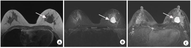
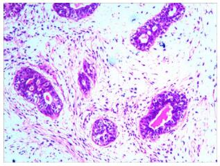
ObjectiveTo explore the differences between the breast phyllodes tumor and the fibroadenoma in MRI. MethodsA case of breast phyllodes tumor misdiagnosed as fibroadenoma in MRI was analyzed. The differences in MRI and the cause of misdiagnosis were analyzed. ResultsThe MRI performance of breast phyllodes tumor was easily mistaken for fibroadenoma. The higher signal area of fissured T2WI was seen in the breast phyllodes tumor. The characteristic expression of breast fibroadenoma was the low signal interval of T1WI and T2WI which were not strengthened internally. In addition, ADC value was also helpful to distinguish breast fibroadenoma from breast phyllodes tumor. This case was benign phyllodes tumor of breast. There was no typical fissured T2WI higher signal area on MRI image. The overall image showed benign tumor. The diagnosis doctor had considered the diagnosis of breast fibroadenoma firstly, which was more common in clinic and leading to misdiagnosis easily. ConclusionThe diagnosis of breast phyllodes tumor in MRI should be made combined with clinical.
2020, 43(3): 512-515.
doi: 10.12122/j.issn.1674-4500.2020.03.30
Abstract:
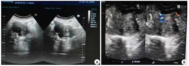
ObjectiveTo explore the value of transabdominal combined with transvaginal color Doppler ultrasound in the differential diagnosis of hysteromyoma. MethodsA total of 245 patients with hysteromyoma and 105 patients with uterine adenomyoma in the hospital between May 2015 and September 2018 were enrolled. All of them completed transabdominal sonography (TAS) and transvaginal sonography (TVS). Shapes, sizes and echo of the uterus and masses, blood flow distribution in the muscular layer and endometrium were observed. With operation and pathology as the golden standard, accuracy rates of TAS, TVS and combination of the two in the differential diagnosis of hysteromyoma were analyzed. The images and uterine artery blood flow parameters were analyzed. ResultsThe diagnostic accuracy, sensitivity and specificity of TAS combined with TVS (96.57%, 96.19% and 96.73%) were significantly higher than those of TAS or TVS alone (P < 0.05). Most of the hysteromyomas were clear, with pseudocapsules. The echoes mainly were low echoes, and blood flow in lesions was rich, which was ring-shaped or semi-ring-shaped. The shape of uterus with uterine adenomyoma was normal, but the lesion was unclear, without pseudocapsule. Most internal echoes were strong, and blood flow around the lesion was not rich. The hemodynamic parameters such as Resistance index and pulsatility index in patients with hysteromyoma were significantly lower than those with uterine adenomyoma (P < 0.05). ConclusionTVS combined with TAS can improve the accuracy in the differential diagnosis of hysteromyoma, and reduce misdiagnosis and missed diagnosis.
2020, 43(3): 516-519.
doi: 10.12122/j.issn.1674-4500.2020.03.31
Abstract:
ObjectiveTo explore the risk factors of hypoxia during one-lung ventilation undergoing thoracic surgery after using flexible fiberoptic bronchoscopy. MethodsA total of 665 patients underwent thoracic surgery from September 2017 to September 2018 in Renmin Hospital of Wuhan University, with flexible fiberopticbronchoscopy to achieve optimal position of lung isolation devices, were retrospectively analyzed. We analyzed gender, age, BMI, lung history, preoperative comorbidities, height to thyromental and sternomental distance(TMD) ratio, smoking and preoperative alcohol abuse, pulmonary function test, preoperative PaO2, breath experiment, preoperative snoring, operation time, time of one-lung ventilation, left or right double-lumen endotracheal tubes(DLT), urine volume. The patients were divided into hypoxemia group and non-hypoxemia group, according to whether hypoxemia developed during one-lung ventilation. The multi-variate logistic regression was used to stratify the risk factors forhypoxia during one-lung ventilation undergoing thoracic surgery after using flexible fiberoptic bronchoscopy. ResultsFifty-eight patients developed hypoxemia with the total number of 665, and the incidence was 8.72%. Age, smoking, preoperative comorbidities, preoperative PaO2, breath experiment, BMI, TMD ratio were related to hypoxemia. The results of logistic regression analysis showed that smoking, pulmonary function test abnormal, low preoperative PaO2, short time of breath experiment and high height to TMD ratio were independent risk factors for hypoxemia(P < 0.05). ConclusionSmoking, pulmonary function test abnormal, low preoperative PaO2, short time of breath experiment, high height to TMD ratio are independent risk factors for hypoxia during one-lung ventilation undergoing thoracic surgery after using flexible fiberoptic bronchoscopy.
2020, 43(3): 520-524.
doi: 10.12122/j.issn.1674-4500.2020.03.32
Abstract:
ObjectiveTo analyze the application value of high-frequency ultrasound combined with magnetic resonance imaging (MRI) in the early diagnosis of breast cancer. MethodsA retrospective study was performed on 85 patients diagnosed and treated in our hospital due to breast masses from January 2018 to November 2019. All patients underwent high-frequency ultrasound and MRI. Based on pathological results, the positive detection rates of high-frequency ultrasound, MRI and the combination of the two were observed. The value of high-frequency ultrasound, MRI and the combination of the two in the diagnosis of breast cancer and its staging were compared. ResultsThe pathological examination of 85 patients with breast masses revealed a total of 107 lesions, including 59 benign lesions and 48 malignant lesions. High-frequency ultrasound showed that 40 lesions were diagnosed as breast cancer with 8 lesions of missed diagnosis, and 45 lesions were diagnosed as benign masses with 14 lesions of misdiagnosis. MRI showed that 46 lesions were diagnosed as breast cancer with 2 lesions of missed diagnosis, and 53 lesions were diagnosed as benign masses with 6 lesions of misdiagnosis. High-frequency ultrasound combined with MRI showed that 47 lesions were diagnosed as breast cancer with 1 lesion of missed diagnosis, and 56 lesions were diagnosed as benign masses with 3 lesions of misdiagnosis. The accuracy, sensitivity, specificity, positive predictive value and negative predictive value of high-frequency ultrasound combined with MRI in the diagnosis of breast cancer were higher than those of high-frequency ultrasound (P < 0.05). The accuracy of MRI in the diagnosis of breast cancer was higher than that of high-frequency ultrasound (P < 0.05). The differences of the sensitivity, specificity, positive predictive value and negative predictive value compared with those of MRI were not significant (P>0.05). High-frequency ultrasound combined with MRI was superior to high-frequency ultrasound or MRI in the diagnosis of breast cancer with different stages (P>0.05). ConclusionBoth high-frequency ultrasound and MRI can effectively diagnose breast cancer. The combined application of the two can improve the diagnostic value of early diagnosis and staging of breast cancer, and provide effective help for clinical treatment.
2020, 43(3): 525-527.
doi: 10.12122/j.issn.1674-4500.2020.03.33
Abstract:
ObjectiveTo evaluate the diagnostic value of MRI, MRV and DWI in cerebral venous sinus thrombosis at different times. MethodsMRI data of 27 patients with cerebral venous sinus thrombosis diagnosed by DSA or clinical follow-up from 2015 to 2019 were analyzed retrospectively. Eighteen patients underwent DWI examination and 9 patients underwent MRI enhancement. ResultsThe MRI showed that the emplacement signal disappeared and the venous sinus appeared abnormal signal in 27 patients with venous sinus thrombosis. 3 cases of CVST showed an empty triangular sign after MRI enhancement. MRV showed that the venous sinus flow signal was different in varying degrees, Or stage thrombosis with high blood flow signal thinning, interruption-based. DWI is sensitive to acute and subacute cerebral venous sinus thrombosis and secondary venous cerebral infarction. ConclusionMRI, MRV combined with DWI can help the early diagnosis of venous sinus thrombosis and reflect the evolution of thrombus.It has great significance for the clinical treatment of venous sinus thrombosis.
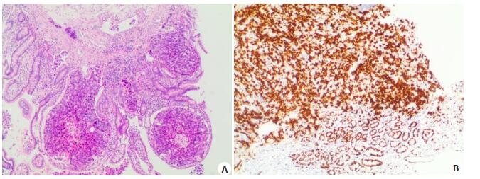
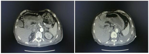
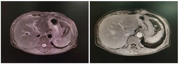
Apatinib combined with chemotherapy for advanced gastric cancer: A case report and literature review
2020, 43(3): 528-532.
doi: 10.12122/j.issn.1674-4500.2020.03.34
Abstract:
Gastric cancer is one of the common digestive tract tumors. Early gastric cancer can be cured by surgery. However, due to the low early diagnosis rate of gastric cancer, most patients have local advanced or metastasis at the time of treatment. The main treatment of advanced gastric cancer is still chemotherapy. The efficacy of chemotherapy alone is limited. With the development of tumor molecular biology and immunology, molecular targeted therapy and immunotherapy have brought new hope for patients with advanced gastric cancer. This article reports one case of apatinib combined with chemotherapy for advanced gastric cancer, providing a reference for targeted combined chemotherapy for advanced gastric cancer.
Gastric cancer is one of the common digestive tract tumors. Early gastric cancer can be cured by surgery. However, due to the low early diagnosis rate of gastric cancer, most patients have local advanced or metastasis at the time of treatment. The main treatment of advanced gastric cancer is still chemotherapy. The efficacy of chemotherapy alone is limited. With the development of tumor molecular biology and immunology, molecular targeted therapy and immunotherapy have brought new hope for patients with advanced gastric cancer. This article reports one case of apatinib combined with chemotherapy for advanced gastric cancer, providing a reference for targeted combined chemotherapy for advanced gastric cancer.
2020, 43(3): 533-536.
doi: 10.12122/j.issn.1674-4500.2020.03.35
Abstract:
ObjectiveTo investigate the effect of ultrasound-guided paravertebral nerve block on cellular immune function during thoracotomy in patients with pulmonary tuberculosis. MethodsSixty patients with elective thoracotomy for pulmonary tuberculosis were randomly divided into the general anesthesia group alone (group G) and the general anesthesia combined with ultrasound-guided paravertebral nerve block group (group P), with 30 patients in each group. Patients in group P underwent a single ultrasound-guided thoracic paravertebral nerve block before anesthesia induction. Both groups were treated with intravenous controlled analgesia and intravenous inhalation combined with general anesthesia for maintenance anesthesia. Operation time and blood loss were recorded in the two groups. VAS scores were recorded at 6 h (T1), 12 h (T2), 24 h (T3) and 72 h (T4) after operation and at cough in both groups. The percentages of CD3+、CD4+、CD8+ and CD4+/ CD8+ of patients in the two groups were recorded one day (T0) before and T1, T2, T3, and T4 after surgery. ResultsUnder the condition of quiet and cough, the VAS of group P was lower than that of group G at 6 h and 12 h after operation (P < 0.05). CD3+, CD4+, CD8+ and CD4+/CD8+ decreased in T1, T2, T3 and T4 compared with the preoperative time points (T0) (P < 0.05).CD3+, CD4+, CD8+ and CD4+/CD8+ in group P were higher at T1 and T2 than in group G (P < 0.05). ConclusionUltrasound-guided thoracic paraspinal nerve block can reduce the early pain after thoracotomy in patients with pulmonary tuberculosis and reduce the cellular immunosuppression after thoracotomy.