


Diagnostic value of high-resolution CT combined with detection of lung cancer serum tumor markers in early lung cancer
-
摘要:
目的探究高分辨率CT(HRCT)联合血清肿瘤标志物检测对早期肺癌的诊断价值。 方法选取2016年3月~2019年11月本院80例早期肺癌患者作为肺癌组,同期选取80例肺良性病变者作为良性组。所有患者均进行HRCT检查及肺癌血清标志物(癌胚抗原、CA125、神经元特异性烯醇化酶、细胞角蛋白19片段)检测,比较两组HRCT影像结果及血清标志物水平,以病理结果为金标准,分析HRCT、血清标志物及二者联合检查的诊断效能。 结果肺癌组与良性组影像特征中分叶征、毛刺征、界面、囊状透亮影比较差异有统计学意义(P < 0.05),肺癌组血清癌胚抗原、CA125、神经元特异性烯醇化酶、细胞角蛋白19片段水平均高于良性组(P < 0.05)。以病理结果为金标准,HRCT单独诊断肺癌灵敏度、特异度、准确度分别为75.00%、92.50%、83.75%,血清标志物分别为85.00%、78.75%、81.75%,二者联合分别为96.25%、77.50%、86.87%;HRCT、血清肿瘤标志物及联合检测诊断早期肺癌的受试者工作特征曲线下面积分别为0.838、0.819、0.875(P < 0.001)。 结论HRCT与肺癌血清标志物对早期肺癌均有一定诊断价值,且二者联合检查能进一步提高诊断灵敏度及准确度,值得临床应用。 Abstract:ObjectiveTo explore the diagnostic value of high-resolution CT (HRCT) combined with detection of serum tumor markers on early lung cancer. MethodsFrom March 2016 to November 2019, 80 patients with early lung cancer and 80 patients with benign lung lesions in the hospital were selected. HRCT examination and detection of lung cancer serum markers [carcinoembryonic antigen (CEA), carbohydrate antigen CA 125, neuron-specific enolase (NSE), cytokeratin 19 fragment (CYFRA21-1)] were performed on all patients. The HRCT imaging results and serum markers levels were compared between the two groups. The pathological results were taken as the gold standards to analyze the diagnostic efficacy of HRCT, serum markers and the combination of the two. ResultsThe differences of the imaging features of lobulation sign, spicule sign, interfaces and cystic lucency shadow between lung cancer group and benign group were significant (P < 0.05). The levels of serum CEA, CA125, NSE and CYFRA21-1 in lung cancer group were significantly higher than those in benign group (P < 0.05). With the pathological results as the gold standards, the sensitivity, specificity and accuracy of HRCT in the diagnosis of lung cancer were75.00%, 92.50% and 83.75%, the above indexes of serum markers were 85.00%, 78.75% and 81.75% respectively, and above indexes of the combination of the two were 96.25%, 77.50% and 86.87%. The values of area under the receiver operating characteristic curve (ROC curve) of HRCT, serum tumor markers and combined detection of early lung cancer were 0.838, 0.819 and 0.875(P < 0.001). ConclusionHRCT and lung cancer serum markers have certain diagnostic value on early lung cancer. The combined examination of the two can further improve the diagnostic sensitivity and accuracy. -
Key words:
- early lung cancer /
- high-resolution CT /
- serum tumor markers /
- diagnosis /
- ROC curve
-

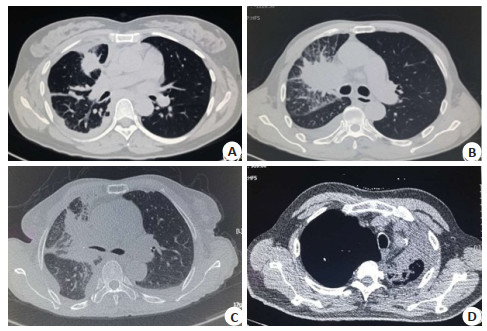
图 1 肺癌患者典型CT影像特征图片
A:女, 47岁, 右肺上叶前段见2.5 cm左右占位, 形状不规则; B:男, 65岁, 右上肺门、肺叶肿块, 周围可见毛刺征; C:女, 72岁, 右上肺叶占位, 形状不规则, 病灶与周围不张肺组织、右上肺动脉、静脉分界不清; D:男, 61岁, 右肺下叶外侧基底段0.3 cm小结节, 可见空泡征.
Figure 1. Typical CT imaging features of lung cancer patients.

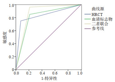
图 2 HRCT、血清肿瘤标志物及联合检测诊断早期肺癌的ROC曲线
Figure 2. ROC curves of HRCT, serum tumor markers and combined detection in the diagnosis of early lung cancer
表 1 肺癌组与良性组HRCT影像特征比较
Table 1. Comparison of HRCT imaging features between lung cancer group and benign group[n=80, n(%)]
影像特征 肺癌组 良性组 t/χ2 P 病灶直径(cm, Mean±SD) 2.25±0.78 2.11±0.54 1.320 0.189 分叶征 51.601 0.000 有 57 (71.25) 12(15.00) 无 23(28.75) 68(85.00) 毛刺征 29.565 0.000 有 51(63.75) 17(21.25) 无 29(36.25) 63(78.75) 形状 3.750 0.053 不规则形 54(67.50) 42(52.50) 圆形或类圆形 26(32.50) 38(47.50) 界面 32.918 0.000 模糊 17(21.25) 49(61.25) 清楚光整 22(27.50) 20(25.00) 清楚毛糙 41(51.25) 11(13.75) 空泡征 1.290 0.256 有 21(26.25) 15(18.75) 无 59(73.75) 65(81.25) 支气管征 3.620 0.057 有 49(61.25) 37(46.25) 无 31(38.75) 43(53.75) 囊状透亮影 17.348 0.000 有 29(36.25) 7(8.75) 无 51(63.75) 73(91.25) 胸膜凹陷征 3.025 0.082 有 46(57.50) 35(43.75) 无 34(42.50) 45(56.25) 血管集束征 2.525 0.112 有 41(51.25) 31(38.75) 无 39(48.75) 49(61.25)  下载: 导出CSV
表 2 肺癌组与良性组血清标志物水平比较
Table 2. Comparison of serum markers levels between lung cancer group and benign group (n=80, Mean±SD)
组别 肺癌组 良性组 t P CEA (ng/mL) 17.41±5.02 3.05±0.73 25.319 0.000 CA125 (U/mL) 48.82±12.21 12.15±3.69 25.714 0.000 NSE (ng/mL) 20.62±5.13 10.47±3.96 14.009 0.000 CYFRA21-1 (ng/mL) 7.28±2.51 2.25±0.75 17.276 0.000
下载: 导出CSV
表 3 HRCT、血清肿瘤标志物的诊断效能
Table 3. Diagnostic efficacy of HRCT and serum tumor markers (n=80)
病理结果 HRCT 血清标志物 二者联合 恶性 良性 恶性 良性 恶性 良性 恶性 60 20 68 12 77 3 良性 6 74 17 63 18 62
下载: 导出CSV
表 4 HRCT、血清肿瘤标志物及联合检测的ROC曲线
Table 4. ROC curves of HRCT, serum tumor markers and combined detection
指标 AUC 标准误 P 95%CI 上限 下限 HRCT 0.838 0.034 0.000 0.771 0.904 血清标志物 0.819 0.035 0.000 0.750 0.888 二者联合 0.875 0.030 0.000 0.816 0.934
下载: 导出CSV
-
[1] 中华医学会, 中华医学会肿瘤学分会, 中华医学会杂志社.中华医学会肺癌临床诊疗指南(2018版)[J].中华肿瘤杂志, 2018, 40(12): 935-64. doi: 10.3760/cma.j.issn.0253-3766.2018.12.012 [2] Masaoutis C, Mihailidou C, Tsourouflis G, et al. Exosomes in lung cancer diagnosis and treatment. From the translating research into future clinical practice[J]. Biochimie, 2018, 151: 27-36. doi: 10.1016/j.biochi.2018.05.014 [3] Hata A, Yanagawa M, Honda O, et al. Effect of matrix size on the image quality of ultra-high-resolution CT of the lung: comparison of 512×512, 1024×1024, and 2048×2048[J]. Acad Radiol, 2018, 25 (7): 869-76. doi: 10.1016/j.acra.2017.11.017 [4] 石海蓉, 张影, 陆颖, 等.血清肿瘤标志物在肺癌骨转移诊断中的价值[J].中华核医学与分子影像杂志, 2018, 38(5): 331-5. doi: 10.3760/cma.j.issn.2095-2848.2018.05.006 [5] Herbst RS, Morgensztern D, Boshoff C. The biology and management of non-small cell lung cancer[J]. Nature, 2018, 553(7689): 446-54. doi: 10.1038/nature25183 [6] Li CL, Lv Y, Shao CY, et al. Tumor-derived exosomal lncRNA GAS5 as a biomarker for early-stage non-small-cell lung cancer diagnosis[J]. J Cell Physiol, 2019, 234(11): 20721-7. doi: 10.1002/jcp.28678 [7] Ueno R, Nemoto M, Uegami W, et al. Pembrolizumab-induced pneumonitis with a perilymphatic nodular pattern in a lung cancer patient: a radio-pathologic correlation[J]. Respir Med Case Rep, 2019, 26: 168-70. http://www.ncbi.nlm.nih.gov/pubmed/30671338 [8] 周环, 满凤媛, 张卫红, 等.多层螺旋CT在肺癌诊断中的应用价值[J].癌症进展, 2019, 17(18): 2131-3, 2195. http://www.cnki.com.cn/Article/CJFDTotal-AZJZ201918007.htm [9] 王炜华, 孙希文, 袁明远.高端高分辨薄层CT在早期肺腺癌诊断筛查中的应用[J].检验医学与临床, 2019, 16(7): 865-9, 873. doi: 10.3969/j.issn.1672-9455.2019.07.001 [10] 蓝美红, 高明明, 侯代伦.超高分辨率CT靶扫描与CT靶重建在肺磨玻璃样结节定性诊断中的价值[J].中国防痨杂志, 2018, 40(7): 702-6. doi: 10.3969/j.issn.1000-6621.2018.07.007 [11] 祁闻, 赵红, 王龙胜, 等.肺部磨玻璃结节的高分辨率CT征象对结节良恶性的鉴别诊断价值[J].安徽医学, 2019, 40(4): 402-5. doi: 10.3969/j.issn.1000-0399.2019.04.013 [12] 徐锋, 张祎捷, 韩纪昌, 等. 64例周围型肺癌的CT影像表现特点分析[J].中国CT和MRI杂志, 2018, 16(7): 7-9, 54. doi: 10.3969/j.issn.1672-5131.2018.07.003 [13] Liu JP, Zhang W, Gu M, et al. Serum SP70 is a sensitive predictor of chemotherapy response in patients with advanced nonsmall cell lung cancer[J]. Cancer Med, 2018, 7(7): 2925-33. doi: 10.1002/cam4.1555 [14] Muley T, Rolny V, He Y, et al. The combination of the blood based tumor biomarkers cytokeratin 19 fragments (CYFRA 21-1) and carcinoembryonic antigen (CEA) as a potential predictor of benefit from adjuvant chemotherapy in early stage squamous cell carcinoma of the lung (SCC)[J]. Lung Cancer, 2018, 120: 46-53. doi: 10.1016/j.lungcan.2018.03.015 [15] 黄琳惠, 黄奕江. CT联合血清CEA、ADAM8检测在老年肺癌诊断中的价值[J].中国医药导报, 2018, 15(19): 147-50. http://d.old.wanfangdata.com.cn/Periodical/yycyzx201819037 [16] Ying L, Wu J, Zhang D, et al. Preoperative serum CA125 is an independent predictor for prognosis in operable patients with non-small cell lung cancer[J]. Neoplasma, 2015, 62(4): 602-9. doi: 10.4149/neo_2015_072 [17] Cheng M, Sun XS, Liu GF, et al. Comprehensive analysis of marker gene detection and computed tomography for the diagnosis of human lung cancer[J]. Oncol Lett, 2018, 16(4): 4400-6. doi: 10.3892/ol.2018.9211 [18] Chen F, Li J, Qi X, et al. Diagnostic value of CYFRA 21-1 and carcinoembryonic antigen in diagnosis of operable lung cancer from benign lung disease[J]. J Cancer Res Ther, 2018, 14(Supplement): S400-4. http://www.ncbi.nlm.nih.gov/pubmed/29970696 [19] 李诚, 伍小平, 陈仲, 等.血清肿瘤标志物在肺癌诊断及预后评估中的应用价值[J].江苏医药, 2019, 45(10): 981-5. http://d.old.wanfangdata.com.cn/Periodical/jsyy201910003 [20] 石毅, 唐昌连, 王燕.肺癌高危人群中低剂量CT联合血清肿瘤标志物对肺癌早期诊断的可行性分析[J].分子影像学杂志, 2016, 39(3): 280-2. doi: 10.3969/j.issn.1674-4500.2016.03.25 -

 点击查看大图
计量
- 文章访问数: 771
- HTML全文浏览量: 318
- PDF下载量: 9
- 被引次数: 0