


High-frequency ultrasound combined with magnetic resonance imaging can improve the early diagnostic value of breast cancer
-
摘要:
目的分析高频超声联合MRI在乳腺癌早期诊断的价值。 方法回顾2018年1月~2019年11月因乳腺肿块于我院诊治的85例患者,所有患者均接受高频超声和MRI检查,以病理结果为标准,观察高频超声、MRI检查以及二者联合检测的阳性检出率,比较高频超声、MRI检查以及二者联合诊断乳腺癌及其分期的价值。 结果85例乳腺肿块患者进行病理学检查,共发现病灶107个,其中良性59个,恶性48个;高频超声检查显示,40个病灶诊断为乳腺癌,其中8个病灶漏诊;45个病灶诊断为良性肿块,其中14个病灶误诊;MRI检查显示,46个病灶诊断为乳腺癌,其中2个病灶漏诊;53个病灶诊断为良性肿块,其中6个病灶误诊;高频超声联合MRI检查显示,47个病灶诊断为乳腺癌,其中1个病灶漏诊;56个病灶诊断为良性肿块,其中3个病灶误诊;高频超声联合MRI诊断乳腺癌的准确性、敏感度、特异度、阳性预测值以及阴性预测值高于高频超声(P < 0.05),MRI诊断乳腺癌的准确性高于高频超声(P < 0.05),敏感度、特异度、阳性预测值以及阴性预测值与MRI比较,差异无统计学意义(P>0.05);高频超声联合MRI诊断不同分期乳腺癌优于高频超声和MRI,但差异无统计学意义(P>0.05)。 结论高频超声和MRI均可有效诊断乳腺癌,二者联合应用可提高对乳腺癌早期诊断及分期的诊断价值,为临床治疗提供有效帮助。 Abstract:ObjectiveTo analyze the application value of high-frequency ultrasound combined with magnetic resonance imaging (MRI) in the early diagnosis of breast cancer. MethodsA retrospective study was performed on 85 patients diagnosed and treated in our hospital due to breast masses from January 2018 to November 2019. All patients underwent high-frequency ultrasound and MRI. Based on pathological results, the positive detection rates of high-frequency ultrasound, MRI and the combination of the two were observed. The value of high-frequency ultrasound, MRI and the combination of the two in the diagnosis of breast cancer and its staging were compared. ResultsThe pathological examination of 85 patients with breast masses revealed a total of 107 lesions, including 59 benign lesions and 48 malignant lesions. High-frequency ultrasound showed that 40 lesions were diagnosed as breast cancer with 8 lesions of missed diagnosis, and 45 lesions were diagnosed as benign masses with 14 lesions of misdiagnosis. MRI showed that 46 lesions were diagnosed as breast cancer with 2 lesions of missed diagnosis, and 53 lesions were diagnosed as benign masses with 6 lesions of misdiagnosis. High-frequency ultrasound combined with MRI showed that 47 lesions were diagnosed as breast cancer with 1 lesion of missed diagnosis, and 56 lesions were diagnosed as benign masses with 3 lesions of misdiagnosis. The accuracy, sensitivity, specificity, positive predictive value and negative predictive value of high-frequency ultrasound combined with MRI in the diagnosis of breast cancer were higher than those of high-frequency ultrasound (P < 0.05). The accuracy of MRI in the diagnosis of breast cancer was higher than that of high-frequency ultrasound (P < 0.05). The differences of the sensitivity, specificity, positive predictive value and negative predictive value compared with those of MRI were not significant (P>0.05). High-frequency ultrasound combined with MRI was superior to high-frequency ultrasound or MRI in the diagnosis of breast cancer with different stages (P>0.05). ConclusionBoth high-frequency ultrasound and MRI can effectively diagnose breast cancer. The combined application of the two can improve the diagnostic value of early diagnosis and staging of breast cancer, and provide effective help for clinical treatment. -
Key words:
- high-frequency ultrasound /
- magnetic resonance imaging /
- breast cancer /
- diagnosis /
- staging
-

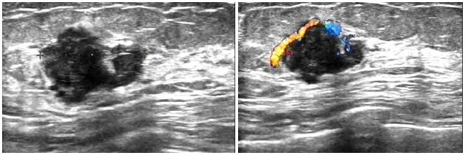
图 2 高频超声显示左乳实性低回声肿物,形态不规则,边缘不清晰,呈现“毛刺状”
Figure 2. High-frequency ultrasound showed a solid hypoechoic mass in the left breast, with irregular shape and unclear edges, showing a "burr-like" shape.

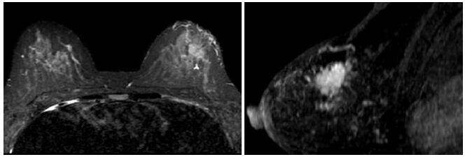
图 3 MRI显示左侧乳腺肿块合并左乳内淋巴结结节,肿块形态不规则,边缘表现为蟹足状改变,T1WI表现低信号,增强扫描可见早期强化
Figure 3. MRI showed the left breast mass with left intramammary lymph node nodules, irregular mass. The edges showed crab-shaped changes, the T1WI showed low signal and the enhanced scan showed early enhancement.
表 1 高频超声和MRI检查诊断乳腺癌结果与病理结果比较
Table 1. Comparison of results of high-frequency ultrasound and MRI diagnosis of breast cancer with pathological results (n)
检查方法 病理结果 合计 + - 高频超声 + 40 14 54 - 8 45 53 MRI + 45 6 51 - 3 53 56 高频超声联合MRI + 47 3 50 - 1 56 57 合计 48 59 107  下载: 导出CSV
表 2 高频超声和MRI检查诊断乳腺癌结果比较
Table 2. Comparison of results of high-frequency ultrasound and MRI in the diagnosis of breast cancer (%)
检查方法 准确性 敏感度 特异度 阳性预测值 阴性预测值 高频超声 79.44 83.33 76.27 74.07 84.91 MRI 91.59 93.75 89.83 88.24 94.64 高频超声联合MRI 96.26 97.92 94.92 94.00 98.25 χ2 16.611 7.091 9.695 8.725 7.729 P 0.000 0.042 0.008 0.013 0.028
下载: 导出CSV
表 3 高频超声和MRI检查诊断不同分期乳腺癌结果比较
Table 3. Comparison of results of high-frequency ultrasound and MRI in the diagnosis of breast cancer in different stages (n)
检查方法 分期 病理结果 合计 Ⅰ Ⅱ Ⅲ Ⅳ 高频超声 Ⅰ 2 2 0 0 4 Ⅱ 1 23 2 1 27 Ⅲ 1 0 10 1 12 Ⅳ 0 0 1 4 5 MRI Ⅰ 3 1 0 0 4 Ⅱ 1 24 1 1 27 Ⅲ 0 0 11 1 12 Ⅳ 0 0 1 4 5 高频超声联合MRI Ⅰ 3 0 0 0 3 Ⅱ 1 25 0 0 26 Ⅲ 0 0 12 1 13 Ⅳ 0 0 1 5 6 合计 4 25 13 6 48
下载: 导出CSV
-
[1] Wu F, Mo M, Qin XX, et al. Cost-effectiveness of multiple screening modalities on breast cancer in Chinese women from Shanghai[J]. Chin J Epidemiol, 2017, 38(12): 1665-71. http://www.wanfangdata.com.cn/details/detail.do?_type=perio&id=zhlxbx201712017 [2] Rebolj M, Assi V, Brentnall A, et al. Addition of ultrasound to mammography in the case of dense breast tissue: systematic review and meta-analysis [J]. Br J Cancer, 2018, 118(12): 1559-70. https://www.ncbi.nlm.nih.gov/pubmed/29736009/ [3] Cummins T, Eliahoo P, Kirk Shung K. High-frequency ultrasound array designed for ultrasound-guided breast biopsy[J]. IEEE Trans Ultrason Ferroelectr Freq Control, 2016, 63(6): 817-27. http://www.wanfangdata.com.cn/details/detail.do?_type=perio&id=d6ca6fce29b200546cc02d6f36333900 [4] 石燕清, 乌兰, 王红春, 等.高频彩色多普勒超声和弹性成像对微小乳腺癌和乳腺良性结节的鉴别诊断价值[J].癌症进展, 2019, 17(15): 1774-6, 1820. http://d.old.wanfangdata.com.cn/Periodical/azjz201915011 [5] Mustafi D, Zamora M, Fan XB, et al. MRI accurately identifies early murine mammary cancers and reliably differentiates between in situ and invasive cancer: correlation of MRI with histology[J]. NMR Biomed, 2015, 28(9): 1078-86. https://www.ncbi.nlm.nih.gov/pubmed/26152557 [6] 魏雪菲, 许守林, 冯雪凤.血清肿瘤标记物在乳腺癌临床分期中的诊断价值[J].实用临床医药杂志, 2017, 21(9): 227-8. http://d.old.wanfangdata.com.cn/Periodical/jslcyxzz201709077 [7] Mahoney MC, Gatsonis C, Hanna L, et al. Positive predictive value of BI-RADS MR imaging[J]. Radiology, 2012, 264(1): 51-8. http://www.wanfangdata.com.cn/details/detail.do?_type=perio&id=14828899c97fc1d6e097a7d098ac830a [8] Chavez-MacGregor M, Mittendorf EA, Clarke CA, et al. Incorporating tumor characteristics to the American joint committee on cancer breast cancer staging system[J]. Oncologist, 2017, 22 (11): 1292-300. https://www.ncbi.nlm.nih.gov/pubmed/28592619 [9] 周丽.乳腺简化MRI筛查技术研究进展[J].临床放射学杂志, 2018, 37(10): 1766-9. http://d.old.wanfangdata.com.cn/Periodical/lcfsxzz201810043 [10] Leblond MA, Duchesne N, Provencher L, et al. Is contralateral breast ultrasound worthwhile in preoperative staging of breast cancer?[J]. J Clin Ultrasound, 2019, 47(4): 195-200. http://www.wanfangdata.com.cn/details/detail.do?_type=perio&id=10.1002/jcu.22693 [11] Daugherty MW, Niell BL. Utility of routine axillary ultrasound surveillance in breast cancer survivors with previously diagnosed metastatic axillary adenopathy[J]. J Biogeogr, 2019, 1(1): 25-31. https://academic.oup.com/jbi/article/1/1/25/5379099 [12] 刘伟.高频超声、1.5T磁共振(MRI)诊断乳腺癌的临床价值[J].中国CT和MRI杂志, 2015, 13(12): 28-30. http://www.wanfangdata.com.cn/details/detail.do?_type=perio&id=zgcthmrizz201512012 [13] 刘巧珍, 唐英杰, 崔志利, 等.高频超声评估乳腺癌新辅助化疗疗效的价值分析[J].肿瘤学杂志, 2018, 24(7): 696-9. http://d.old.wanfangdata.com.cn/Periodical/zlxzz201807010 [14] 阳君, 赵欣, 苏丹柯, 等.钼靶和超声及MRI对乳腺癌的诊断价值多中心研究及卫生经济学评价[J].放射学实践, 2018, 33(6): 579-81. http://d.old.wanfangdata.com.cn/Periodical/fsxsj201806008 [15] Patel BK, Hilal T, Covington M, et al. Contrast-enhanced spectral mammography is comparable to MRI in the assessment of residual breast cancer following neoadjuvant systemic therapy[J]. Ann Surg Oncol, 2018, 25(5): 1350-6. http://www.wanfangdata.com.cn/details/detail.do?_type=perio&id=c01ee685fb69336dee44a2f9e1629ee5 [16] Mann RM, Kuhl CK, Moy L. Contrast-enhanced MRI for breast cancer screening[J]. J Magnc Reson Imaging, 2019, 50(2): 377-90. http://d.old.wanfangdata.com.cn/OAPaper/oai_pubmedcentral.nih.gov_2360541 [17] 卢东霞, 李文华, 王殿峰, 等.高频超声与高场磁共振在乳腺癌中的联合应用价值分析[J].实用医学影像杂志, 2018, 19(5): 43-4. http://d.old.wanfangdata.com.cn/Periodical/syyxyxzz201805015 [18] 王勇, 王喜平, 石茜, 等.全数字化平板乳腺检查在乳腺癌临床分期诊断中的应用价值[J].临床医学研究与实践, 2019, 4(23): 145-6, 163. http://d.old.wanfangdata.com.cn/Periodical/lcyxyjysj201923060 [19] 张怡.临床触诊阴性乳腺癌患者超声及MRI诊断分析[J].中国CT和MRI杂志, 2019, 17(3): 30-2. http://d.old.wanfangdata.com.cn/Periodical/zgcthmrizz201903010 [20] 王月爱, 阳力, 陈晓琼, 等.彩色多普勒超声与MRI对乳腺癌的联合诊断价值[J].中国中西医结合影像学杂志, 2018, 16(6): 647-9. http://d.old.wanfangdata.com.cn/Periodical/zgzxyjhyxxzz201806034 [21] Kim Y, Kim SH, Song BJ, et al. Early prediction of response to neoadjuvant chemotherapy using dynamic contrast-enhanced MRI and ultrasound in breast cancer[J]. Korean J Radiol, 2018, 19(4): 682-91. http://www.wanfangdata.com.cn/details/detail.do?_type=perio&id=3cf909ab478cc948489daeadcd246139 [22] 司爽, 张伟, 王慧颖, 等.乳腺X线摄影、超声及磁共振诊断乳腺癌的性能评价[J].中国临床医学影像杂志, 2020, 31(1): 20-3. http://d.old.wanfangdata.com.cn/Periodical/zglcyxyxzz202001006 [23] 田林, 李玉娜, 胡啸, 等. 6种影像学技术在致密型乳腺诊断中的应用[J].分子影像学杂志, 2020, 43(1): 7-11. doi: 10.12122/j.issn.1674-4500.2020.01.02 -

 点击查看大图
计量
- 文章访问数: 665
- HTML全文浏览量: 285
- PDF下载量: 5
- 被引次数: 0