


Transabdominal combined with transvaginal color Doppler ultrasound can improve the accuracy in the differential diagnosis of hysteromyoma
-
摘要:
目的研究经腹部联合经阴道彩色多普勒超声检查鉴别诊断子宫肌瘤的价值。 方法选择2015年5月~2018年9月本院收治的子宫肌瘤患者(n=245)和子宫腺肌瘤患者(n=105),均在本院进行经腹部超声(TAS)和经阴道超声(TVS)检查,扫查子宫以及肿块的形态、大小、回声等情况,并观察肌层、内膜血流分布情况。以手术病理学为金标准,分析TAS、TVS以及两者联合鉴别诊断子宫肌瘤的准确度,并分析两者声图像表现,比较两者子宫动脉血流参数。 结果TAS联合TVS诊断的准确度为96.57%、敏感度为96.19%、特异度为96.73%,均高于单纯TAS或TVS诊断(P < 0.05);子宫肌瘤大部分病灶较为清晰,且有假包膜,病灶回声以低回声为主,病灶部位血流较为丰富,呈环形或半环形;子宫腺肌瘤子宫形态较为正常,病灶部位模糊不清,且无假包膜,内部回声多见强回声,病灶部位周围血流不丰富;子宫肌瘤患者阻力指数、血流搏动指数等血流动力学参数指标水平均低于子宫腺肌瘤患者(P < 0.05)。 结论TVS联合TAS检查可提高子宫肌瘤鉴别诊断的准确率,降低误诊、漏诊现象,有较好的临床诊断价值。 Abstract:ObjectiveTo explore the value of transabdominal combined with transvaginal color Doppler ultrasound in the differential diagnosis of hysteromyoma. MethodsA total of 245 patients with hysteromyoma and 105 patients with uterine adenomyoma in the hospital between May 2015 and September 2018 were enrolled. All of them completed transabdominal sonography (TAS) and transvaginal sonography (TVS). Shapes, sizes and echo of the uterus and masses, blood flow distribution in the muscular layer and endometrium were observed. With operation and pathology as the golden standard, accuracy rates of TAS, TVS and combination of the two in the differential diagnosis of hysteromyoma were analyzed. The images and uterine artery blood flow parameters were analyzed. ResultsThe diagnostic accuracy, sensitivity and specificity of TAS combined with TVS (96.57%, 96.19% and 96.73%) were significantly higher than those of TAS or TVS alone (P < 0.05). Most of the hysteromyomas were clear, with pseudocapsules. The echoes mainly were low echoes, and blood flow in lesions was rich, which was ring-shaped or semi-ring-shaped. The shape of uterus with uterine adenomyoma was normal, but the lesion was unclear, without pseudocapsule. Most internal echoes were strong, and blood flow around the lesion was not rich. The hemodynamic parameters such as Resistance index and pulsatility index in patients with hysteromyoma were significantly lower than those with uterine adenomyoma (P < 0.05). ConclusionTVS combined with TAS can improve the accuracy in the differential diagnosis of hysteromyoma, and reduce misdiagnosis and missed diagnosis. -

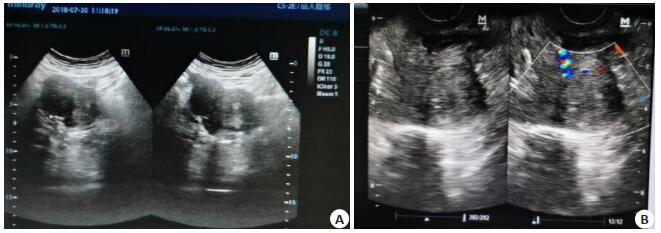
图 1 经腹部及阴道超声检查
A:经腹部超声检查, 子宫宫体局限性隆起, 病灶结构清晰; B:经阴道超声检查, 病灶部位血流丰富, 呈现半环形, 前后壁肌间和宫底部查见多个弱回声肌瘤.
Figure 1. Trans-abdominal and transvaginal sonography.
表 1 TVS和TAS诊断效能对比
Table 1. Comparison of diagnostic efficiency between TVS and TAS[n(%)]
金标准 TAS TVS 联合诊断 阳性 阴性 阳性 阴性 阳性 阴性 子宫肌瘤(n=245) 213 (86.94) 32 (13.06) 227 (92.65) 18 (7.35) 237 (96.73)ab 8 (3.27) 子宫腺肌瘤(n=105) 89 (84.76) 16 (15.24) 93 (88.57) 12 (11.43) 101 (96.19)ab 4 (3.81) 合计 302 (86.29) 48 (13.71) 320 (91.43) 30 (8.57) 338 (96.57)ab 12 (3.43) aP < 0.05 vs TAS; bP < 0.05 vs TVS; 阳性表示正确检出, 阴性表示误诊或漏诊. TAS: Transabdominal sonography; TVS: Transvaginal sonography  下载: 导出CSV
表 2 两种疾病患者血流参数比较
Table 2. Comparison of blood flow parameters between patients with the two diseases (Mean±SD)
组别 RI PI Vs Vd 子宫肌瘤(n=245) 0.56±0.15 1.21±0.16 61.23±2.43 4.62±1.03 子宫腺肌瘤(n=105) 0.73±0.12 1.39±0.13 70.47±2.21 5.70±0.86 t 10.285 10.175 33.476 9.426 P 0.000 0.000 0.000 0.000
下载: 导出CSV
-
[1] Islam MS, Ciavattini A, Petraglia F, et al. Extracellular matrix in uterine leiomyoma pathogenesis: a potential target for future therapeutics [J]. Hum Reprod Update, 2018, 24(1): 59-85. https://academic.oup.com/humupd/article/24/1/59/4652921 [2] McWilliams MM, Chennathukuzhi VM. Recent advances in uterine fibroid etiology[J]. Semin Reprod Med, 2017, 35(2): 181-9. http://www.wanfangdata.com.cn/details/detail.do?_type=perio&id=97a7967d2d05319e3aba811f45dab039 [3] 王蓓玉. 268例围绝经期妇女子宫肌瘤流行病学调查及危险因素分析[J].中国妇幼保健, 2018, 33(16): 3737-40. http://d.old.wanfangdata.com.cn/Periodical/zgfybj201816045 [4] Guang Y, Ying D, Sheng Y, et al. Early Doppler Ultrasound in the Superior Mesenteric Artery and the Prediction of Necrotizing Enterocolitis in Preterm Neonates[J]. J Ultrasound Med. 2019, 38 (12): 3283-9. https://www.ncbi.nlm.nih.gov/pubmed/31218729 [5] 孟璐.超声鉴别诊断子宫肌瘤和子宫腺肌瘤研究进展[J].中国介入影像与治疗学, 2015, 12(9): 577-9. http://www.wanfangdata.com.cn/details/detail.do?_type=perio&id=zgjryxyzlx201509014 [6] 熊羽佳.阴道超声与腹部超声对可疑子宫腺肌症的准确度、灵敏度和特异度分析[J].中国性科学, 2019, 28(7): 89-93. http://d.old.wanfangdata.com.cn/Periodical/zgxkx201907025 [7] Frank ML, Schäfer SD, Möllers M, et al. Importance of Transvaginal Elastography in the Diagnosis of Uterine Fibroids and Adenomyosis. Stellenwert der transvaginalen Elastografie in der Diagnose von uterinen Myomen und Adenomyose[J]. Ultraschall Med, 2016, 37 (4): 373-8. [8] Guerriero S, Saba L, Pascual MA, et al. Transvaginal ultrasound vs magnetic resonance imaging for diagnosing deep infiltrating endometriosis: systematic review and meta-analysis[J]. Ultrasound Obstet Gynecol, 2018, 51(5): 586-95. doi: 10.1002/uog.18961 [9] Ng SSM, Jorge S, Malik M, et al. A-kinase anchoring protein 13 (AKAP13) augments progesterone signaling in uterine fibroid cells [J]. J Clin Endocrinol Metab, 2019, 104(3): 970-80. http://www.wanfangdata.com.cn/details/detail.do?_type=perio&id=55af2146147d6f8dff2273f578635cce [10] Cui YM, Dong YY, Guo BC, et al. Effect of HIFU on endometrial receptivity and sex hormone level in uterine fibroid patients and analysis of influencing factors for its treatment rate[J]. Exp Ther Med, 2019, 17(3): 2291-7. doi: 10.3892/etm.2019.7194/download [11] Laughlin-Tommaso SK, Hesley GK, Hopkins MR, et al. Clinical limitations of the International Federation of Gynecology and Obstetrics (FIGO) classification of uterine fibroids[J]. Int J Gynaecol Obstet, 2017, 139(2): 143-8. https://mayoclinic.pure.elsevier.com/en/publications/clinical-limitations-of-the-international-federation-of-gynecolog [12] 孟璐, 赵一婷, 牛旺, 等.实时超声弹性成像技术鉴别诊断子宫肌瘤和子宫腺肌瘤[J].中国医学影像技术, 2016, 32(6): 919-22. http://www.wanfangdata.com.cn/details/detail.do?_type=perio&id=zgyxyxjs201606027 [13] 赵庆红, 石华, 杨菁, 等.实时超声弹性成像定量参数分析对子宫肌瘤和子宫腺肌瘤的诊断价值[J].中华全科医师杂志, 2015, 14(3): 223-5. http://www.wanfangdata.com.cn/details/detail.do?_type=perio&id=zhqkyszz201503017 [14] Kröncke T, David M. Uterine Artery Embolization (UAE) for Fibroid Treatment - Results of the 7th Radiological Gynecological Expert Meeting[J]. Rofo, 2019, 191(7): 630-4. https://www.ncbi.nlm.nih.gov/pubmed/31137043 [15] 侯庆香, 丁晓萍, 冯莉, 等.超声引导下射频消融治疗子宫腺肌瘤878例临床分析[J].中国超声医学杂志, 2015, 31(8): 763-5. http://d.old.wanfangdata.com.cn/Periodical/zgcsyxzz201508033 [16] Hossain MZ, Rahman MM, Ullah MM, et al. A comparative study of magnetic resonance imaging and transabdominal ultrasonography for the diagnosis and evaluation of uterine fibroids[J]. Mymensingh Med J, 2017, 26(4): 821-7. https://pubmed.ncbi.nlm.nih.gov/29208870/ [17] 李洁凤.彩色多普勒超声经腹与经阴道联合诊断484例子宫肌瘤的临床效果[J].检验医学与临床, 2014, 32(2): 249-50. http://d.old.wanfangdata.com.cn/Periodical/jyyxylc201402054 [18] 卢艳明.腹部超声联合经阴道超声在妇科急腹症中的临床应用价值[J].实用临床医药杂志, 2015, 19(15): 189-90. http://d.old.wanfangdata.com.cn/Periodical/jslcyxzz201515066 [19] 杜明祯, 张龙月, 龙海军, 等.经阴道与经腹部超声在卵泡监测中的应用价值比较[J].中国医疗设备, 2015, 30(3): 53-5. http://d.old.wanfangdata.com.cn/Periodical/ylsbxx201503015 [20] Jayakumaran JS, Egan, Tatikola M, et al. Transvaginal ultrasound is superior to transabdominal ultrasound in the identification of a short cervix[J]. Am J Obstet Gynecol, 2019, 221(4): 365-7. https://www.ncbi.nlm.nih.gov/pubmed/31351584 [21] 何宗珊, 邹秋果, 黄燕.经腹及经阴道超声联合应用扫查子宫肌瘤对比分析[J].中国性科学, 2016, 25(4): 48-50. http://d.old.wanfangdata.com.cn/Periodical/zgxkx201604016 [22] 段红波, 邱淑琴.经腹和经阴道超声在子宫肌瘤诊断中的临床价值[J].医学影像学杂志, 2015, 25(8): 1498-500. http://d.old.wanfangdata.com.cn/Periodical/yxyxxzz201508061 [23] Marigliano C, Panzironi G, Molisso L, et al. First experience of realtime elastography with transvaginal approach in assessing response to MRgFUS treatment of uterine fibroids [J]. Radiol Med, 2016, 121 (12): 926-34. -

 点击查看大图
图(1) / 表(2)
计量
- 文章访问数: 1060
- HTML全文浏览量: 350
- PDF下载量: 6
- 被引次数: 0