Find Duplicates
 Check Document
 Submission(new)
 Experts Office
 Editorial Office
2020 Vol. 42, No. 2
Display Method:
2020, 43(2): 183-187.
doi: 10.12122/j.issn.1674-4500.2020.02.01
Abstract:
Medical imaging is an important basis for radiologists to make medical diagnosis. However, with the rapid development of medical imaging technology, considerable and complicated medical imaging information brings great challenges to radiologists. Deep learning, as the most popular research field in artificial intelligence, has advantages of processing big data and extracting effective information. Therefore, deep learning has gradually become the top choice for researchers to analyze medical images. Firstly, this paper explains the concept of deep learning. It briefly summarizes the common implementation models of deep learning, including convolutional neural networks, recurrent neural networks, deep belief networks, and automatic encoders. The basic structure of a convolutional neural network is a convolutional layer, a pooling layer, and a fully connected layer. Recurrent neural network is composed of input layer, hidden layer and output layer. The foundation of the deep belief network is the Boltzmann machine. The autoencoder includes coding layer, hidden layer and decoding layer. Then, the classification of CT pulmonary nodules and MRI brain diseases shows that deep learning has high accuracy in automatic disease classification. Deep learning is consistent with artificial segmentation in medical image segmentation by segmenting the left ventricle, paravertebral muscles, and liver. Deep learning is relatively mature in the detection of lung nodules and breast cancer. Finally, the problems of small labeled sample and overfitting are proposed, and we hope to solve this problem by sharing image data. The application of deep learning in medical imaging has bright prospects and great significance on the clinical doctors' work.
Medical imaging is an important basis for radiologists to make medical diagnosis. However, with the rapid development of medical imaging technology, considerable and complicated medical imaging information brings great challenges to radiologists. Deep learning, as the most popular research field in artificial intelligence, has advantages of processing big data and extracting effective information. Therefore, deep learning has gradually become the top choice for researchers to analyze medical images. Firstly, this paper explains the concept of deep learning. It briefly summarizes the common implementation models of deep learning, including convolutional neural networks, recurrent neural networks, deep belief networks, and automatic encoders. The basic structure of a convolutional neural network is a convolutional layer, a pooling layer, and a fully connected layer. Recurrent neural network is composed of input layer, hidden layer and output layer. The foundation of the deep belief network is the Boltzmann machine. The autoencoder includes coding layer, hidden layer and decoding layer. Then, the classification of CT pulmonary nodules and MRI brain diseases shows that deep learning has high accuracy in automatic disease classification. Deep learning is consistent with artificial segmentation in medical image segmentation by segmenting the left ventricle, paravertebral muscles, and liver. Deep learning is relatively mature in the detection of lung nodules and breast cancer. Finally, the problems of small labeled sample and overfitting are proposed, and we hope to solve this problem by sharing image data. The application of deep learning in medical imaging has bright prospects and great significance on the clinical doctors' work.
2020, 43(2): 188-192.
doi: 10.12122/j.issn.1674-4500.2020.02.02
Abstract:
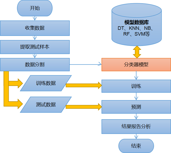
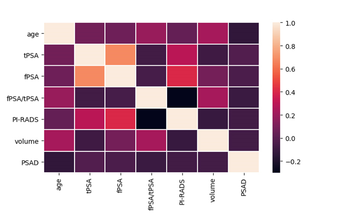
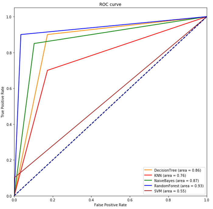
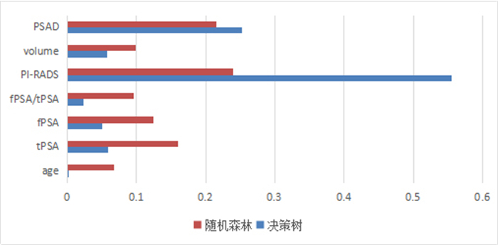
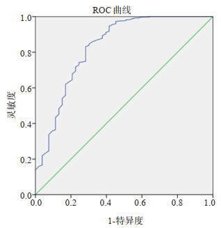
ObjectiveTo establish five machine learning models including decision tree, k-nearest neighbor, Naive Bayes, random forest and support vector machine (SVM) combined with multi-parameter MRI PI-RADS v2.1 score and clinical data, to evaluate the diagnostic value of these models for prostate cancer. MethodsA total of 242 patients who received MR examination in our hospital were analyzed retrospectively. PI-RADS v2.1 score, age, total prostate specific antigen, free prostate specific antigen, free prostate antigen ratio, volume and prostate specific antigen density were recorded into five machine learning models for diagnosis. The diagnostic value of machine learning model was evaluated by F1 value and ROC curve, and the proportion of characteristic variables was calculated. ResultsThe AUC of the random forest model was the largest (AUC=0.93), the AUC of the decision tree and the Naive Bayes model was also higher (AUC=0.86 and 0.87, respectively), and the support vector machine was the worst (AUC=0.55). The importance of each characteristic variable was calculated by random forest and decision tree model, and PI-RADS score accounted for the largest proportion, followed by PSA density and prostate volume, and age contributed the least to model classification. Conclusionrandom forest, naive bayes and decision tree regression model are all good in the diagnosis of prostate cancer, among which random forest is a better diagnosis model, and PI-RADS v2.1 and PSAD account for an important proportion in the diagnosis model of prostate cancer.
2020, 43(2): 193-196.
doi: 10.12122/j.issn.1674-4500.2020.02.03
Abstract:
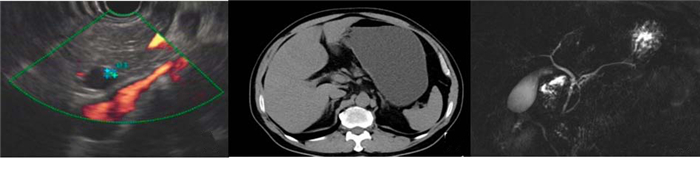
In recent years, the application of indocyanine green molecular fluorescence imaging in hepato-biliary and pancreatic surgery is more extensive. The application of indocyanine green molecular fluorescence imaging is more and more mature. It plays an indispensable role in the perioperative period of hepato-biliary and pancreatic surgery. The application of indocyanine green in liver surgery mainly includes preoperative evaluation and intraoperative detection of small cancer foci, tumor boundary definition and left and right hemiliver definition, which can make liver resection more accurate and safe, especially in tumor resection can effectively reduce the risk of surgery and improve the R0 resection rate. It is widely used in biliary surgery for intrahepatic cholangiocarcinoma, cholecystectomy and pancreatic tumor, which can avoid the injury of biliary tract and improve the diagnosis and treatment effect of pancreatic tumor. Therefore, by referring to the recent domestic and foreign research results on the application of indocyanine green in hepato-biliary and pancreatic surgery, this paper comprehensively expounds the application of ICG molecular fluorescence imaging technology in hepato-biliary and pancreatic surgery.
In recent years, the application of indocyanine green molecular fluorescence imaging in hepato-biliary and pancreatic surgery is more extensive. The application of indocyanine green molecular fluorescence imaging is more and more mature. It plays an indispensable role in the perioperative period of hepato-biliary and pancreatic surgery. The application of indocyanine green in liver surgery mainly includes preoperative evaluation and intraoperative detection of small cancer foci, tumor boundary definition and left and right hemiliver definition, which can make liver resection more accurate and safe, especially in tumor resection can effectively reduce the risk of surgery and improve the R0 resection rate. It is widely used in biliary surgery for intrahepatic cholangiocarcinoma, cholecystectomy and pancreatic tumor, which can avoid the injury of biliary tract and improve the diagnosis and treatment effect of pancreatic tumor. Therefore, by referring to the recent domestic and foreign research results on the application of indocyanine green in hepato-biliary and pancreatic surgery, this paper comprehensively expounds the application of ICG molecular fluorescence imaging technology in hepato-biliary and pancreatic surgery.
2020, 43(2): 197-202.
doi: 10.12122/j.issn.1674-4500.2020.02.04
Abstract:
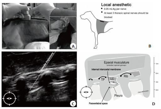
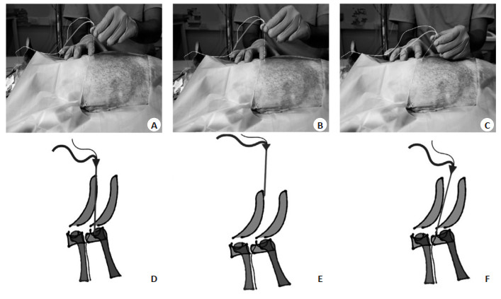
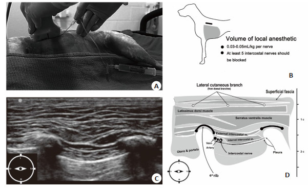
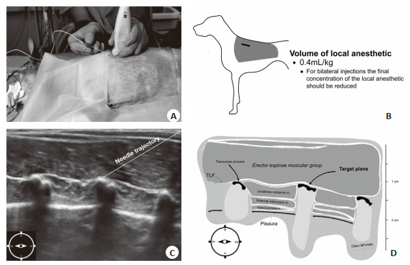
In recent years, the research of regional anesthesia in big animals has been greatly developed. A variety of new techniques of thoracic block have emerged. The routine implementation of technique in veterinary clinical practice provides a valuable reference for Multi-mode analgesic strategy. It reduces the consumption of systemic analgesics during perioperative period. At the same time, these technologies have also made great contributions to the establishment of big animal models and the development of human medical research. This paper combined the relevant anatomy, localization methods of nerve stimulators and accurate ultrasound-assisted technology to sort out the literatures published with big animals chest near the spinal nerve block methods in recent years. We reviewed thoracic paravertebral blocks, intercostal nerve blocks and erector spinae plane blocks, so as to provide appropriate reference materials for regional anesthesia in big animals and ultimately serve the development of scientific research.
In recent years, the research of regional anesthesia in big animals has been greatly developed. A variety of new techniques of thoracic block have emerged. The routine implementation of technique in veterinary clinical practice provides a valuable reference for Multi-mode analgesic strategy. It reduces the consumption of systemic analgesics during perioperative period. At the same time, these technologies have also made great contributions to the establishment of big animal models and the development of human medical research. This paper combined the relevant anatomy, localization methods of nerve stimulators and accurate ultrasound-assisted technology to sort out the literatures published with big animals chest near the spinal nerve block methods in recent years. We reviewed thoracic paravertebral blocks, intercostal nerve blocks and erector spinae plane blocks, so as to provide appropriate reference materials for regional anesthesia in big animals and ultimately serve the development of scientific research.
2020, 43(2): 203-206.
doi: 10.12122/j.issn.1674-4500.2020.02.05
Abstract:
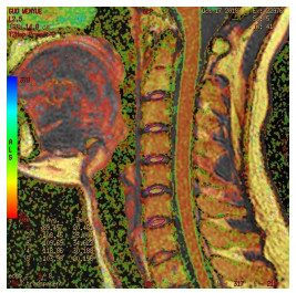
The ultrasonography, as a method of placental implantation, has many limitations, such as too deep placenta, too little amniotic fluid and too fat pregnant women. Magnetic resonance imaging is increasingly used in the diagnosis of placenta accreta, especially for patients with poor ultrasonic evaluation. In recent years, fast scanning sequences and functional imaging of MRI, such as SSTSE, HASTE, DWI and MRS, play a great role in the diagnosis of placenta accreta. The direct signs of MR diagnosis of placental implantation are: thin myometrium, discontinuous myometrium and unclear boundary between myometrium and placenta. Indirect signs of placental implantation: heterogeneous signal in placenta; T2WI like low signal band in placenta; abnormal distribution of blood vessels in placenta, tortuosity and coarseness of blood vessels; local expansion of placenta; local bulge of uterus; invasion of adjacent organs, etc. The possibility of abnormal placenta increased with the number of imaging features of placenta invasion, and was related to the risk factors of patients such as cesarean section, placenta previa and so on. Some scholars have pointed out that the occurrence of placenta dark belt is the strongest evidence of placenta implantation. This article reviews the application and progress of MRI in placenta implantation.
The ultrasonography, as a method of placental implantation, has many limitations, such as too deep placenta, too little amniotic fluid and too fat pregnant women. Magnetic resonance imaging is increasingly used in the diagnosis of placenta accreta, especially for patients with poor ultrasonic evaluation. In recent years, fast scanning sequences and functional imaging of MRI, such as SSTSE, HASTE, DWI and MRS, play a great role in the diagnosis of placenta accreta. The direct signs of MR diagnosis of placental implantation are: thin myometrium, discontinuous myometrium and unclear boundary between myometrium and placenta. Indirect signs of placental implantation: heterogeneous signal in placenta; T2WI like low signal band in placenta; abnormal distribution of blood vessels in placenta, tortuosity and coarseness of blood vessels; local expansion of placenta; local bulge of uterus; invasion of adjacent organs, etc. The possibility of abnormal placenta increased with the number of imaging features of placenta invasion, and was related to the risk factors of patients such as cesarean section, placenta previa and so on. Some scholars have pointed out that the occurrence of placenta dark belt is the strongest evidence of placenta implantation. This article reviews the application and progress of MRI in placenta implantation.
2020, 43(2): 207-212.
doi: 10.12122/j.issn.1674-4500.2020.02.06
Abstract:
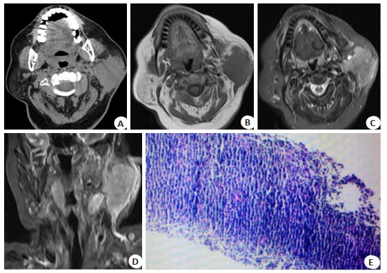
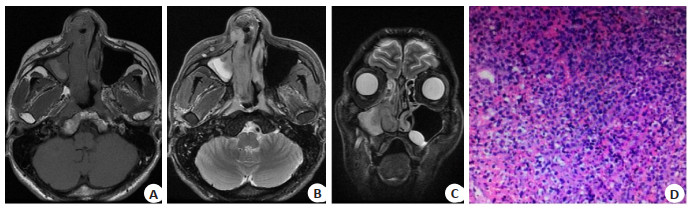
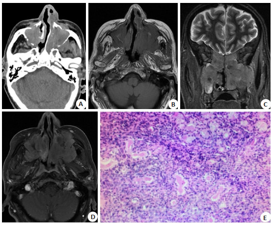
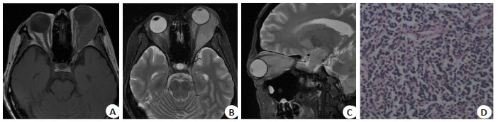
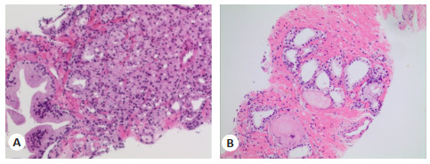
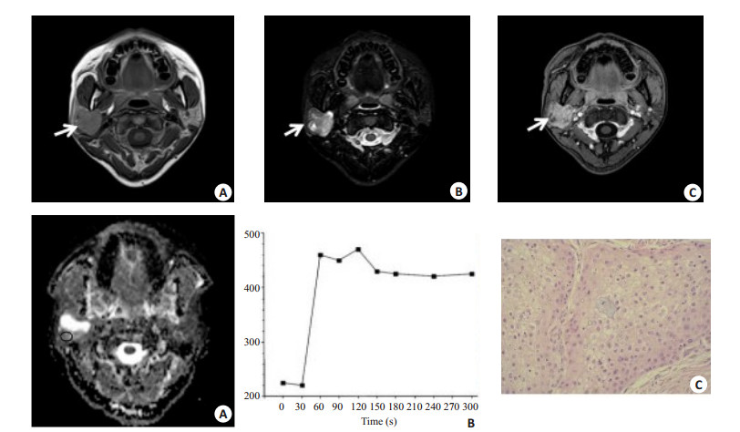
ObjectiveTo evaluate the difference of the pathology and imaging features of non-Hodgkin lymphoma (NHL) in different regions of the head and neck. MethodsThe pathology and imaging(CT and MRI)features of 49 patients with primary extranodal NHL confirmed by pathology were analyzed retrospectively. Twenty-one cases were located in Waldeyer's Ring (WR) and 28 cases were located in extra-WR. Five patients underwent CT scan, 16 patients underwent MRI scan, 28 patients underwent both CT and MR scans, and contrast CT or MRI scans were performed in 20 patients. The characteristics of pathology subtypes and imaging features of mass morphology, local structure invasion and the rate of nodes involvement were evaluated. The imaging features of NHL in WR and extra-WR were compared by using Chi-square test. ResultsWR was the most common region of diffuse large B-cell lymphoma (DLBCL) (n=16, 76.2%), nasal cavity was the most common extraWR site (n=17, 34.7%) and NK/T cell lymphoma was the most histological subtype (n=12, 70.6%). WR NHL was commonly in masses, and extra-WR NHL commonly in diffuse infiltration growth (χ2=9.926, P=0.002). WR NHL was less locally aggressive than extra-WR NHL(χ2=25.126, P=0.000). WR NHL was more commonly combined with cervical nodes involvement (χ2=21.778, P=0.000). ConclusionCorresponding NHL subtypes have predilections for different regions in the head and neck. The mass morphology, local structure invasion and the rate of nodes involvement between WR and extra-WR NHL are different.

2020, 43(2): 213-216.
doi: 10.12122/j.issn.1674-4500.2020.02.07
Abstract:
Photodynamic therapy (PDT) is an emerging tumor therapy method in recent years. After the photosensitizer injected into the body, it specifically accumulates in the tumor tissue and then illuminate the tumor site with a specific wavelength of light, causing the photodynamic reaction、tumor cell organelle damage、tumor cell apoptosis and necrosis and tumor immunity to kill the malignancy. Recently, as the key factors of photodynamic therapy, new photosensitizer and light source have been continuously explored and applied in clinical practice. There are more clinical studies on photodynamic therapy for malignant tumors, especially head and neck malignancies. With the introduction of new photosensitizers such as hypericin and associated light sources and the combination of photodynamic techniques with other treatments, the main observation indicator of the effect of photodynamic therapy, "survival time", has been extended. The related adverse reactions such as solar dermatitis and drug allergic reaction have been reduced. In addition, its preservation of vital organ function integrity and cosmetic function is satisfactory. This article reviews the clinical application of PDT in head and neck malignant tumors in recent years. We focused on the advantages of good effect, preserving functional integrity of important organs, beauty and less side effects in photodynamic treatment of nasopharyngeal carcinoma, sinus carcinoma, laryngeal cancer, oral cancer, ficial skin cancer and other common head and neck malignancies. It could better understand the development of photodynamic technology and express the need for better photosensitizer and light source and photodynamic therapy combined with other treatment methods.
Photodynamic therapy (PDT) is an emerging tumor therapy method in recent years. After the photosensitizer injected into the body, it specifically accumulates in the tumor tissue and then illuminate the tumor site with a specific wavelength of light, causing the photodynamic reaction、tumor cell organelle damage、tumor cell apoptosis and necrosis and tumor immunity to kill the malignancy. Recently, as the key factors of photodynamic therapy, new photosensitizer and light source have been continuously explored and applied in clinical practice. There are more clinical studies on photodynamic therapy for malignant tumors, especially head and neck malignancies. With the introduction of new photosensitizers such as hypericin and associated light sources and the combination of photodynamic techniques with other treatments, the main observation indicator of the effect of photodynamic therapy, "survival time", has been extended. The related adverse reactions such as solar dermatitis and drug allergic reaction have been reduced. In addition, its preservation of vital organ function integrity and cosmetic function is satisfactory. This article reviews the clinical application of PDT in head and neck malignant tumors in recent years. We focused on the advantages of good effect, preserving functional integrity of important organs, beauty and less side effects in photodynamic treatment of nasopharyngeal carcinoma, sinus carcinoma, laryngeal cancer, oral cancer, ficial skin cancer and other common head and neck malignancies. It could better understand the development of photodynamic technology and express the need for better photosensitizer and light source and photodynamic therapy combined with other treatment methods.
2020, 43(2): 217-220.
doi: 10.12122/j.issn.1674-4500.2020.02.08
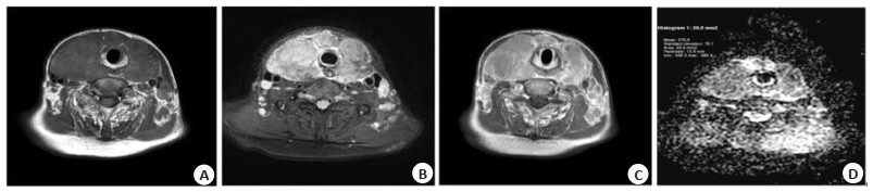
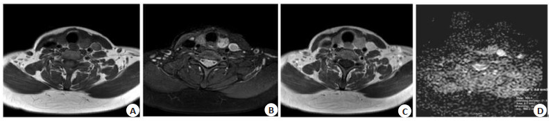
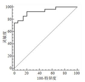
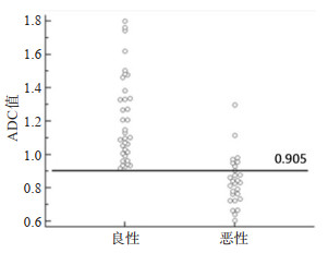
Abstract:
ObjectiveTo assess the ability of diffusion-weighted imaging (DWI) on 3.0 T MRI in preoperatively differentiating malignant thyroid nodules from benign nodules. MethodsFrom October 2017 to November 2018, 56 patients with thyroid nodules(50 benign nodules and 35 malignant nodules)were included. All lesions were received surgical resection and confirmed by pathology. There were 12 males and 44 females, with the age from 22 to 79 years old (average 49±15.69). Prior surgery conventional MR and diffusion-weighted imaging (DWI) were examined on 3.0 T MRI scanner with head and neck coils. The b values of 0 and 1000 s/mm2 were used. The independent sample t test was used to compare the apparent diffusion coefficient (ADC) value between benign and malignant thyroid nodules. The receiver operating characteristic (ROC) analysis was performed to assess the diagnostic efficiency. ResultsThe ADC values of benign and malignant thyroid nodules were significantly different (P < 0.001). The ADC value of benign thyroid nodule was (1.22±0.26)×10-3 mm2/s, which was significantly higher than that of malignant thyroid nodules [(0.84±0.15)×10-3 mm2/s]. The ADC thresholds of 0.91×10⁻³ s/mm2 showed the best discriminative value, with the sensitivity of 74.1%, specificity of 100%, and the area under the ROC curve (AUC) of 0.93. ConclusionThe ADC value on 3.0 T MRI was a promising parameter for diagnosing malignant thyroid nodules from benign thyroid nodule.

2020, 43(2): 221-224.
doi: 10.12122/j.issn.1674-4500.2020.02.09
Abstract:
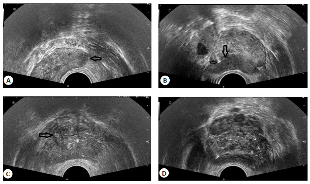
ObjectiveTo explore the clinical value of prostate biopsy in diagnosing benign and malignant prostate lesions under the guidance of TRUS. MethodsFrom February 2019 to January 2020, 62 patients with clinically suspected prostate cancer who underwent transperineal prostate biopsy in our Hospital were included. The patients received a freehand transperineal biopsy of the prostate. According to the pathological results, the patients were divided into two groups. The prostate cancer group(25 cases, males, 55-82 years old) and prostatic hyperplasia group (37 cases, males, 54-83 years old). The prostate size, abnormal echoes, systematic biopsy and targeted biopsy were recorded. The ultrasonographic features were compared between two groups. ResultThe pathological types of prostate cancer group were prostate adenocarcinoma (including acinar carcinoma). The prostatic hyperplasia group showed normal prostatic tissue, gland and interstitial hyperplasia, muscle and fibrous tissue. A total of 145 needle malignant tissues were obtained by puncture in prostate cancer group, and 108 needle (75%) were located in the prostatic peripheral zone. There were 81 needle tissues which malignant tissue accounting for more than 50% of the tissue length. A total of 52 abnormal echoes were found by TRUS. The sizes of abnormal echoes and pathological components were significant different between 2 groups. ConclusionTRUS-guided transperineal biopsy has advantages over the prostatic peripheral zone. It can directly and accurately puncture the suspected lesion area, and make it important for the diagnosis of benign and malignant prostatic lesions.
2020, 43(2): 225-229.
doi: 10.12122/j.issn.1674-4500.2020.02.10
Abstract:
Artificial intelligence (AI) has been used in the daily medical care of Urology field. For example, through the establishment of neural network, the prognosis of urolithiasis can be predicted from the CT results. The stone clearance rate and the amount of intraoperative bleeding can be predicted, so that doctors can better choose the treatment plan. Through the study of CT texture, we can distinguish the benign and malignant renal tumors accurately, improve the detection rate of clear cell carcinoma, and greatly reduce the rate of missed diagnosis and misdiagnosis. The radiology and texture analysis are used to study bladder cancer, distinguish between low-level and high-level tumors, so that doctors can choose the surgical methods with less trauma to patients. The algorithm is used to predict the response to treatment and the recurrence rate of tumor, so as to prolong the survival rate of patients. To develop Gleason score prediction, MRI computer- aided algorithm diagnosis, surgical results and biochemical recurrence prediction for prostate cancer, and provide individualized treatment programs for different patients. It is found that these methods are superior to the traditional statistical methods. The purpose of this paper is to review the development of imaging system application of AI in urology in recent years, and provide a broader idea for clinical application of AI in the future.
Artificial intelligence (AI) has been used in the daily medical care of Urology field. For example, through the establishment of neural network, the prognosis of urolithiasis can be predicted from the CT results. The stone clearance rate and the amount of intraoperative bleeding can be predicted, so that doctors can better choose the treatment plan. Through the study of CT texture, we can distinguish the benign and malignant renal tumors accurately, improve the detection rate of clear cell carcinoma, and greatly reduce the rate of missed diagnosis and misdiagnosis. The radiology and texture analysis are used to study bladder cancer, distinguish between low-level and high-level tumors, so that doctors can choose the surgical methods with less trauma to patients. The algorithm is used to predict the response to treatment and the recurrence rate of tumor, so as to prolong the survival rate of patients. To develop Gleason score prediction, MRI computer- aided algorithm diagnosis, surgical results and biochemical recurrence prediction for prostate cancer, and provide individualized treatment programs for different patients. It is found that these methods are superior to the traditional statistical methods. The purpose of this paper is to review the development of imaging system application of AI in urology in recent years, and provide a broader idea for clinical application of AI in the future.
2020, 43(2): 230-234.
doi: 10.12122/j.issn.1674-4500.2020.02.11
Abstract:
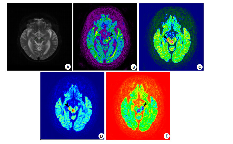
ObjectiveTo explore the clinical value of Diffusion Kurtosis Imaging (DKI) in nigrostriatal region of idiopathic Parkinson's disease and analyze the correlation with clinical progress. MethodsThirty-five patients diagnosed of Parkinson's disease in Xinjiang Second Clinical Hospital were selected as the case group, and 20 healthy control patients in our hospital during the same period were collected as the control group. All subjects underwent MRI routine scanning and DKI sequence scanning on a 3.0 T magnetic resonance imaging machine. The mean Kurt Osis (MK) values of bilateral substantia nigra were measured on the postprocessed DKI sequence respectively. The average values of MK, Anisotropic fraction (FA), axial kurtosis (AK) and radial kurtosis (RK) in the brain of the case group and the control group between 2 groups were compared. Patients in the case group were divided into early Parkinson's disease group (n=13) and advanced Parkinson's disease group (n=22). ResultsThe mean values of MK in the bilateral substantia nigra of the case group were significantly higher than those of the control group (P < 0.05), while the FA values were lower than those of the control group (P < 0.05). The AK and RK values between 2 groups had no significant difference (P>0.05). No significant correlation was found between MK and FA values of bilateral substantia nigra in early PD group and advanced PD group (P>0.05). ConclusionMK and FA values in DKI parameters can be used as early biomarkers of neurodegeneration. MK and FA values in DKI are not suitable for evaluating the progress of PD patients.
2020, 43(2): 235-241.
doi: 10.12122/j.issn.1674-4500.2020.02.12
Abstract:
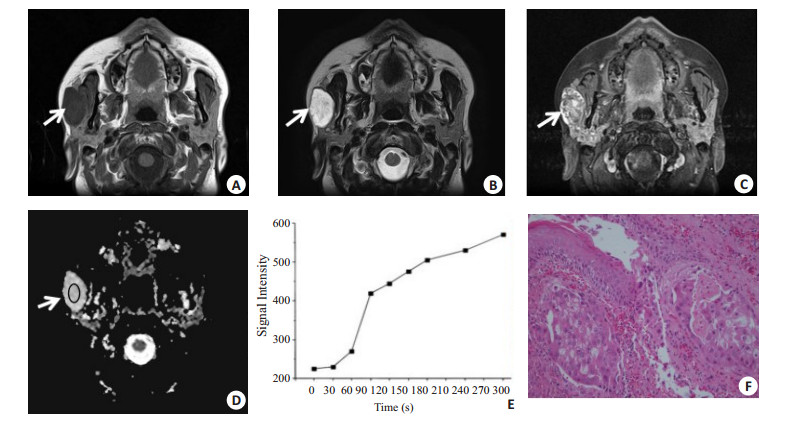
ObjectiveTo explore the diagnostic value of the combined application of magnetic resonance dynamic enhanced scanning (DCE-MRI) and diffusion weighted imaging (DWI) in the differentiation of benign and malignant tumors of the parotid gland. MethodsForty patients with parotid gland tumors confirmed by surgical pathology were retrospectivly analyzed. The patients aged from 28 to 80 years old (average age at 46 years old), with 25 males and 15 females. The patients were performed MR plain scan, dynamic enhancement and DWI scan before surgery to draw tumors. The dynamic enhancement time-signal intensity curve (TIC) and related parameters (peak time Tpeak, clearance rate WR at 600 s) were displayed, and the measured ADC value of the tumor was displayed on the ADC map. Two independent sample t tests were used to compare benign and malignant parotid tumors. Differences between TIC parameters and ADC values were analyzed by single-factor analysis of variance for pleomorphic adenoma, adenolymphoma, and malignant tumors. ResultsTwenty-eight of the 40 cases of parotid gland tumors were benign, mainly pleomorphic adenoma and adenolymphoma, and 12 were malignant with many pathological types. Polymorphic adenoma was dominated by A-type (slow-rising type) and C-type (fastforward slow- declining type) curves, and the A-type curve only existed in pleomorphic adenoma, of which 1 case was malignant pleomorphic adenoma. Distinguish pleomorphic adenoma from other benign tumors and malignant tumors; adenolymphomas were dominated by B-type (rapid ascent and descent) curves, which were more characteristic. Malignant tumors were dominated by C-type curves, but it also existed in some benign tumors. There was a partial overlap between them. The ADC value of benign tumors of the parotid gland was significantly higher than that of malignant tumors (1.13× 10⁻³ mm2/s vs 0.84 × 10⁻³ mm2/s). The differences of ADC values between pleomorphic adenoma, malignant tumor, and adenolymphoma were significant (P < 0.01). The difference of ADC values between adenolymphoma and malignant tumor was not significant (P>0.05). ConclusionMR routine scanning combined with dynamic enhancement provides valuable information for the qualitative diagnosis of parotid gland tumors. Combined with ADC value can further improve the differential diagnosis of benign and malignant tumors.

2020, 43(2): 242-246.
doi: 10.12122/j.issn.1674-4500.2020.02.13
Abstract:
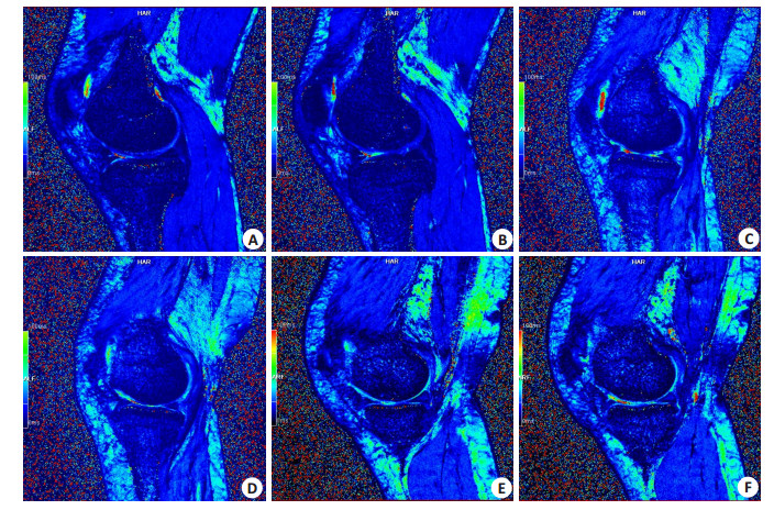
ObjectiveTo evaluate the diagnostic value of T2* mapping-assisted magnetic resonance imaging (MRI) in patients with different degree of OA cartilage injury of knee joint. MethodsA total of 46 patients with knee OA admitted to our hospital from June 2016 to June 2019 were selected as pathological group. There were 32 males and 14 females in pathological group, with the age from 22-69 years old (41.19 ± 15.53). Another 7 healthy volunteers were taken as the control group, including 5 males and 2 females with the age from 23 to 64 years old (42.05±15.28). Two groups underwent routine MRI and T2* mapping sequence scans of the knee. The T2* values of cartilage in different areas were compared. In addition, T2* values in the loading zone and non-loading zone of the medial and lateral condyles of the femur were analyzed for the patients in the lesion group with different degree of cartilage damage, shallow and deep cartilage in each region. ResultsThe T2* values of medial femoral condyle, lateral femoral condyle, medial tibial plateau and lateral tibial plateau in the lesion group were significantly higher than those in the control group (P < 0.05). The T2* values of medial femoral condyle, lateral femoral condyle, medial tibial plateau and lateral tibial plateau in the severe group were significantly higher than those in the mild group (P < 0.05). The T2* values of the medial femoral condyle, the lateral femoral condyle, the medial tibial plateau and the superficial cartilage of the lateral tibial plateau in the lesion group were significantly higher than those in the deep cartilage (P < 0.05). The T2* values in the loading zone of medial femoral condyle and lateral condyle in the lesion group were significantly higher than that in the loading zone (P < 0.05). ConclusionT2* mapping-assisted MRI has a high value in the diagnosis of patients with different degree of OA cartilage injury of knee joint. It can provide reliable basis for the judgment of patients' conditions.
2020, 43(2): 247-252.
doi: 10.12122/j.issn.1674-4500.2020.02.14
Abstract:
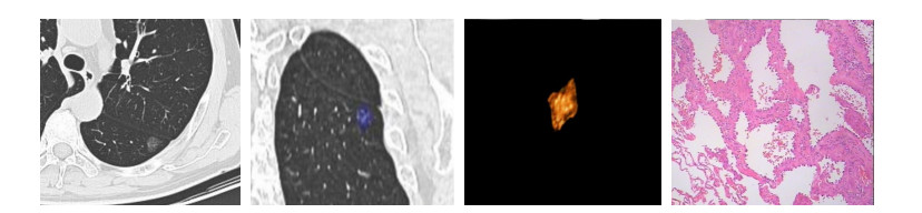
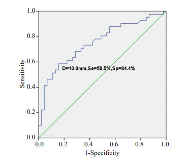
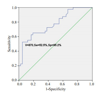
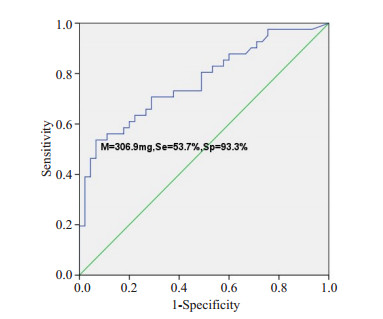
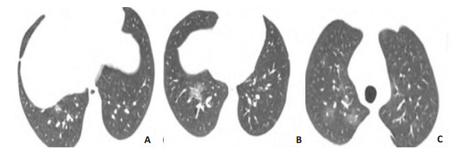
ObjectiveTo analyze the high resolution computed tomography findings, clinicopathological features and genetic characteristics of pure ground-glass opacities of the lung. MethodsThe retrospective study included 86 patients with pGGOs who underwent surgical resection in the thoracic surgery department of hainan hospital of PLA general hospital from April 2014 to April 2019, including 30 males and 56 females, with the age from 21 to 78 years old. The perioperative data were analyzed to distinguish HRCT imaging features, histopathological features and gene mutations in the invasive adenocarcinoma group (IA) and the control group (MIA, AIS and AAH) according to the different survival. ResultsA total of 86 patients with pGGOs were enrolled, including 41 in group IA and 45 in control group, including 25 in MIA, 14 in AIS and 6 in AAH. The difference of general data between the two groups was not significant(P>0.05). However, there was a significant difference in surgical procedure (P=0.001). Patients in the IA group were more likely to receive lobectomy, while those in the control group were more likely to receive sublobectomy. The differences of diameter, volume and mass of pGGOs between the two groups were significant (P < 0.001), but no significant differences in mean CT value and conversion density (P>0.05). According to ROC curve analysis, the lung IA group and the control group were identified with a critical value of 10.8 mm, with a sensitivity of 58.5%, specificity of 84.4% and AUC of 0.753. The critical values of volume and AUC were 870 mm3 and 0.773, respectively. The diagnostic sensitivity was 52.5% and the specificity was 95.2%. The critical value of mass was 306.9 mg, the AUC value was 0.769, the diagnostic sensitivity was 53.7% and the specificity was 93.3%. There were significant differences in lobules and burrs, but no significant differences in pGGOs shape, pleural traction and vascular cluster. In terms of gene mutation status, 2 of the 8 patients in the control group had EGFR gene mutation, and 9 of the 20 patients in the IA group had EGFR mutation. The difference of gene mutation between the two groups was not significant (P>0.05). ConclusionIn summary, the pathological manifestations of pure ground glass nodules in lungs are diverse. Combining HRCT image features and image analysis can help to make a qualitative diagnosis and guide subsequent follow-up strategies and formulate surgical plans. For pure ground glass nodules with a diameter greater than 10.8 mm, a volume greater than 870 mm3, and a mass greater than 306.9 mg, with malignant imaging features such as lobular signs and burr signs, minimally invasive surgery should be actively performed. The surgical method is based on imaging and patient wishes. Sublobar resection combined with lymph node biopsy can achieve satisfactory prognosis.
2020, 43(2): 253-258.
doi: 10.12122/j.issn.1674-4500.2020.02.15
Abstract:
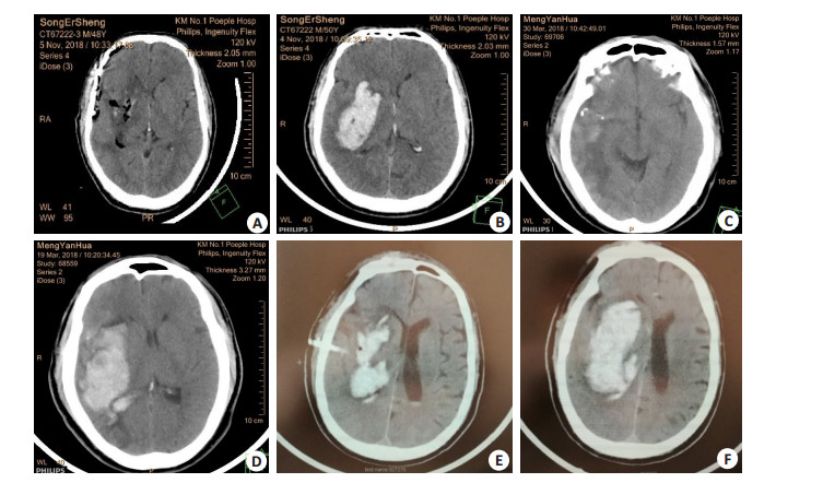
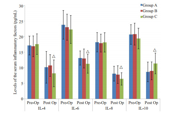
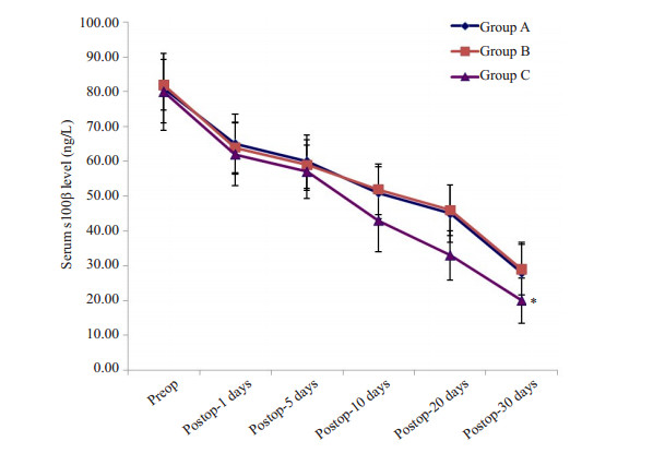
ObjectiveTo compare and evaluate the efficacy and safety of the neuroendoscopic surgery with the small bone window craniotomy and the burr hole, urokinase infusion, and catheter drainage for the treatment of evacuation in the hematoma of HICH patients. MethodsA total of 90 patients with HICH were randomized into three groups. Group A: underwentsmall bone window craniotomy (n=30). Group B: underwentburr hole + urokinase infusion + catheter drainage surgery (n=30). Group C: underwent neuroendoscopic surgery (n=30). The hematoma evacuation rate, mortality rate, glasgow coma scale (GCS), glasgow outcome score (GOS), and complications including rebleeding, pneumonia, and intracranial infection were compared in the three groups. The levels of IL4, IL6, IL8, IL10 and serum S100β protein in the peripheral blood of all HICH patients were determined by ELISA method. ResultsAll surgical approaches were successfully performed. The hematoma evacuation percentage was significantly different in the three groups, which were 74.5%, 43.1% and 88.2%, respectively (P < 0.05). Operation time, rebleeding rate, pneumonia rate, and mortality rate in Group C was lower than Group A and Group B, respectively (P < 0.05).The GOS score 6 months after the surgical approach showed that the number of Grade I and Grade II in Group C was significantly higherthan that in Group A and B (P < 0.05), while the number of Grade III, Grade IV and Grade V in Group C was significantly lower than Group A and B (P < 0.05). The postoperative IL10 level of patients in Group C were elevated and significantly higher than that in Group A and Group B at 72 hours, while the IL-4, IL-6 andIL-8 were decreased and significantly lower than that in Group A and B. The level of S100β in Group C was significantly lower than that in Group A and B at 28 days after operation (P < 0.05). ConclusionNeuroendoscopic technical has minimal invasive, direct vision, higher hematoma evacuation rate, lower incidence rate of complications, and significant improvement of clinical outcomes. It might be a more promising approach for the treatment of HICH.
2020, 43(2): 259-263.
doi: 10.12122/j.issn.1674-4500.2020.02.16
Abstract:
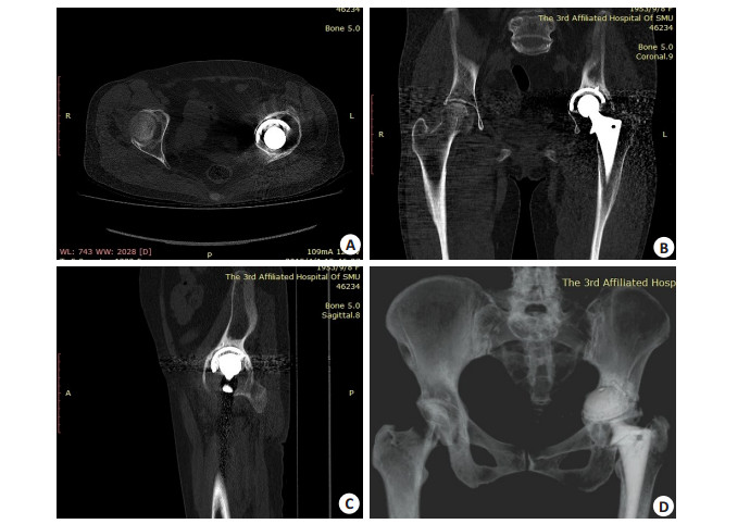
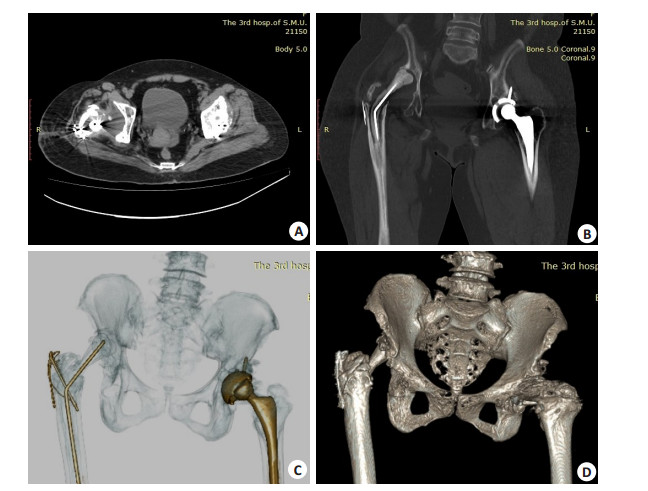
ObjectiveTo explore the application of multi-row spiral CT in the preoperative assessment of revision surgery after artificial hip replacement. MethodsFrom March 2014 to May 2019, 21 patients (21 hips) of revision surgery after artificial hip replacement in the Third Affilated Hospital of Southern Medical University were included. There were 7 males and 14 female, with median age of 62 years old. The reasons of revision surgery included 13 hips of prosthesis loosening, 2 hips of prosthesis dislocation, 2 hips of periprosthesis fracture, and 4 hips of two-stage exchange. CT examinations were performed to all hips before revision surgery. Two radiologists reviewed the CT images to analyze the radiologic sign of failure of prosthesis and develop a proposal for the revision surgery. ResultsAmong 13 hips of prosthesis loosening, there were 10 hips with artificial acetabulum loosening, which reperent prosthesis shifting upward and inward of all 10 hips, obvious lucent areas of periprosthesis of 4 hips, massive soft tissues filling in the acetabulum of 2 hips, bony defect of the acetabulum of 2 hips. There were 6 hips with artificial femoral loosening, which repesented as prosthesis shifting of 5 hips, obvious lucent areas of periprosthesis of 2 hips, and no definitely abnormal findings of 1 hip. Addtionally, 1 hip with prosthesis loosening coexisted lining abrasion, manifesting as vanish of interval of artificial acetabulum and femoral. There were 2 hips with dislocation of prosthesis, which represented as femoral heads dislocating out of acetabulum laterally, superiorly, posteriorly. There were 2 hips with periprosthesis fractures, which represented as broken bones round the middle segment or the distal of the prosthesis. The both hips were suffered from prosthesis loosening. There were 4 hips of two-stage exchange, which showed residual prosthesis, spacers, soft tissues in the acetabulum on CT images, among which obvious bony defect were found in the acetabulum of 3 hips. ConclusionMulti-row spinal CT could reveal factors of prosthesis failure, and develop a proposal for the revision surgery after artificial hip replacement.
2020, 43(2): 264-269.
doi: 10.12122/j.issn.1674-4500.2020.02.17
Abstract:
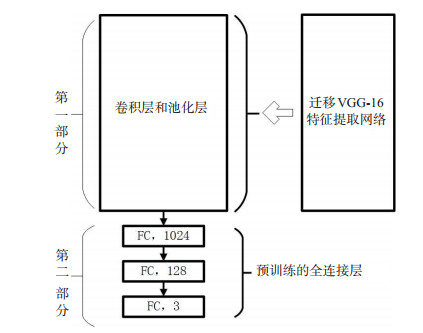
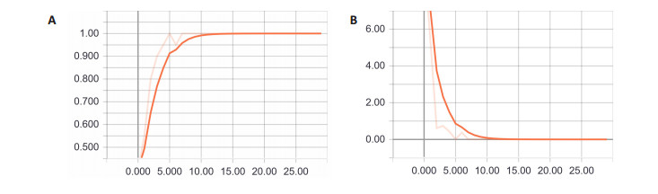
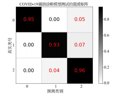
ObjectiveTo investigate the value of deep learning technology in the diagnosis of pneumonitis associated with new coronavirus infection in combination with CT imaging features of patients with new coronavirus infected pneumonia (COVID- 19). MethodsWe collected CT image data of patients diagnosed as COVID-19 in Central South Hospital of Wuhan University and Tongji Medical College of Huazhong University of Science and Technology to construct a small sample COVID-19 data set. VGG-16 was taken to extract high- level abstract features and design the full The connection layer together constitutes the preliminary COVID-19 intelligent auxiliary diagnosis model based on transfer learning. The COVID-19 training set was used to iteratively train the diagnosis model, continuously optimize the parameters of the fully connected layer network. We finally trained a migration based on the VGG-16 convolutional neural network. Learning COVID-19 intelligent assistant diagnosis model. ResultsOn the samples of the three categories of early, advanced and severe stages in the COVID-19 test set, the sensitivities of the COVID-19 intelligent auxiliary diagnostic model test were 0.95, 0.93 and 0.96, and the F1 Scores were 0.98, 0.95 and 0.92, respectively. The comprehensive diagnostic accuracy rate reached 94.59%. ConclusionThe COVID-19 assisted diagnosis model trained with transfer learning technology on a small sample data set has high reliability. It can provide doctors with reference opinions for diagnosis and improve doctors' work efficiency in the critical period of epidemic prevention and control.
2020, 43(2): 270-273.
doi: 10.12122/j.issn.1674-4500.2020.02.18
Abstract:
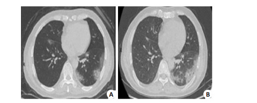
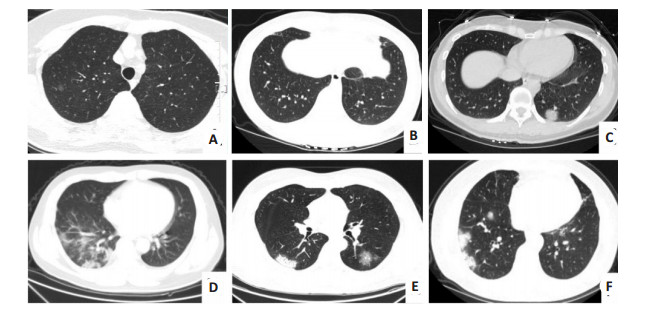
ObjectiveTo analyze the clinical manifestations and CT distribution characteristics of imported COVID-19 in Pudong, Shanghai. MethodsWe retrospectivly analyzed the clinical and imaging data of 12 cases of imported COVID-19 patients who were positive for viral nucleic acid examination. The number, distribution, morphology, density, boundary, mediastinum and pleura of the lesions were observed by CT. ResultsEleven cases had a history of exposure to the epidemic area and 1 case was unknown. Ten cases had fever and 9 with cough. Leukocyte counts, neutrophil counts and lymphocyte counts were normal in 10 cases. The monocyte counts was elevated in 7 cases, eosinophilia was decreased in 10 cases. Creactive protein was elevated in 2 cases, and serum amyloid was elevated in 3 cases. CT distribution features: all the 12 cases had multiple lesions, including 3 cases involving a single lobe, 3 cases involving two lobes, and 6 cases involving multiple lobes. The lower lobe of the lung was mainly involved, including 10 cases. Peripheral distribution was observed in 5 cases and peripheral distribution and central distribution in 7 cases. Pleural involvement in 11 patients, 12 patients did not see pleural effusion and mediastinal lymph node enlargement. We found 1 case of halo, 2 cases of interlobular septal thickening, 1 case of paving stone, 7 cases of inner bronchial wall thickening, 7 cases of blood vessel thickening, and 3 cases of air bronchogram. ConclusionThe clinical manifestations of COVID-19 patients are mainly fever and cough. In the laboratory, the leukocyte counts, neutrophil counts, lymphocyte counts are more normal and eosinophil counts decrease. The CT distribution is characterized by multiple ground hyaline shadows in the periphery of the lower lobe of the lung.
2020, 43(2): 274-277.
doi: 10.12122/j.issn.1674-4500.2020.02.19
Abstract:
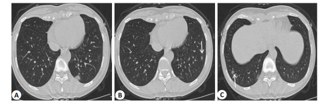
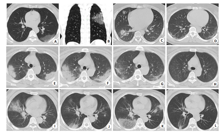
ObjectiveTo explore the changes and final outcome of chest CT in patients with common COVID-19 during the course of disease. MethodsThirty-three patients with clinical common COVID-19 were mainly treated with antiviral therapy and symptomatic treatment of related basic diseases during hospitalization, including 17 males and 16 females, with the age from 18 to 74 years old (48.60±11.85 years old). According to the changes of condition, the patients were reexamined by chest CT, at intervals of 2 to 10 days and 14 days after discharge. The clinical and imaging data of the patients were collected. The location, number, shape and density of the lesions and the signs of pleural thickening and pleural effusion were observed. The characteristics and interval time of the first diagnosis and reexamination were counted. The dynamic changes of CT and the correlation between prognosis and time were analyzed. ResultsAt first diagnosis, 3 cases showed negative CT. In other 30 cases, 75.8% showed multiple ground glass shadow in both lungs, and 45.5% were accompanied with subconsolidation or consolidation. The lesions were mainly located under the pleura. Thirty-three patients were reexamined for 1-4 times during hospitalization, and 21 cases showed aggravation or decline in the course of the disease. All of them were obviously absorbed. After discharge, 17 cases were completely absorbed, 10 cases had a small amount of residue, 60% of them were complicated with underlying diseases, and 6 cases had not been reexamined so far. Thirty-three patients entered the recovery stage after the onset of symptoms or nucleic acid test positive 15.79 ±3.79 days. The recovery period of 17 patients with complete absorption were 21.70±5.61 days, and the total course of disease were 37.89 ±7.15 days. ConclusionThe prognosis of clinical common type COVID-19 is good. The dynamic changes of imaging manifestations and their correlation with time have a certain rule, which has a certain value in guiding clinical treatment.
2020, 43(2): 278-281.
doi: 10.12122/j.issn.1674-4500.2020.02.20
Abstract:
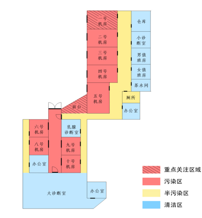
The epidemic of COVID-19 spreads quickly and widely, which seriously endangers people's health. Early detection and isolation of suspected or confirmed patients is essential to control the development of the epidemic. The screening of COVID-19 is inseparable from diagnostic imaging. This paper analyzes the possible infection factors in the process of examination. We put forward the emergency management strategy of medical imaging department during the epidemic situation of COVID-19, including establishment of epidemic control management team, division and strict administration of infection control area, protection level management and supplies configuration, classification management of radiation examination, enhancement of the disease differentiation ability, standardized management of patients, environment, devices and staff, and hoping to be helpful to the prevention and control of the epidemic.
The epidemic of COVID-19 spreads quickly and widely, which seriously endangers people's health. Early detection and isolation of suspected or confirmed patients is essential to control the development of the epidemic. The screening of COVID-19 is inseparable from diagnostic imaging. This paper analyzes the possible infection factors in the process of examination. We put forward the emergency management strategy of medical imaging department during the epidemic situation of COVID-19, including establishment of epidemic control management team, division and strict administration of infection control area, protection level management and supplies configuration, classification management of radiation examination, enhancement of the disease differentiation ability, standardized management of patients, environment, devices and staff, and hoping to be helpful to the prevention and control of the epidemic.
2020, 43(2): 282-285.
doi: 10.12122/j.issn.1674-4500.2020.02.21
Abstract:
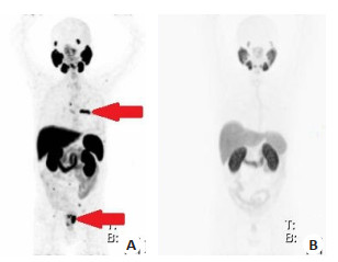
ObjectiveTo analyze the adverse drug reaction (ADR) of 18F-PSMA-1007 as a new tracer in PET/CT examination of prostate cancer, and propose a specific solution according to the characteristics of this tracer. MethodsFifty-eight patients with 18F-PSMA-1007 PET/CT from May 2019 to August 2019 were selected as the observation group according to the inclusion criteria. The use of 18F-PSMA-1007 tracer added the inspection procedure of pre-screening and tracer dilution before injection. A total of 58 patients using the routine examination procedure were randomly selected as the control group. The patients in the observation group were 25-75 years old, with an average age of 52.65±2.77 years old. The patients in the control group were 29-79 years old, with an average age of 62.91±2.35 years old. The patients in the two groups were all males, and there was no significant difference of other general data between two groups. The incidence of similar ADR was compared between the two groups. ResultsAmong the 58 patients in the control group, there were 16 patients (27.59%) with pain caused by tracer injection. Discomfort after injection of tracer was found in 3 patients, accounting for 5.17%. There were 2 cases (3.45%) of the patients with low specific uptake of tracer, whose image contrast could not meet the diagnostic requirements. The number and incidence of the ADR events in the observation group were: 2 cases (3.45%), 1 case (1.72%), 0 case (0%). The occurrence probability of each adverse event in the control group was lower than that in the observation group (P < 0.05). ConclusionApplication of pre-screening and pre-injection dilution of the tracer can greatly reduce the incidence of ADR in 18F-PSMA-1007 PET/CT examination of prostate cancer patients. It ensure the success rate of the examination.
2020, 43(2): 286-290.
doi: 10.12122/j.issn.1674-4500.2020.02.22
Abstract:
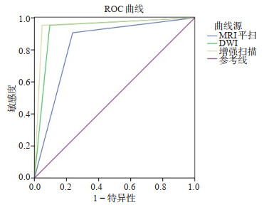
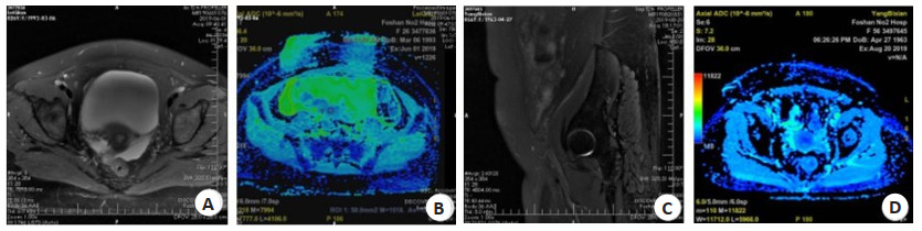
ObjectiveTo explore the diagnostic efficacy of pelvic MRI plain scan, gadopentetate dimeglumine enhanced scan and diffusion-weighted imaging (DWI) on patients with pelvic abscess. MethodsSixty-four patients with possible pelvic abscess who were admitted from January 2018 to June 2019 were selected. All patients underwent MRI plain scan, enhanced scan and DWI. The biopsy or surgical pathology was used as the gold standard, and the results of MRI plain scan, enhanced scan and DWI examination were evaluated in the diagnosis of pelvic abscess. The efficacy indicators (sensitivity, specificity, accuracy rate, positive predictive value, negative predictive value) of each examination method were compared. ROC curve was drawn to compare the AUC of each examination method, and the MRI findings of pelvic abscess were observed. ResultsThe differences in the sensitivity, specificity and accuracy rate between enhanced scan and DWI compared with those of MRI plain scan were not significant (P > 0.05). The differences in the sensitivity, specificity and accuracy rate between enhanced scan and DWI were not significant (P > 0.05). The AUC value of ROC of DWI and enhanced scan was higher than that of MRI plain scan (P < 0.05). The difference of the AUC between enhanced scan and DWI was not significant (P > 0.05). MRI plain scan accurately diagnosed 39 cases, most of which were cystic solid masses, with irregular shape, clear boundary, visible capsule and obvious separation. T1WI sequence showed that the masses were mostly with low signal, and T2WI sequence showed the masses were with uneven high signal and slightly higher signal. DWI examination showed that most lesions were with high signal, low ADC value and obviously limited diffusion. Enhanced scan showed that most lesions showed progressive enhancement and some lesions showed different degrees of enhancement on the dividing wall. ConclusionPelvic MRI, gadopentetate dimeglumine enhanced scan and DWI all have good efficacy in the diagnosis of pelvic abscess. The enhanced scan has the highest efficacy.
2020, 43(2): 291-295.
doi: 10.12122/j.issn.1674-4500.2020.02.23
Abstract:
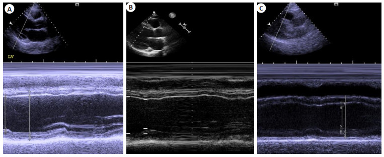
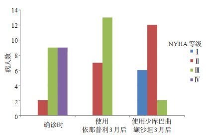
ObjectiveTo investigate the short- term functionality efficacy of Sacubitril/Valsartan in heart failure patients with reduced ejection fraction (HFrEF) on echocardiography, biochemical and clinical. MethodsTwenty HFrEF patients who were first diagnosed in our hospital from June 2018 to September 2019 were selected, with age of 51.4±9.923 years old. Patients were regularly treated with ACEI (angiotensin-converting enzyme inhibitor) Enalapril for 3 months and then switched to Sacubitril/ Valsartan for 3 months. The effect of the two medication between the baseline and 3 months after Enalapril treatment, and 3 months after switching to Sacubitril/Valsartan for N-terminal brain natriuretic peptide, left ventricular ejection fraction measured by transthoracic echocardiography, and 6 min walking test and New York Heart Association(NYHA)scale assessment were compared. ResultsAfter 3 months of treatment with Sacubitril/Valsartan, the increasing amplitude of left ventricular ejection fraction in patients was greater than the before (P < 0.05). The decreasing amplitude of N-terminal brain natriuretic peptide after 3 months of treatment with Sacubitril/Valsartan was greater than that after 3 months of treatment with Enalapril (P < 0.05). The difference in the improvement of distance walked on the 6 min walk test and NYHA scale significant (P < 0.05). ConclusionThe short-term (3 months) functionality efficacy of Sacubitril/Valsartan in HFrEF patients is better than that of Enalapril.
2020, 43(2): 296-299.
doi: 10.12122/j.issn.1674-4500.2020.02.24
Abstract:
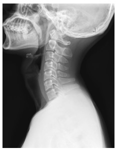
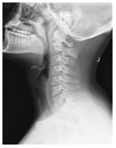
ObjectiveTo explore the effect of straight cervical curvature on early cervical disc degeneration in college students using magnetic resonance T2-mapping. MethodsFifty-six college students in Shantou area were recruited for lateral X-ray and T2-mapping. They were divided into straight cervical curvature group (n=43) and normal cervical curvature group (n=13) according to arc string distance of cervical spine. Spearman correlation was used for analyzing the correlation between the arc string distance of cervical curvature and T2 values of cervical disc nucleus pulposus. The T2 values between the two groups were compared using Wilcoxon's rank sum test. ResultsPositive correlation was observed between T2 value of the cervical disc nucleus pulposus and the arc string distance of cervical curvature (r=0.277, P < 0.05). The results showed that the differences of the T2 value of cervical nucleus pulposus in C2/3 and C3/4 segments between straight cervical curvature group and normal cervical curvature group were significant (P < 0.05), with no significant different in other segments (P > 0.05). ConclusionStraight cervical curvature may be one of the factors, which can accelerate the degeneration of cervical disc nucleus pulposus in college students. T2-mapping can detect changes of biochemical components in the early cervical disc degeneration, which will provide basis for the prevention and early intervention of cervical disc degeneration.
2020, 43(2): 300-303.
doi: 10.12122/j.issn.1674-4500.2020.02.25
Abstract:
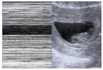
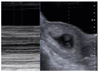
ObjectiveTo explore the predictive effect of fetal heart rate in early pregnancy on pregnancy outcome in patients with RSA by ultrasonography. MethodsThe pregnant women with 42-69 days of gestation who were examined by ultrasound were collected. Among them, 255 pregnant women with RSA history and 201 pregnant women without RSA history were enrolled. According to the pregnancy outcome during 42-196 days (Pregnancy 6-28 weeks), the patients were divided into three groups: RSA abnormal pregnancy group: spontaneous abortion occurred during the pregnancy of the pregnant women with previous RSA history; RSA normal pregnancy group: the pregnancy of the pregnant women with previous RSA history was more than 28 weeks; non RSA normal pregnancy group: the pregnancy of the pregnant women without previous RSA history was more than 28 weeks. The size of gestational sac and the length of head and buttock (CRL) were measured by transabdominal or transvaginal ultrasound, and the fetal heart rate was measured by M sampling and amplification mode. The mean value was measured three times continuously, and the size of gestational sac, CRL and fetal heart rate were recorded. The data of each group were followed up to 28 weeks of gestation. The data of each group were compared. ResultsIn 255 cases of RSA, 83.0% of the pregnancies were terminated 8 weeks ago. The lower the fetal heart rate, the higher the rate of embryo termination. There was no significant difference between RSA normal pregnancy group and non RSA normal pregnancy group (P > 0.05). In RSA abnormal pregnancy group and non RSA normal pregnancy group, there was significant difference between the mean fetal heart rate at 7 and 8 weeks of gestation (P < 0.05), and there was no significant difference between the mean fetal heart rate at 6 and 9 weeks of gestation (P > 0.05). The fetal heart rate was used as the test variable, the pregnancy outcome was the gold standard, the diagnostic value of fetal heart rate was predicted, the area of the curve was 0.831, the results were statistically significant (P < 0.05). ConclusionThe slow fetal heart rate in early pregnancy of patients with RSA has a certain predictive value for the adverse pregnancy outcome.
2020, 43(2): 304-308.
doi: 10.12122/j.issn.1674-4500.2020.02.26
Abstract:
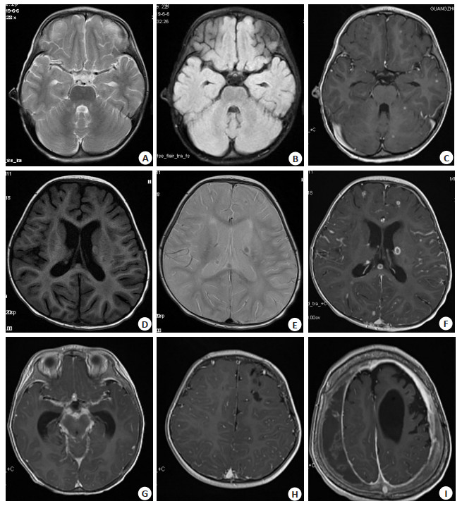
ObjectiveTo explore the value of MRI enhanced scan of head and cerebrospinal fluid examination in early diagnosis of intracranial tuberculosis in infants and children. MethodsThirty- one cases of the intracranial tuberculosis in infants and children were selected, which were diagnosed in clinic in the guangzhou chest hospital from January to December, 2018. All cases underwent MRI plain, enhanced scan and cerebrospinal fluid examination. The MRI imaging features and cerebrospinal fluid changes of the cases were analyzed. ResultsThe results of MRI showed 7 cases with meningeal tuberculosis, 7 cases with solid brain tuberculosis, 16 cases with mixed intracranial tuberculosis, and 1 case with simple hydrocephalus. Meningeal thickening 23 cases(74.19%), 16 cases occurred in the basal cistern, 14 cases of pial meningeal thickening, and 1 case of thickening of epedymal membrane. Nine cases with tuberculosis nodules (29.32%), 7 cases with tuberculoma (22.58%), 10 cases with tuberculous encephalitis (32.26%), 1 case of tuberculous cerebral infarction and 5 cases with softening of the brain. The MRI of intracranial tuberculosis showed hypervascularization of the brain surface in the 17 cases (54.84%). The typical change of cerebrospinal fluid Examination was found in 14 cases (45.16%), and in 9 cases of partial change of cerebrospinal fluid examination. ConclusionThe MRI enhanced scan can detect early the lesion, especially when the blood vessels on the brain surface were increased or enlarged, early signs of intracranial tuberculosis infection were significant. The changes of cerebrospinal fluid examination, the history of close contact with tuberculosis and the extracranial tuberculosis are combined. It can greatly improve the early diagnosis of intracranial tuberculosis in infants and children.
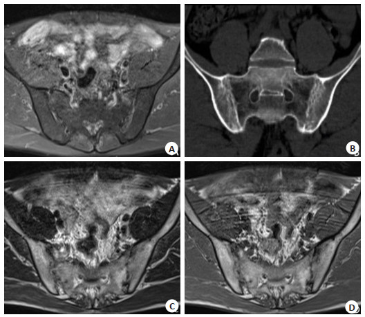
Imaging features of CT and MRI diagnosis of sacroiliac joint lesions in early ankylosing spondylitis
2020, 43(2): 309-312.
doi: 10.12122/j.issn.1674-4500.2020.02.27
Abstract:
ObjectiveTo observe the imaging features of CT and MRI in the diagnosis of sacroiliac joint lesions in early ankylosing spondylitis. MethodsFrom November 2018 to November 2019, 60 patients with early as sacroiliac joint disease in our hospital were selected, including 36 males and 24 females, with the age from 20 to 60 years old (37.28±6.33 years old). The course of disease was from 4 months to 10 years (4.80±1.17 years). All patients were examined by CT and MRI. The imaging characteristics and sensitivity of the two detection methods were compared. ResultsThe sensitivity of level Ⅰ as sacroiliac joint lesions in MRI was higher than that in CT (P < 0.05). The difference of the sensitivity of level 0, level Ⅱ, level Ⅲ and level Ⅳ as sacroiliac joint lesions in CT and MRI was not significant (P > 0.05). The detection rate of articular surface osteocystic degeneration and articular surface erosion in MRI was higher than that in CT (P < 0.05). The two groups had widened and narrowed joint space, osteosclerosis and osteosclerosis under key surfaces. There was no significant difference in the detection rate of articular cartilage swelling(P > 0.05). CT examination showed that the articular surface was serrated, with multiple small cysts, narrowed and blurred joint space. The most of the affected parts were in the middle and lower part of the joint. The joint effusion showed long T1 and T2 signals, the arthritic edema T1 showed signals, T2 showed high signals. The destruction of articular soft bone T1 showed low signals, T2 showed high signals, and the intensity was uneven. ConclusionMRI can clearly show the early as sacroiliac joint lesions. The abnormal detection rate and early diagnosis rate are higher than CT.
2020, 43(2): 313-316.
doi: 10.12122/j.issn.1674-4500.2020.02.28
Abstract:
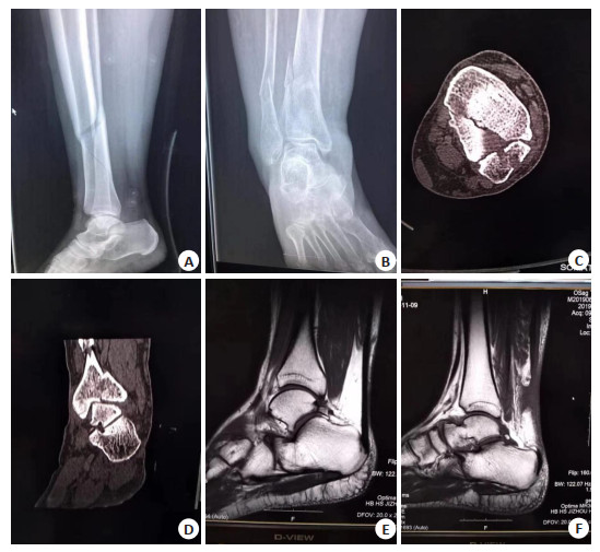
ObjectiveTo explore the significance of X-ray, CT and MRI combined with Weber typing in the differential diagnosis of injury degree of patients with tibiofibular syndesmosis. MethodsSeventy-five patients with tibiofibular syndesmosis injury were treated in our hospital from October 2017 to October 2018. Weber classification: 31 patients of type A, 25 patients of type B and 19 patients of type C. There were 39 cases of right ankle fracture and 36 cases of left ankle fracture. According to Lauge-Hansen classification, there were 9, 5 and 8 cases of pronation and abduction type Ⅰ, Ⅱ, and Ⅲ respectively; 6, 7, 9 and 5 cases of pronation and external rotation Ⅰ, Ⅱ, Ⅲ and Ⅳ respectively; 9, 4, 2 and 3 cases of supination and external rotation Ⅰ, Ⅱ, Ⅲ and Ⅳ respectively. There were 3 cases of supinational adduction type Ⅰ and 5 cases of supinational adduction type Ⅱ. X-ray, CT and MRI examination were carried out for the patients respectively. The results of MRI examination were taken as the gold standard. The consistency of X- ray, CT and MRI examination results and the difference between the sensitivity of combined diagnosis and single diagnosis were analyzed. ResultsThirty-nine patients with anterior ligament injury and 36 patients with deep ligament injury were found by MRI. As a gold standard, 41 patients with shallow ligament injury were found by CT diagnosis combined with Weber classification, 35 of which were consistent with MRI, 34 of which were consistent with MRI. The consistency of CT diagnosis combined with Weber typing and MRI diagnosis was stronger (P < 0.05). The consistency of X-ray combined with Weber typing and MRI diagnosis was stronger (P < 0.05). The sensitivity of combined CT and X-ray combined with Weber typing was significantly higher than that of single detection (P < 0.05). ConclusionThe sensitivity of X-ray, CT, examination combined with Weber classification in the diagnosis of the injury degree of tibiofibular syndesmosis is improved. The consistency with MRI is high, so it is recommended to promote the clinical application.
2020, 43(2): 317-320.
doi: 10.12122/j.issn.1674-4500.2020.02.29
Abstract:
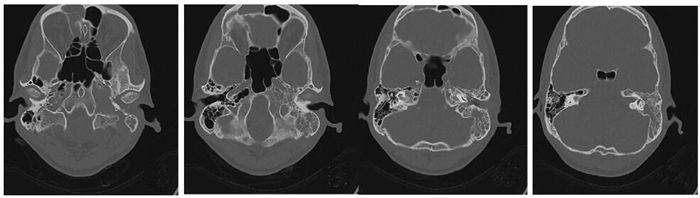
ObjectiveTo explore the value of CT plain scan and CT reconstruction (including volume reconstruction and multiplanar reconstruction) in the diagnosis of ossicular chain lesions. MethodsThe clinical data of 120 patients with ossicular chain lesions admitted to the author's hospital from October 2017 to December 2018 were retrospectively analyzed. The images of CT plain scan and CT reconstruction were discussed and analyzed. The Kappa evaluation was used to analyze the consistency of CT diagnosis. The diagnostic accuracy, sensitivity and specificity of axial and coronal images in CT plain scan and the CT reconstruction images for ossicular chain lesions were compared. ResultsCoincidence rate of CT reconstruction with the intraoperative results in the diagnosis of the structure of the ossicular chain lesion was higher. The Kappa value of the anvil exceed 0.75, and the Kappa value of the malleus, topside structure of stapes, incudomalleolar joint, and ankle joint all exceed 0.40. CT had good consistency in the diagnosis of the above lesions. The diagnostic sensitivity, specificity and accuracy of axial and coronal images in CT plain scan for ossicular chain lesions were 85.00%, 84.17% and 86.67%, respectively. The sensitivity, specificity and accuracy of CT reconstruction in the diagnosis of ossicular chain lesions were 88.33%, 87.50% and 89.17%, respectively. The difference was not significant (P>0.05). The sensitivity, specificity and accuracy of axial and coronal images in CT plain scan combined with CT reconstruction were 98.33%, 98.33% and 99.17% respectively, which were significantly higher than those of axial and coronal images in CT plain scan and CT reconstruction techniques alone in the diagnosis of ossicular chain lesions (P < 0.05). ConclusionCT plain scan and CT reconstruction can effectively display the imaging characteristics of ossicular chain lesions. But the combination of the two techniques can effectively improve the sensitivity, specificity and accuracy in the diagnosis of ossicular chain lesions.
2020, 43(2): 321-324.
doi: 10.12122/j.issn.1674-4500.2020.02.30
Abstract:
ObjectiveTo compare the clinical value of of EUS, MRCP and CT in the diagnosis of chololithiasis. MethodsEUS, MRCP and CT were used to diagnose 165 patients with suspected cholelithiasis. The diagnosis results were compared with the results of pathological diagnosis. The sensitivity, specificity, accuracy, PPV, NPV and detectionrate of stones with different diameterss of three diagnostic methods were analyzed and compared. ResultsIn the diagnosis of cholelithiasis, the sensitivity, specificity, accuracy, PPV and NPV of EUS and MRCP were significantly higher than CT (P < 0.05). The sensitivity and accuracy of EUS were significantly higher than MRCP (P < 0.05). For the stones < 4 mm, the detection rate of EUS was significantly higher than MRCP and CT (P < 0.05), while the detection rate of MRCP was significantly higher than CT (P < 0.05). ConclusionsIn the diagnosis of cholelithiasis, EUS and MRCP are significantly better than CT. EUS has more advantages over MRCP, especially for stones < 4 mm in the lower segment of the bile duct.
2020, 43(2): 325-329.
doi: 10.12122/j.issn.1674-4500.2020.02.31
Abstract:
ObjectiveTo compare the clinical effect and safety of different drugs combined with anesthesia for painless gastroscopy in elderly patients. MethodsA total of 160 elderly patients who planned to undergo painless gastroscopy were randomly divided into RP group (remifentanil+propofol+lidocaine) and DRE group (dizocine+remifentanil+etomidate), with 80 casesin each group. RP group included 44 males and 36 females, with the average age of 65.48±5.98 years old. DRE group included 40 males and 40 females with the average age of 66.19±5.13 years old. The two groups were compared in terms of the time of endoscopic implantation, the time of gastroscopy examination, the time of patients' waking up and the time of patients' leaving hospital. Systolic blood pressure (SBP), diastolic blood pressure (DBP), heart rate (HR), respiratory rate (RR) and blood oxygen saturation (SpO2) of the two groups before gastroscopy (T0), during gastroscopy (T1) and after gastroscopy (T2) were compared. Plasma C-reactive protein (CRP), cortisol (Cor) and norepinephrine (NE) levels were measured before (T0) and after (T2) gastroscopy. The anaesthesia effect and adverse reactions during and after operation were compared between the two groups. Operator satisfaction, anesthesiologist satisfaction, and patient comfort were recorded. ResultsThere were no significant differences in the time of endoscopic implantation, time of gastroscopy, time of patient awakening and time of patient leaving hospital between the two groups (P>0.05). SBP and DBP in RP group were lower than RE group at T1 and T2 (P < 0.05). HR at T2 was lower than RE group (P < 0.05). SpO2 at T1 was lower than RE group (P < 0.05). Compared with T0, CRP Cor and NE levels increased in the two groups at T2, but the increase in DRE group was lower than that in RP group (P < 0.05). There was no significant difference in anesthetic effect between the two groups (P>0.05). No serious adverse reactions such as laryngospasm and allergy occurred in the two groups. The incidence of hypotension in DRE group was lower than that in RP group (P < 0.05). The incidence of muscle fibrillation was higher than that in RP group (P < 0.05). There was no significant difference in operator satisfaction and patient comfort between the two groups (P>0.05). The satisfaction of anesthesiologists in DRE group was higher than that in RP group(P < 0.05). ConclusionEtomidate combined with remifentanil and low-dose dizocine are used for painless gastroscopy in elderly patients, which is better than propofol combined with remifentanil and lidocaine in stabilizing hemodynamics, reducing stress response and improving patient comfort.
2020, 43(2): 330-334.
doi: 10.12122/j.issn.1674-4500.2020.02.32
Abstract:
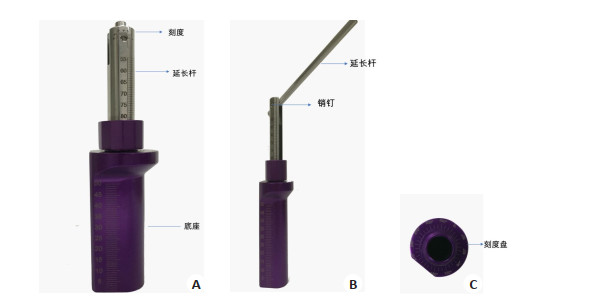
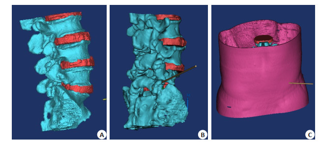
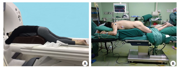
ObjectiveTo determine the combined preoperative application of three-dimensional reconstruction technique with the percutaneous transforaminal puncture locator whether can improve the accuracy of lumbar disc removal operation. MethodsPatients with lumbar disc herniation were divided into 12 in the locator used groups and 10 in the conventional surgery groups. Six patients in the locator used groups were performed by experienced and skilled operators and 6 were performed by beginners. In the conventional surgery groups, 5 patients were performed by the beginners and 5 by experienced and skilled operators. During the operation, the number of perspectives and the perspective time of reaching the first landing point and the second landing point were compared. ResultsIn the conventional surgery groups, the first landing time of beginners and skilled persons were 8.50±1.64 and 3.30±0.52 mins, times of perspectives were 18.50±2.35 and 7.70±0.82. The second landing time were 9.80±2.79 and 3.70±1.03 mins. Times of perspectives were 9.00±3.52 and 4.70±2.07. The operation time of the first landing point of the locator used groups were 3.00±0.71 and 2.60±0.55 mins, and times of perspectives were 5.20±1.10 and 4.80±1.10. The second landing time were 7.00±0.71 and 3.40±0.55 mins. Times of perspectives were 6.40±0.89 and 5.60±1.67. ConclusionThe combination of three-dimensional reconstruction technique and puncture locator can reduce the number of punctures and shorten the puncture time during intervertebral foramen
2020, 43(2): 335-339.
doi: 10.12122/j.issn.1674-4500.2020.02.33
Abstract:
ObjectiveTo explore the value of CT images in identifying the properties of tracheobronchial foreign bodies. MethodsSixty-nine patients with tracheobronchial foreign bodies diagnosed from August 2014 to April 2019 were collected. The patients were divided into peanut group(n=36) and non-peanut group (n=33) according to the type of foreign bodies. Peanut group includes 25 males and 11 females. The differences of clinical characteristics (the history of foreign body aspiration, age, gender and place of residence), features of fiberoptic bronchoscope (location of foreign bodies, granulation formation) and chest CT images(CT value, boundary definition of foreign bodies, bronchial occlusion, length and width of foreign bodies, emphysema, bronchoconstriction, pneumonia, mediastinal displacement, mediastinal emphysema, atelectasis, and bronchiectasis) between the two groups were compared. ResultsFor clinical characteristics, the history of foreign body aspiration in the peanut group was more common than that in the non-peanut group. The difference of the history of foreign body aspiration between the two groups was significant(P=0.044). The age, sex and place of residence were not found significant differences between both groups (P>0.05). For the fiberoptic bronchoscope, the differences of the location of foreign bodies (P=0.361) and granuloma formation (P=0.543) between the two groups were not significant. For the chest CT images, the CT value of the peanut group was lower than that of the non-peanut group (P < 0.001). The boundary of foreign bodies in the peanut group was more blurred than that in the non-peanut group (P=0.005). The bronchial occlusion was more common in the peanut group than that in the non-peanut group (P < 0.001). There were no significant differences in other features (P>0.05) on CT images. ConclusionCombined with the clinical history of foreign body aspiration, chest CT images can identify the properties of tracheobronchial foreign bodies.
2020, 43(2): 340-344.
doi: 10.12122/j.issn.1674-4500.2020.02.34
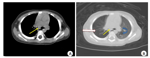
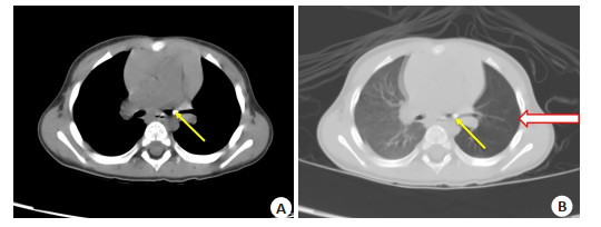
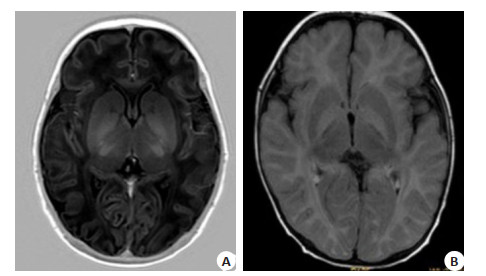
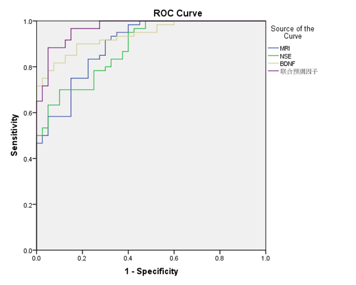
Abstract:
ObjectiveTo explore the clinical significance of MRI combined with serum brain-derived neurotrophic factor (BDNF) and neuron-specific enolase (NSE) in the diagnosis and outcome of newborns with hyperbilirubinemia. MethodsSixtycases of newborns with hyperbilirubinemia in our hospital from March 2017 to August 2019 were selected as the research group, including 34 males and 26 females, with the age of 38.4±1.6 weeks. In the same period, 40 newborns in our hospital were selected as the control group, including 19 males and 21 females, with the age of 37.8±1.5 weeks. The serum BDNF and NSE levels of two groups were compared. According to MRI imaging findings, patients in research group was divided into MRI abnormal group and MRI normal group. The serum BDNF and NSE levels of the subgroup were compared. Finally, the diagnostic value of MRI, BDNF and NSE as the diagnosis of hyperbilirubinemia were analyzed. ResultsThe total effective rate of clinical outcomes in the hyperbilirubinemia group was 50.00%, which was lower than that in the control group (75.00%). The difference of the effective rates between two groups was significant(P < 0.05). In the hyperbilirubinemia group, 54 children with abnormal MRI were accounting for 90.00%. Six patients in the control group with abnormal MRI were accounting for 15.00%, and the difference between two subgroups was significant (P < 0.05). The level of serum NSE and BDNF in the research group were significantly higher than those in the control group (P < 0.05). Serum NSE and BDNF levels in children with abnormal MRI were significantly higher than those in normal MRI (P < 0.05). The diagnostic effect of MRI combined with serum BDNF and NSE on hyperbilirubinemia was higher than single factor diagnosis. ConclusionMRI combined with serum NSE and BDNF can provide observational basis for the early diagnosis and outcome of children with hyperbilirubinemia.
2020, 43(2): 345-348.
doi: 10.12122/j.issn.1674-4500.2020.02.35
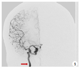
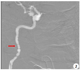
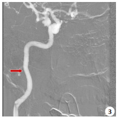
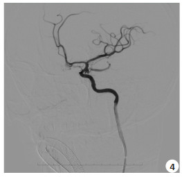
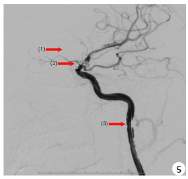
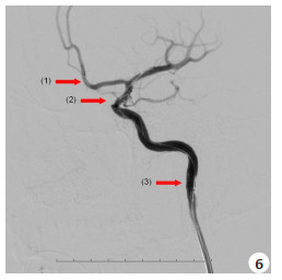
Abstract:
ObjectiveTo investigate the efficacy and safety of nimodipine combined with tirofiban in the treatment of acute thrombosis in cerebrovascular intervention. MethodsFrom May 2016 to May 2019, 152 patients received interventional examination and treatment in Xiaolan People's Hospital affiliated to Southern Medical University. Ninteen cases of cerebral vasospasm and thrombosis were found during operation, including 8 males and 11 females, 16 ruptured aneurysms and 3 unruptured aneurysms, 12 stent-assisted embolizations and 7 spring coil embolizations. The age of these patients ranged from 28 to 81 years, with an average of 48.0 years. During the angiography or treatment process, the blood vessels were found to have sausage-like changes, such as small lumen, slow blood flow, filling defect, distal blood vessels not developed, considering vasospasm and acute thrombosis.The nimodipine was immediate injected through the guiding catheter to relieve vasospasm. The sequential tirofiban was used to prevent thrombosis and dissolve thrombus. ResultsNineteen patients underwent a bolus injection of nimodipine in combination with tirofiban. Immediate angiography showed that the vasospasm was reversed and the distal vascular development was fine. Only two cases of lacunar cerebral infarction were confirmed by postoperative CT scans. CT scans of other 17 cases showed no abnormalities. All patients were followed up for 1 month to 1 year without cerebral infarction. ConclusionThe application of nimodipine combined with tirofiban in acute thrombosis in cerebrovascular intervention is effective and safe.
2020, 43(2): 349-351.
doi: 10.12122/j.issn.1674-4500.2020.02.36
Abstract:
ObjectiveTo investigate the efficacy of short-term mometasone furoate nasal spray combined with saline lavage in patients with chronic sinusitis after functional nasal endoscopy. MethodsEighty-six patients with chronic sinusitis with functional nasal endoscopy were selected. They were randomly divided into observation group and control group, with 43 cases in each group. After the operation, the control group was given a saline wash and mometasone furoate nasal spray, while the observation group was given a wash of mometasone furoate. The vas and Kennedy scores of the two groups were compared before operation. LKES and SNOT22 scores of the two groups were compared 3 days after operation and after lavatory treatment. The adverse reactions of the two groups were analyzed. ResultsThe difference of VAS score and Kennedy score between the two groups was not significant (P>0.05). The LKES score and SNOT score of the two groups were significantly lower than those of the three days after the operation (P < 0.05). The LKES score and SNOT score of the observation group were lower than that of the control group (PP < 0.05). In the observation group, there was 1 case of active bleeding in the nasal cavity after operation, which could be stopped after compression and hemostasis. The other patients in the two groups did not have any obvious adverse reactions. It could successfully complete the treatment plan. ConclusionShort-term washing with mometasone furoate in patients with chronic rhionosinusitis with nasai polyps after functional nasal endoscopy can help the recovery of nasal mucosa and better alleviate the clinical symptoms of patients.
2020, 43(2): 352-355.
doi: 10.12122/j.issn.1674-4500.2020.02.37
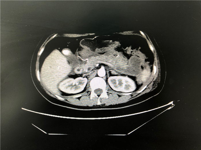
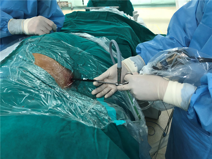
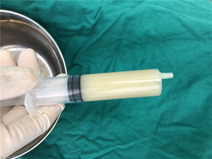
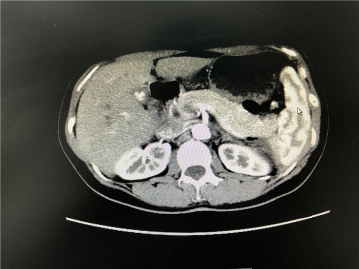
Abstract:
ObjectiveTo explore the operation method of "one-step" debridement under CT guided retroperitoneal hardpipe endoscope in the treatment of severe acute pancreatitis with peripancreatic infection. MethodsWe retrospectively analyzed the clinical data of 4 patients with severe acute pancreatitis complicated with peripancreatic infection who underwent CT-guided retroperitoneal hard-pipe endoscope "one-step" debridement from January 2017 to August 2019 in our Hospital. Three patients were male and one was female. The age was 47.5±10.4 years. The relevant literatures at home and abroad were reviewed. ResultsOne operation was performed in 3 cases, the other one was due to the expansion of necrotic area. On the 8th day after operation, hard mirror debridement through the original drainage tube sinus was performed, and the postoperative symptoms were relieved. The hospital stay after operation was 19.75 ± 7.2 d. Four patients had no complications such as bleeding and gastrointestinal fistula after operation. There were no complications such as pseudocysts in the follow-up. ConclusionCT-guided retroperitoneal hard-pipe endoscope "one-step" debridement is safe and feasible for the treatment of severe acute pancreatitis peripancreatic infection.It can control the development of inflammation in time, shorten the hospitalization time and save the hospitalization cost, but more clinical practice and high-quality research are needed to provide evidence to support it.