Find Duplicates
 Check Document
 Submission(new)
 Experts Office
 Editorial Office
2020 Vol. 43, No. 4
column
Display Method:
2020, 43(4): 557-562.
doi: 10.12122/j.issn.1674-4500.2020.04.01
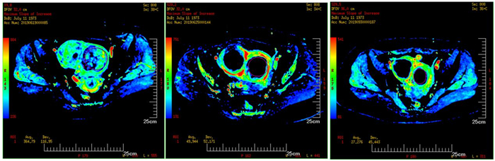
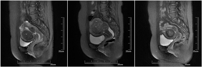
Abstract:
ObjectiveTo compare the efficacy of IVIM-DWI and DCE-MRI in the treatment of uterine fibroids with highintensity focused ultrasound scalpel. Methods60 patients with hysteromyoma underwent high intensity focused ultrasound ablation. IVIM-DWI and DCE-MRI were performed in all patients 1 week before operation, 48 hours after operation and 3 months after operation respectively. The differences between IVIM-DWI and DCE-MRI in ablated and UN ablated areas were compared between IVIM-DWI and DCE-MRI through relevant parameters (ADC, eADC, standardABC, f value, D value, D*value, MSI, SER). The correlation between the two scanning parameters was analyzed. ResultsThe values of D* and F related with IVIM-DWI and the MSI, SER value of DCE-MRI at 48 h and 3 months after operation were significantly different from those before operation (P < 0.05). And there was a significant positive correlation between the values of F and D* of IVIMDWI and MSI and SER of DCE-MRI. ConclusionCompared with DCE-MRI, IVIM-DWI can not distinguish the ablation boundary of hysterical, but the parameters of IVIM-DWI can still provide some reference value for the blood supply and perfusion of uterine leiomyoma in the early stage after HIFU.
2020, 43(4): 563-567.
doi: 10.12122/j.issn.1674-4500.2020.04.02
Abstract:
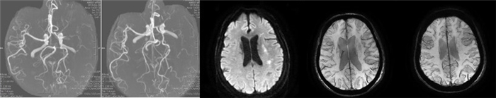
Arterial spin labeling (ASL) is an absolute quantitative perfusion method that measures the perfusion of cerebral blood flow by labeling hydrogen protons in arterial blood for perfusion assessment. It can not only generate perfusion images for qualitative judgment, but also quantitatively calculate the characteristic parameters of perfusion, that is, cerebral blood flow.3D-ASL features fast imaging, perfusion uniformity, high signal-to-noise ratio, low SAR value, and is currently the most recommended perfusion technique in ASL white papers. As a new ASL-derived technology, TASL in blood supply area can select the artery of interest for labeling, obtain the perfusion image of the artery of interest supply area, and obtain the cerebral blood flow of the corresponding brain area, which is of great significance in evaluating the establishment of collateral circulation in patients with cerebrovascular diseases. ASL is therefore currently widely used in the study of neurological diseases. Acute cerebral infarction (ACI) is a disease caused by various reasons (such as cerebral arteriosclerosis thrombosis, Takayasu arteritis, etc.), resulting in acute ischemia and hypoxia of the central nervous system followed by tissue ischemic necrosis, and adverse consequences. The most effective treatment of ACI includes thrombolysis, embolectomy, etc., so as to achieve the purpose of reperfusion in the ischemic area, hoping to expand the treatment time window and save the tissue in the non-core infarct area as much as possible. Therefore, clinical multi-modal imaging examination of acute ischemic stroke, including earlier detection of abnormal cerebral blood flow perfusion, assessment of the degree and extent of perfusion reduction and diseased vessels, has a higher expectation. With a large number of studies in recent years, ASL and its derivative technology have shown great value in these aspects. This article reviews the imaging principle and derivative technology of ASL, the application progress of ASL in ACI and the advantages and disadvantages of ASL in ACI disease.
Arterial spin labeling (ASL) is an absolute quantitative perfusion method that measures the perfusion of cerebral blood flow by labeling hydrogen protons in arterial blood for perfusion assessment. It can not only generate perfusion images for qualitative judgment, but also quantitatively calculate the characteristic parameters of perfusion, that is, cerebral blood flow.3D-ASL features fast imaging, perfusion uniformity, high signal-to-noise ratio, low SAR value, and is currently the most recommended perfusion technique in ASL white papers. As a new ASL-derived technology, TASL in blood supply area can select the artery of interest for labeling, obtain the perfusion image of the artery of interest supply area, and obtain the cerebral blood flow of the corresponding brain area, which is of great significance in evaluating the establishment of collateral circulation in patients with cerebrovascular diseases. ASL is therefore currently widely used in the study of neurological diseases. Acute cerebral infarction (ACI) is a disease caused by various reasons (such as cerebral arteriosclerosis thrombosis, Takayasu arteritis, etc.), resulting in acute ischemia and hypoxia of the central nervous system followed by tissue ischemic necrosis, and adverse consequences. The most effective treatment of ACI includes thrombolysis, embolectomy, etc., so as to achieve the purpose of reperfusion in the ischemic area, hoping to expand the treatment time window and save the tissue in the non-core infarct area as much as possible. Therefore, clinical multi-modal imaging examination of acute ischemic stroke, including earlier detection of abnormal cerebral blood flow perfusion, assessment of the degree and extent of perfusion reduction and diseased vessels, has a higher expectation. With a large number of studies in recent years, ASL and its derivative technology have shown great value in these aspects. This article reviews the imaging principle and derivative technology of ASL, the application progress of ASL in ACI and the advantages and disadvantages of ASL in ACI disease.
2020, 43(4): 568-571.
doi: 10.12122/j.issn.1674-4500.2020.04.03
Abstract:
Rs- fMRI is a scientific method to explore brain activity in clinical acupuncture which assists researchers to draw research conclusions and provide feedback in clinical practice.There were three kinds of RS fMRI analysis methods commonly used in acupuncture clinical research in recent 10 years: Regional Homogeneity (ReHo), Amplitude Of Low Frequency Fluctuation (AlFF) and Functional Connectivity(FC). In acupuncture research, ReHo could be used to analyse the activation or inhibition of a acupoint in a certain or several brain regions, so as to acknowledge the target effect brain area of the acupoint, and lay a foundation for the study of "acupoint-brain-disease". ALFF could directly reflect the intensity of spontaneous synchrony of local neurons, and to a certain extent, reflect the interaction and neural network connections of relevant brain regions. FC obtained the functional connections between brain regions, namely the "highway" of brain regions, which was easy to understand and the results are direct. Different analysis methods showed the brain activity of acupuncture at different acupoints, such as the synchronization of local nerve activity, the connectivity between adjacent brain areas, the spontaneous fluctuation and functional activity of the brain.This paper elaborates the applicable research directions of ReHo, ALFF and FC analysis methods in acupuncture research.
Rs- fMRI is a scientific method to explore brain activity in clinical acupuncture which assists researchers to draw research conclusions and provide feedback in clinical practice.There were three kinds of RS fMRI analysis methods commonly used in acupuncture clinical research in recent 10 years: Regional Homogeneity (ReHo), Amplitude Of Low Frequency Fluctuation (AlFF) and Functional Connectivity(FC). In acupuncture research, ReHo could be used to analyse the activation or inhibition of a acupoint in a certain or several brain regions, so as to acknowledge the target effect brain area of the acupoint, and lay a foundation for the study of "acupoint-brain-disease". ALFF could directly reflect the intensity of spontaneous synchrony of local neurons, and to a certain extent, reflect the interaction and neural network connections of relevant brain regions. FC obtained the functional connections between brain regions, namely the "highway" of brain regions, which was easy to understand and the results are direct. Different analysis methods showed the brain activity of acupuncture at different acupoints, such as the synchronization of local nerve activity, the connectivity between adjacent brain areas, the spontaneous fluctuation and functional activity of the brain.This paper elaborates the applicable research directions of ReHo, ALFF and FC analysis methods in acupuncture research.
2020, 43(4): 572-576.
doi: 10.12122/j.issn.1674-4500.2020.04.04
Abstract:
Quantitative magnetic resonance imaging has been widely used in the study of various diseases in recent years. The best application of quantitative magnetic resonance imaging will be helpful for early diagnosis and treatment. In this paper, several common quantitative MRI imaging techniques are classified according to the imaging principle. The principles and research progress of chemical exchange saturation transfer, chemical shift imaging, magnetic resonance spectrum, quantitative susceptibility mapping, and relaxation rate mapping technique are introduced. Finally, this paper analyzed the research progress. The analysis results show that there are many kinds of quantitative magnetic resonance imaging techniques and quantitative substances. Moreover, some quantitative magnetic resonance imaging techniques are overlapping. One technology can quantify multiple substances, and many technologies can be used for quantitative research of a disease-related substance. Although relevant studies have been carried out to compare various techniques for quantifying the same substance, the comparison results still need to be further explored. This paper's analysis results are convenient to understand the standard quantitative magnetic resonance imaging technology and its research progress and provide a reference basis for clinical diagnosis and treatment research.
Quantitative magnetic resonance imaging has been widely used in the study of various diseases in recent years. The best application of quantitative magnetic resonance imaging will be helpful for early diagnosis and treatment. In this paper, several common quantitative MRI imaging techniques are classified according to the imaging principle. The principles and research progress of chemical exchange saturation transfer, chemical shift imaging, magnetic resonance spectrum, quantitative susceptibility mapping, and relaxation rate mapping technique are introduced. Finally, this paper analyzed the research progress. The analysis results show that there are many kinds of quantitative magnetic resonance imaging techniques and quantitative substances. Moreover, some quantitative magnetic resonance imaging techniques are overlapping. One technology can quantify multiple substances, and many technologies can be used for quantitative research of a disease-related substance. Although relevant studies have been carried out to compare various techniques for quantifying the same substance, the comparison results still need to be further explored. This paper's analysis results are convenient to understand the standard quantitative magnetic resonance imaging technology and its research progress and provide a reference basis for clinical diagnosis and treatment research.
2020, 43(4): 577-581.
doi: 10.12122/j.issn.1674-4500.2020.04.05
Abstract:
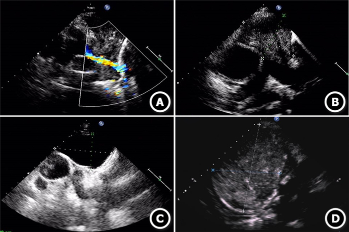
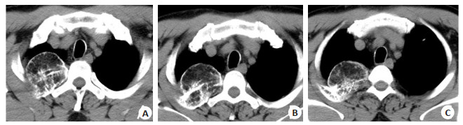
ObjectiveTo evaluate the value of transthoracic echocardiography in the diagnosis of mediastinal tumors. MethodsThe clinical data of patients with mediastinal tumors detected by transthoracic echocardiography from January 1st, 2018 to December 31st, 2019 were analyzed retrospectively. The ultrasonic features of mediastinal tumors in transthoracic echocardiography were summarized and compared with radiographic images (X-ray and CT scan) and histopathology. ResultsTwenty asymptomatic patients with mediastinal tumors were diagnosed by transthoracic echocardiography.Mediastinal tumors were divided into cyst (n=9), thymic tumor (n=5), lymph node enlargement (n=3), teratoma (n=2) and lymphoma (n=1). Compared with X-ray diagnosis, ultrasonic examination of mediastinal tumors has obvious advantages. In addition, the value of ultrasound in the diagnosis of mediastinal tumors was no less than that of CT (Kappa=0.792, P < 0.001). Taking the results of pathological examination as the gold standard, the misdiagnosis rate of transthoracic echocardiography in the diagnosis of mediastinal tumors was 15%, and the sensitivity and specificity for the diagnosis of benign and malignant tumors were 75% and 92%, respectively. ConclusionTransthoracic echocardiography can be used in the diagnosis of mediastinal tumors. It can not only observe the internal structure of mediastinal space occupying lesions, but also judge the benign and malignant properties of mediastinal tumors.
2020, 43(4): 582-586.
doi: 10.12122/j.issn.1674-4500.2020.04.06
Abstract:
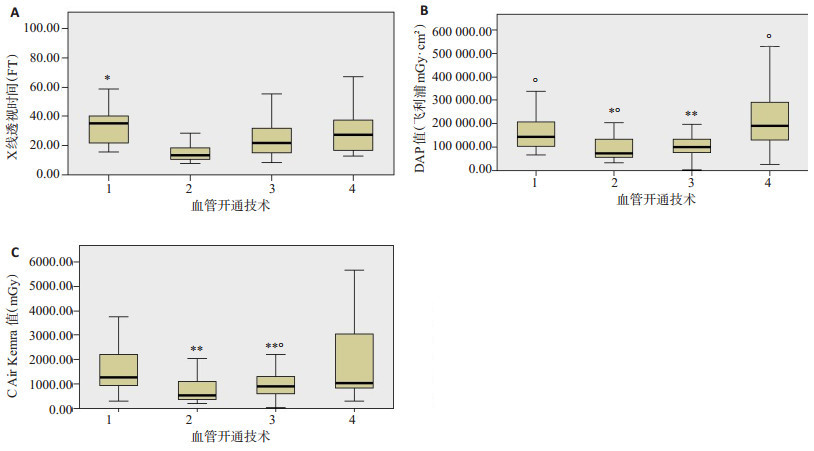
ObjectiveTo analyze the dose of radiation in patients with acute ischemic stroke and to explore the influence of gender, age, lesion location and diagnosis and treatment methods on radiation dose. MethodsA total of 156 patients with acute ischemic stroke who underwent interventional therapy in our hospital were retrospectively analyzed, and 113 cases were finally included. Fluoroscopy time (FT), dose area product (DAP) and air specific kinetic energy (AK) were recorded by a random radiation monitoring system. The effects of gender, age (30-60 years old group, 60-80 years old group and ≥80 years old group), occlusion position (anterior and posterior circulation) and vascular opening technology on radiation dose were analyzed. ResultsThe FT value of the patients ranged from 7.94 to 97.41 min, with an average value of 24.0±14.4 min. The DAP value of the dose area ranged from 1638 to 551 959 mGy · cm2, the average was 137 422.8±107 778.1 mGy · cm2. The AK value of the dose area ranged from 11 to 5726 mGy, the average was 1210.9 ± 1070.8 mGy. Compared with males, females had less radiation dose. The values of DAP and AK had significant difference (P < 0.05). There was no significant difference between the three age groups in the analysis of the variance of each dose (P>0.05). There was no significant difference in the dose received by patients with anterior and posterior circulation occlusion (P>0.05). Patients with single catheter aspiration (ASPI) had the lowest radiation dose. The radiation dose of the catheter aspiration combined with stent retriever (ASPI + SR) and the stent implantation group was larger than that of the catheter aspiration only, and the difference of the three dose values was statistically significant (P < 0.05). ConclusionThe radiation dose of patients with acute ischemic stroke varies greatly, and gender, the way of vascular opening are all important factors affecting the radiation dose. Therefore, more aggressive protective measures may be needed for patients with different characteristics.
2020, 43(4): 587-592.
doi: 10.12122/j.issn.1674-4500.2020.04.07
Abstract:
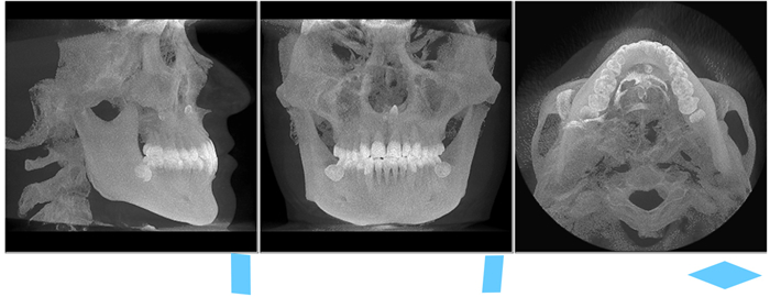
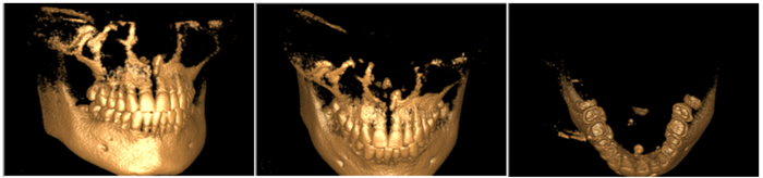
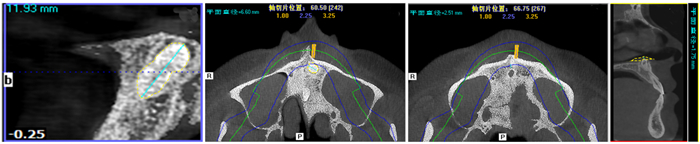
ObjectiveDemonstrates the anatomical characteristics of the supernumerary teeth with the surrounding tissue in the middle maxilla to provide a reference for clinical practice. Methods309 supernumerary teeth in the middle maxilla from 237 patients were selected and diagnosed by CBCT at the Stomatological Hospital of Lanzhou University from November 2011 to April 2016, including 187 males and 50 females, with the age from 6 to 47 years old (average 20 years old). Identified the number, evaluated the size, the shape, 3D space, surrounding bone thickness, the neighboring teeth location, and the adjacency relationship of supernumerary teeth by using CBCT three- dimensional reconstruction of the image and the length measurement tool in the CBCT scanning. ResultsSupernumerary teeth are mostly ambushed in the jaw palatal side of the dental arch, one as 71.4% (n=221), two as 27.5% (n=85), and three as 1.1% (n=3). Located on the 11-21 occupy 89.5% (n=277), near the palatal side 87.5% (n=270), near the lip side 4.5% (n=15), internal arch 8.0% (n=25) and other locations occupy 11.5%. The appearance of the teeth covered comicalness (79.3%, n=245), no deform (9.8%, n=30), and lateral incisor (10.9%, n=34). Status of inverted impacted occupied 77.4% (n=239), vertical 17.0% (n=53), and other angulation 5.6% (n=17). According to the bone wall of type Ⅰ, type Ⅱ and type Ⅲ respectively occupied by 83.1% (n=257), 5.6% (n=17), 11.3% (n=35). Around 86.7% (n= 268) contact with permanent or germ teeth, among 13.3% (n=41) distance greater than 1 mm. Teeth average length is 11.92 mm and the largest diameter of the crown is 6.06 mm. ConclusionAnatomic morphology and spatial position of supernumerary teeth in the upper anterior region are relatively stable. CBCT can display the neighboring tissue in a three-dimensional way to provide an experimental basis for early diagnosis and early intervention into a dental clinic.
2020, 43(4): 593-596.
doi: 10.12122/j.issn.1674-4500.2020.04.08
Abstract:
Cardiovascular disease is a common disease that seriously threatens human health, and occupies the first place in the death caused by various diseases. Studies have shown that cardiac diastolic function has been impaired even if there are no obvious clinical symptoms in the early stage of many cardiovascular diseases. From the view of early prevention, clinical diagnosis and late intervention treatment, it is of positive clinical significance to understand the changes of cardiac diastolic function as early as possible. At present, echocardiography is commonly used in clinical non- invasive evaluation of cardiac diastolic function, which can achieve the purpose of early detection of cardiac diastolic function, and has important value for early diagnosis, curative effect evaluation and prognosis of the disease. By reviewing the related literature at home and abroad, this paper reviews the research status of pulsed Doppler, color M-mode echocardiography, tissue Doppler, Real- time threedimensional echocardiography, Tei index and Two-dimensional speckle tracking technique commonly used in echocardiography to evaluate cardiac diastolic function. As a new technique, two-dimensional speckle tracking technique has been widely studied and applied in the evaluation of left ventricular diastolic function. Its independent characteristic makes it simpler, more feasible and more accurate than other methods. In addition, the advantages and disadvantages of common methods for evaluating cardiac diastolic function in echocardiography were also analyzed. In conclusion, ultrasonic technology for evaluating cardiac diastolic function will become more and more advanced with the improvement of science and technology, and will play an irreplaceable role in medical imaging and even the whole field of ultrasound medicine in the future.
Cardiovascular disease is a common disease that seriously threatens human health, and occupies the first place in the death caused by various diseases. Studies have shown that cardiac diastolic function has been impaired even if there are no obvious clinical symptoms in the early stage of many cardiovascular diseases. From the view of early prevention, clinical diagnosis and late intervention treatment, it is of positive clinical significance to understand the changes of cardiac diastolic function as early as possible. At present, echocardiography is commonly used in clinical non- invasive evaluation of cardiac diastolic function, which can achieve the purpose of early detection of cardiac diastolic function, and has important value for early diagnosis, curative effect evaluation and prognosis of the disease. By reviewing the related literature at home and abroad, this paper reviews the research status of pulsed Doppler, color M-mode echocardiography, tissue Doppler, Real- time threedimensional echocardiography, Tei index and Two-dimensional speckle tracking technique commonly used in echocardiography to evaluate cardiac diastolic function. As a new technique, two-dimensional speckle tracking technique has been widely studied and applied in the evaluation of left ventricular diastolic function. Its independent characteristic makes it simpler, more feasible and more accurate than other methods. In addition, the advantages and disadvantages of common methods for evaluating cardiac diastolic function in echocardiography were also analyzed. In conclusion, ultrasonic technology for evaluating cardiac diastolic function will become more and more advanced with the improvement of science and technology, and will play an irreplaceable role in medical imaging and even the whole field of ultrasound medicine in the future.
2020, 43(4): 597-600.
doi: 10.12122/j.issn.1674-4500.2020.04.09
Abstract:
ObjectiveTo investigate the correlation between arterial hemidominance and prominent vessel sign (PVS) after unilateral middle cerebral artery (MCA) occlusion in M1 segment. Methods48 patients with acute ischemic stroke with unilateral MCA M1 segment occlusion from January 2018 to March 2020 were collected. Among them, there were 27 males and 21 females which the average age was 52.8±7.7 years. All patients were divided into two groups: lateral dominance group (n=26)and control group (n=22) according to the unilateral dominance of ACA and/or PCA revealed by three-dimension time of flight MRA (3D-TOF MRA). The difference of PVS shown by SWAN sequence was observed between the two groups. The patients were divided into PVS positive group(n=27) and negative group (n=21) according to SWAN sequence. On the day of admission and one week after admission, the hemidominance group and the control group, PVS negative and PVS positive groups were scored with the National Institutes of Health Stroke rating scale (NIHSS), and the differences were analyzed. ResultsAmong the 48 patients, the incidence of PVS in the unilateral dominance group was 42.31% (11/26), while that in the control group was 72.73% (16/22), the control group was significantly higher than the lateral dominant group, the difference was statistically significant (P < 0.05). The NIHSS scores of patients in the hemiplegia group and PVS negative group were lower than those in the control group and PVS positive group on the day of admission and one week later, and the difference was statistically significant. ConclusionAfter unilateral MCA M1 segment occlusion, the unilateral dominance of cerebral artery and negative PVS indicate the establishment of collateral circulation and good perfusion state, which is closely related to the short-term prognosis of the patients.
2020, 43(4): 601-605.
doi: 10.12122/j.issn.1674-4500.2020.04.10
Abstract:
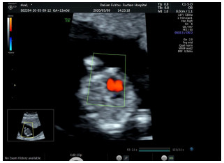
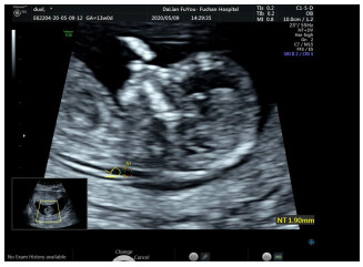
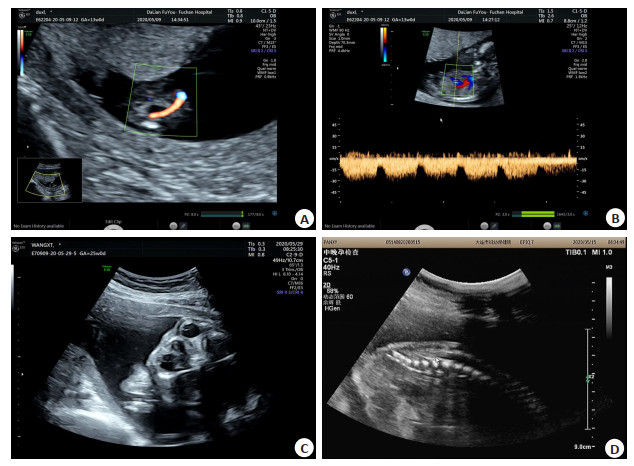
ObjectiveTo research the application value of nuchal translucency thickness (NT) ultrasound in the first trimester of pregnancy combined with color Doppler ultrasound in the second trimester of pregnancy in prenatal fetal malformation screening. MethodA total of 2417 pregnant women who underwent fetal malformation screening in our hospital from August 2017 to December 2019 were selected as the research objects. All pregnant women received NT ultrasound examination at 11-14 weeks of gestation, and received two-dimensional and four-dimensional color Doppler ultrasound examination in 22-28 weeks of pregnancy. Taking the results of induced labor or delivery were regarded as "gold standard", the NT value and abnormal rate of NT in abnormal fetus and normal fetus were compared, the results of NT ultrasound, two-dimensional and four-dimensional color Doppler ultrasound were compared, the diagnostic rates of NT ultrasound, two-dimensional and fourdimensional color Doppler ultrasound and their combination in the diagnosis of fetal malformation were compared. Results88 cases were diagnosed with abnormal fetus in 2417 cases of pregnant women, the incidence rate was 3.64%. The NT value of abnormal fetus was significantly higher than that of normal fetus, and the abnormal rate of NT (93.18%) was significantly higher than that of normal fetus (1.33%), the differences were significant (P < 0.05). There were 82 abnormal fetuses detected by NT ultrasound, the diagnostic accuracy rate was 93.18%; 85 abnormal fetuses were detected by two-dimensional combined with four-dimensional color Doppler ultrasound, and the diagnostic accuracy rate was 96.59%. The diagnostic accuracy of two-dimensional combined with four-dimensional color Doppler ultrasound was slightly higher than that of NT ultrasound, the difference was not significant (P>0.05). The sensitivity, specificity and accuracy of NT ultrasound combined with two-dimensional and four-dimensional color Doppler ultrasound in the diagnosis of fetal malformation were 100.00%, 99.57% and 99.59%, respectively, which were higher than those of NT ultrasound, two-dimensional and four-dimensional color ultrasound (93.18%, 98.67%, 98.47% and 96.59%, 99.06%, 98.97% respectively), and the difference between three groups were significant (P < 0.05). ConclusionNT ultrasound in early pregnancy and color Doppler ultrasound in second trimester have advantages in prenatal screening of fetal malformations, and all of them have good diagnostic value. The combined application of them can further improve the detection rate of fetal malformations, which has great significance for the early termination of pregnancy and the reduction of abnormal fetal birth.
2020, 43(4): 606-609.
doi: 10.12122/j.issn.1674-4500.2020.04.11
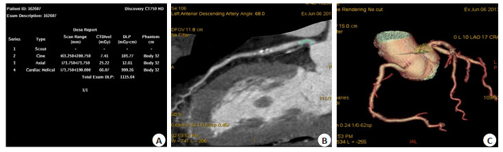
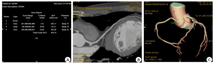
Abstract:
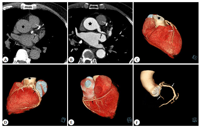
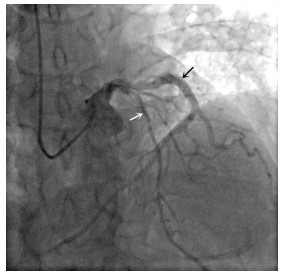
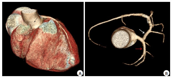
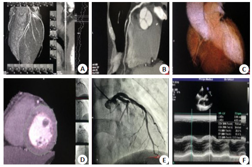
ObjectiveTo summarize the clinical characteristics and coronary CT angiography (CCTA) findings of congenital absence of the right coronary arteries (RCA). MethodsThirteen patients with congenital absence of RCA diagnosed by selective coronary angiography (SCA) in our hospital from January 2014 to June 2019 were retrospectively reviewed with emphasis placed upon the patients' clinical manifestations and CCTA findings. ResultsThe clinical manifestations of the 13 patients were of not specific, and most of them showed symptoms of cardiovascular diseases. Non-enhanced CCTA scans showed high density calcified plaques in the left main, left anterior descending and/or left circumflex coronary arteries areas. Enhanced CCTA scans showed different degrees of narrowness in the left main, left anterior descending and/or left circumflex, and no RCA were given off in the ascending aorta or right coronary sinus areas. The left main, left anterior descending and left circumflex were enlarged in different degrees, and the enlarged left circumflex extended to the posterior surface of the right ventricle, where it gave off its branches to supply the right atrium and right ventricle. Besides that, findings on selective coronary angiography (SCA) were almost identical to those of CCTA. ConclusionCongenital absence of the RCA is rare, and it's clinical manifestations are not specific, which make it difficult to diagnosis by the clinical manifestations alone. However, CCTA combined with SCA can be used to confirm the diagnosis.
2020, 43(4): 610-614.
doi: 10.12122/j.issn.1674-4500.2020.04.12
Abstract:
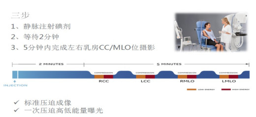
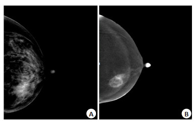
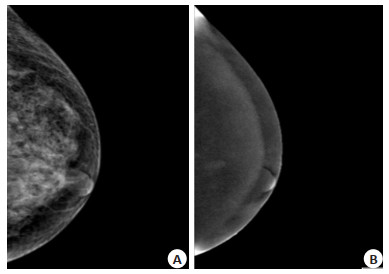
ObjectiveTo investigate the diagnostic value of comparing conventional mammography (MG) and contrastenhanced spectralmammography (CESM) for benign and malignantbreast diseases, using pathological results as the gold standard. MethodsSeventy-four patients who completed CESM and MG tests and obtained pathological diagnosis from May 2018 to April 2020 were included in the study. All patients were female, aged 22-56 years, with an average age of 41.02±8.24 years old. Using the American GE Stenographe Essential fully digital mammary gland machine, through the upper limb after intravenous injection of iodine contrast agent USES the end (CC) and internal and external oblique (MLO), by a pressure high and low to exposure, we obtain low energy image and subtraction angiography image of high energy subtracting low energy by special algorithm. Three radiology breast specialist performed quality analysis of the images and made imaging diagnosis. Taking pathology as the standard, the difference in the diagnosis of benign and malignant breast diseases between cesm and Mg was compared, and the diagnostic coincidence rate of the two methods for the same tumor grade was further compared, and the diagnostic efficacy of the two methods was analyzed and compared. ResultsThe sensitivity, specificity, positive predictive value, negative predictive value and accuracy of MG in the diagnosis of benign and malignant breast lesions were 77.50%, 80.95%, 79.48%, 70.27% and 79.07%, respectively. The sensitivity, specificity, positive predictive value, negative predictive value and accuracy of CESM were 100%, 90.47%, 90.90%, 100% and 95.12% respectively (P < 0.05). In the study, the diagnostic coincidence rate of MG in grade 3-5 breast tumor grade was 79.00%, 66.70%, 86.70%, 85.70% and 100% respectively, while CESM was 100%, 92.30%, 87.50%, 90% and 100% (P < 0.05). ConclusionCESM is superior to conventional mammography in the diagnosis of benign and malignant breast diseases, especially in the diagnosis of grade 3and 4A tumors.
2020, 43(4): 615-620.
doi: 10.12122/j.issn.1674-4500.2020.04.13
Abstract:
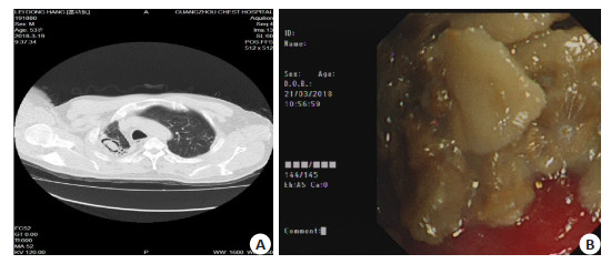
ObjectiveSummarize and analyze the etiology and clinical characteristics of hemoptysis of patients with previous history of pulmonary tuberculosis to provide reference for the diagnosis and treatment of them. MethodsThe clinical data of 182 inpatients with a history of pulmonary tuberculosis and hemoptysis were retrospectively analyzed in Guangzhou Chest Hospital from January to December 2018, including imaging changes, bronchoscopy, etiology and other results. And the etiology and clinical characteristics were summarized. A total of 182 patients were included, 145 males and 37 females, with an average age at 55.13±13.73 years old. ResultsThe average time from cure of tuberculosis to hemoptysis was 10.13±8.29 years. The main causes of hemoptysis were pulmonary tuberculosis posterior cavity secondary aspergilloma (23.63%), followed by bronchiectasis and pulmonary tuberculosis recurrence (23.08%), pneumonia (19.23%) and NTM lung disease (8.79%). A few of them were lung tumor (1.65%) and bronchial foreign body (0.55%). ConclusionMost of hemoptysis in patients with a history of pulmonary tuberculosis is related to the structural damage of the lung. The recurrence of pulmonary tuberculosis is only one of the causes. The diagnosis of pulmonary tuberculosis recurrence can not just base on the past history of pulmonary tuberculosis and imaging changes. It is easy to cause misdiagnosis, missed diagnosis and mistreatment. It is suggested that comprehensive analysis of clinical data should be applied, starting from common causes, the existing medical technology should be used to find the evidence of diagnosis, and at the same time, some rare causes should be watched out for.
2020, 43(4): 621-624.
doi: 10.12122/j.issn.1674-4500.2020.04.14
Abstract:
Liver cancer is one of the common malignant tumors of the digestive system. Most patients have no clinical symptoms in the early stages of liver cancer and are mostly advanced at the time of consultation. The high mortality rate determines that early diagnosis and early treatment are the most effective methods to improve survival rate.Therefore, how to improve the early diagnosis of minimal liver cancer has become an urgent issue that we need to solve. The key molecules of early neovascularization of micro hepatocellular carcinoma have become a very important step in early diagnosis. Therefore, by reviewing relevant domestic and foreign literature in recent years, this article studied four key molecules of early diagnosis of micro hepatocellular like carcinoma-Prox1, Glypican-3, EpCAM, CXCR4. The current situation and deficiencies were reviewed in order to provide new directions and ideas for the early screening and diagnosis of minimal liver cancer.
Liver cancer is one of the common malignant tumors of the digestive system. Most patients have no clinical symptoms in the early stages of liver cancer and are mostly advanced at the time of consultation. The high mortality rate determines that early diagnosis and early treatment are the most effective methods to improve survival rate.Therefore, how to improve the early diagnosis of minimal liver cancer has become an urgent issue that we need to solve. The key molecules of early neovascularization of micro hepatocellular carcinoma have become a very important step in early diagnosis. Therefore, by reviewing relevant domestic and foreign literature in recent years, this article studied four key molecules of early diagnosis of micro hepatocellular like carcinoma-Prox1, Glypican-3, EpCAM, CXCR4. The current situation and deficiencies were reviewed in order to provide new directions and ideas for the early screening and diagnosis of minimal liver cancer.
2020, 43(4): 625-628.
doi: 10.12122/j.issn.1674-4500.2020.04.15
Abstract:
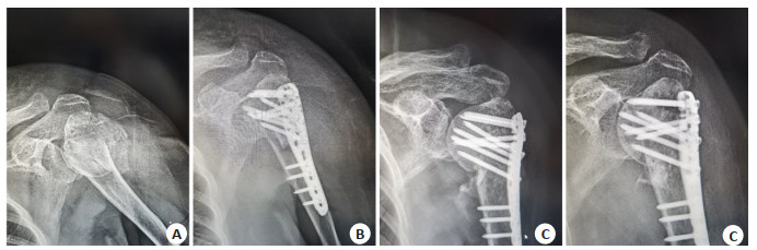
ObjectiveTo explore the correlation between function and imaging indexes after anatomic locking plate (ALP) for proximal humeral fractures (PHF). MethodsA retrospective analysis was performed on the 120 PHF patients who were admitted to the hospital from June 2016 to January 2019. According to Neer classifications, they were divided into two-part fracture group (n=28), three-part fracture group (n=60) and four-part fracture group (n=32). All were treated with ALP. The operation time and intraoperative blood loss were recorded. At 1 year after surgery, loss angle of neck shaft angle, loss height of humeral head and range of motion (ROM) of effected shoulder joints were examined by X-ray films. The shoulder function was evaluated by Constant-murley scores. The postoperative pain was evaluated by scores of VAS. The correlation between postoperative function and imaging indexes in PHF patients was analyzed by Pearson correlation analysis. ResultsWith the increase of Neer classification, intraoperative blood loss was significantly increased in PHF patients (P < 0.05), postoperative loss angle of neck shaft angle and loss height of humeral head were significantly increased (P < 0.05), anteflexion, external and internal rotation angles of shoulder joints and Constant scorewere significantly decreased (P < 0.05), and VAS score was significantly increased (P < 0.05). Pearson correlation analysis showed that postoperative anteflexion, external rotation and internal rotation of shoulder joints, and Constant score were significantly negatively correlated with postoperative loss angle of neck shaft angle and loss height of humeral head (P < 0.05). ConclusionROM of shoulder joints is significantly correlated with changes in neck shaft angle and height of humeral head in PHF patients after ALP.
2020, 43(4): 629-633.
doi: 10.12122/j.issn.1674-4500.2020.04.16
Abstract:
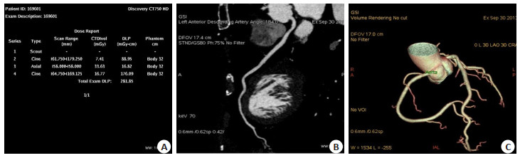
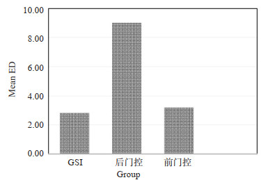
ObjectiveTo explore the advantages and clinical value of coronary angiography in GSI scanning model by comparing the radiation dose and image quality among the GSI model, the traditional front and back door control technique. MethodsA total of 162 patients underwent successfully CT coronary angiography in our hospital from May 2019 to May 2020 were selected as research objects. According to the scanning model, these patients were divided into three groups. Group A was the GSI scanning mode, group B was the backdoor control mode and C group was the front door control mode. The image noise (SD), contrast, SNR (SNR), comparison of noise ratio (CNR), image quality index (FOM), and the effective radiation dose (ED) for these groups were compared. ResultsComparison of image quality: ① SD of A group was significantly lower than that of B group and C group (P < 0.01). ②SNR, CNR and FOM of Group A were significantly higher than B group (P < 0.01) and C group (P < 0.05). ③SNR, CNR and FOM of group C were higher than group B, but without significantly difference (P>0.05). Comparison of ED: ① The mean ED of group A and C were 2.87 ± 0.83 mSv, 3.34 ± 2.36 mSv, respectively, and were both significantly lower than group B (9.04 ± 3.06 mSv) (P < 0.01). ② ED in group A and group C decreased 68.25% and 63.05%, respectively, compared with group B, and ED in group A decreased by about 14.07% compared with group C. ConclusionCompared to the front and back door technique, GSI model scanning technology not only can effectively reduce the radiation dose, but also can significantly improve the quality of image, and it should be popularized in clinic.
2020, 43(4): 634-638.
doi: 10.12122/j.issn.1674-4500.2020.04.17
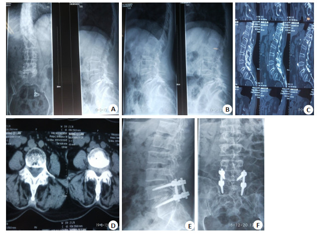
Abstract:
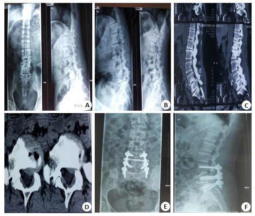
ObjectiveTo explore the clinical effect of posterolateral bone graft fusion (PLF) in the treatment of patients with lumbar spondylolisthesis. MethodsA retrospective study of 118 patients with lumbar spondylolisthesis was conducted in our hospital's Orthopedics Department. Among them, 58 patients were treated with PLF combined with pedicle screw internal fixation (PLF group), and 60 patients were treated with posterior interbody fusion (PLIF) along with pedicle screw internal fixation surgery (PLIF group). Operation time, surgical bleeding volume, bone graft fusion rate using X lines, CT, complications rate, JOA score before and after the operation, intervertebral space height, vertebral body slippage rate, ect. were statistically analyzed in two groups. ResultsThere was no statistically significant difference in operation time, the amount of bleeding, JOA score intervertebral space height, and vertebral spondylolisthesis rate between the PLF group and the PLIF group (P>0.05); the bone graft fusion rate in the PLF group was 84.48% lower than the 96.67% in the PLIF group (P < 0.05); The surgical complication rate had no significant different with 6.90% of PLF group and 3.33% of PLIF group.1 year after operation, JOA score in PLF group was lower than 96.67% in PLIF group (P < 0.05), the vertebral spondylolisthesis rate of patients in PLF group was higher than that of PLIF group (P < 0.05); the height of intervertebral space in PLF group was lower than that of PLIF group 1 month and 1 year after surgery (P < 0.05). ConclusionPLF and PLIF are both good treatment for lumbar spondylolisthesis. PLIF surgery is more beneficial to maintain postoperative lumbar spine function recovery, intervertebral space height, and reduce the incidence of postoperative vertebral spondylolisthesis.
2020, 43(4): 639-642.
doi: 10.12122/j.issn.1674-4500.2020.04.18
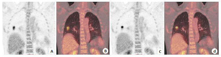
Abstract:
ObjectiveTo compare the effect of Q.STATIC respiratory gated (QSRG) and ungated (UG) scanning on the image quality and quantitative analysis of lung nodules, and evaluate the clinical application value of QSRG. MethodsThis prospective study included patients with suspected lung nodules from November 2019 to May 2020 who agreed to undergo QSRG scan. A total of 65 patients (295 lung nodules) were enrolled, and 4 patients were excluded due to continuous irregular breathing. The maximum standard uptake value (SUVmax), tumor metabolic volume (MTV), mean standard uptake value (SUVmean), standard deviation (SD) value of the aortic arch and clinical scan time of measurable lung nodules under QSRG and UG were recorded. The differences between QSRG and UG PET in the detection of lung nodules, measurable lung nodules, SUVmax, MTV, signal-to-noise ratio (SNR) and clinical scan time were compared. SPSS 22.0 software was used to perform Wilcoxon signed rank test. ResultsAmong QSRG PET and UG PET, the detection of lung nodules were 295 vs 275, measurable lung nodules were 196 vs 182. SUVmax were 6.90±4.40 g/mL vs 6.64±4.28 g/mL (P < 0.05). MTV were 3.23±8.01 cm3 vs 3.44±8.66 cm3 (P < 0.05). SNR were 30.30±20.91 vs 30.22±21.97 (P>0.05) and scan time were 16.45±3.74 min vs 13.21±3.98 min (P < 0.05). Single bed QSRG increased the time of whole body scan to 3 min. ConclusionQSRG did not increase the radiation dose. Compared with UG images, QSRG images had higher detection rate and measurable ability of lung nodule. The quantitative analysis of SUVmax and MTV was more accurate, and the total scanning time was slightly increased.
2020, 43(4): 643-646.
doi: 10.12122/j.issn.1674-4500.2020.04.19
Abstract:
Renal agenesis (RA) is a rare congenital birth defect, which the incidence of single kidney deficiency is about 1‰ and that of double kidney deficiency is about 0.1‰ ~0.3‰. Heredity is the main pathogenic factor, but its pathogenesis is still unclear. Clinically, most RA fetuses do not have typical signs and symptoms, and also lack highly specific diagnostic indicators. At present, the fetus of RA is often found by prenatal ultrasound examination in the second and third trimesters of pregnancy, and a few pregnant women will choose to further improve MRI for diagnosis. Therefore, prenatal diagnosis of RA has obvious limitations in clinical practice. In recent years, with the development of high-throughput sequencing technology, CNV-seq and WES have gradually been widely used in the field of Obstetrics and gynecology. High-throughput sequencing technology for prenatal diagnosis of fetus has the advantages of high maternal-fetal safety, high accuracy and easy operation, which not only provides a new choice for prenatal diagnosis of RA, but also provides a scientific basis for exploring its genetic causes and related mechanisms. This article reviews the normal embryonic kidney development and the application of prenatal diagnostic techniques such as ultrasound, MRI, CNV-seq and WES in RA.
Renal agenesis (RA) is a rare congenital birth defect, which the incidence of single kidney deficiency is about 1‰ and that of double kidney deficiency is about 0.1‰ ~0.3‰. Heredity is the main pathogenic factor, but its pathogenesis is still unclear. Clinically, most RA fetuses do not have typical signs and symptoms, and also lack highly specific diagnostic indicators. At present, the fetus of RA is often found by prenatal ultrasound examination in the second and third trimesters of pregnancy, and a few pregnant women will choose to further improve MRI for diagnosis. Therefore, prenatal diagnosis of RA has obvious limitations in clinical practice. In recent years, with the development of high-throughput sequencing technology, CNV-seq and WES have gradually been widely used in the field of Obstetrics and gynecology. High-throughput sequencing technology for prenatal diagnosis of fetus has the advantages of high maternal-fetal safety, high accuracy and easy operation, which not only provides a new choice for prenatal diagnosis of RA, but also provides a scientific basis for exploring its genetic causes and related mechanisms. This article reviews the normal embryonic kidney development and the application of prenatal diagnostic techniques such as ultrasound, MRI, CNV-seq and WES in RA.
2020, 43(4): 647-650.
doi: 10.12122/j.issn.1674-4500.2020.04.20
Abstract:
Breast cancer has become the most common cancer in Chinese women, and breast magnetic resonance imaging (MRI) has become increasingly important in the diagnosis of breast diseases. MRI has high sensitivity in the diagnosis of breast cancer, but lack specificity. As an important supplementary technique for breast MRI, diffusion weighted imaging (DWI) can improve the specificity and has been widely used in clinic. Apparent diffusion coefficient (ADC) is a measure of the diffusion of water molecules in tissues, which can be calculated using diffusion-weighted images. DWI-ADC value becomes a hot topic in the differentiating benign and malignant breast lesions and evaluating the prognosis, metastasis risk and chemotherapy efficacy of breast cancer. Building upon recent surveys of this field, this article reviews the basic principle, application progress, limitation and prospect of DWI-ADC.
Breast cancer has become the most common cancer in Chinese women, and breast magnetic resonance imaging (MRI) has become increasingly important in the diagnosis of breast diseases. MRI has high sensitivity in the diagnosis of breast cancer, but lack specificity. As an important supplementary technique for breast MRI, diffusion weighted imaging (DWI) can improve the specificity and has been widely used in clinic. Apparent diffusion coefficient (ADC) is a measure of the diffusion of water molecules in tissues, which can be calculated using diffusion-weighted images. DWI-ADC value becomes a hot topic in the differentiating benign and malignant breast lesions and evaluating the prognosis, metastasis risk and chemotherapy efficacy of breast cancer. Building upon recent surveys of this field, this article reviews the basic principle, application progress, limitation and prospect of DWI-ADC.
2020, 43(4): 651-654.
doi: 10.12122/j.issn.1674-4500.2020.04.21
Abstract:
ObjectiveToo discuss the application value of multi- slice spiral CT (MDCT) in the evaluation of elderly coronary heart disease (CHD). Methods110 elderly patients with CHD treated in our hospital from January 2018 to February 2020 were selected. Among them, the MDCT, echocardiography and coronary angiography (CAG) were performed in our hospital. The CAG was used as the "gold standard" to analyze the diagnostic value of MDCT. The left ventricular ejection fraction (LVEF), left ventricular end diastolic volume (LVEDV), left ventricular end systolic volume (LVESV), left ventricular myocardial mass (LVMM), left ventricular stroke volume (LVSV) and left ventricular short axis shortening rate (LVFS) measured by MDCT and echocardiography were compared. ResultsThe sensitivity, specificity, accuracy, positive predictive value and negative predictive value of MDCT diagnosis were 93.80%, 93.49%, 93.73%, 97.98% and 81.77%, respectively. Compared with echocardiography, MDCT showed no significant difference in LVEF, LVEDV, LVESV, LVMM, LVSV and LVFS (P < 0.05). MDCT showed that LVEF, LVSV and LVFS in patients with severe stenosis were significantly lower than those in patients with mild and moderate stenosis (P < 0.05), while LVEDV, LVESV and LVMM were significantly higher than those in patients with mild and moderate stenosis (P < 0.05). Gensini score were negatively correlated with LVEF, LVSV and LVFS (r=-0.433, -0.412 and -0.422, P < 0.05), and positively correlated with LVEDV, LVESV and LVMM (r=0.410, 0.366 and 0.378, P < 0.05). ConclusionMDCT has a good value in the diagnosis of CHD in the elderly, which can accurately evaluate left ventricular function and is related to the degree of coronary artery stenosis.
2020, 43(4): 655-658.
doi: 10.12122/j.issn.1674-4500.2020.04.22
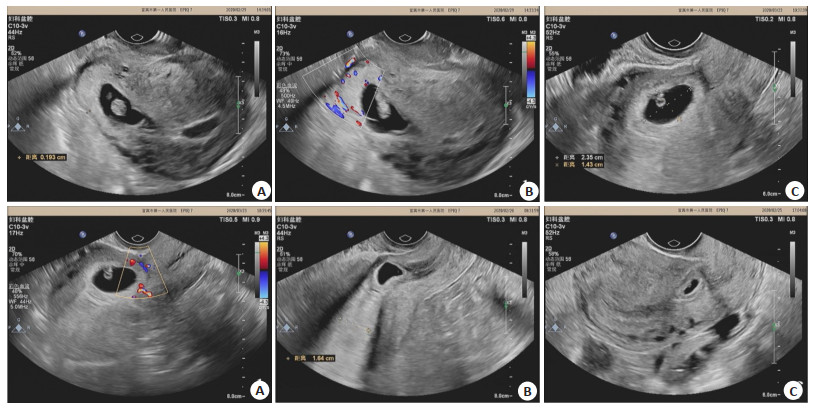
Abstract:
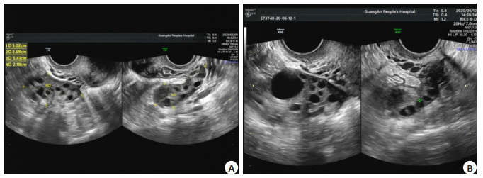
ObjectiveTo analyze the monitoring value of vaginal Doppler ultrasound on ovarian artery hemodynamics and follicular development in infertile patients with polycystic ovary syndrome (PCOS) during treatment process. Methods267 infertile patients with PCOS diagnosed and treated in the hospital from December 2017 to December 2019 were selected as the research subjects. All patients underwent vaginal Doppler ultrasound. And the ovarian artery hemodynamics peak systolic blood flow velocity (PSV), end-diastolic blood flow velocity (EDV), resistance index (RI), pulsatility index (PI)], serum hormones levels [follicle stimulating hormone (FSH), luteinizing hormone (LH), estradiol (E2) and follicular development (follicle number, follicle size, ovarian volume) were compared among the patients before and after treatment. ResultsBefore treatment, the ultrasound manifestations of infertile patients with PCOS showed ovarian enlargement, honeycomb changes of bilateral ovaries and abnormal follicular development. The number of all faceted follicles was ≥12, with diameter less than 10mm in different sizes (mostly of 2-9 mm) and showed a fence-like arrangement, enhanced medulla echo and thickened capsule. And the blood vessels were clear, blood flow was rich and blood flow signal was strip in the ovarian matrix. The blood flow spectrum showed a high-velocity and low-resistance state. After treatment, the ultrasound manifestations showed large ovary volume and a large number of follicles, with dominant follicles present in one side of the ovaries. And ovarian capsule presented a strong echo and there was a richer blood flow in the ovarian matrix. Compared with before treatment, the number of follicles in infertile patients with PCOS increased significantly after treatment (P < 0.05), and the size of follicles and ovarian volume increased significantly (P < 0.05). Compared with before treatment, the ovarian artery hemodynamic indexes of PSV and EDV among infertile patients with PCOS increased significantly after treatment (P < 0.05) while the RI and PI decreased significantly (P < 0.05). Compared with before treatment, the levels of FSH, LH and E2 of infertile patients with PCOS significantly increased after treatment (P < 0.05). ConclusionVaginal Doppler ultrasound can monitor ovarian artery hemodynamics and follicular development in infertile patients with PCOS, and has important clinical value in the guidance treatment of infertile patients with PCOS.

2020, 43(4): 659-663.
doi: 10.12122/j.issn.1674-4500.2020.04.23
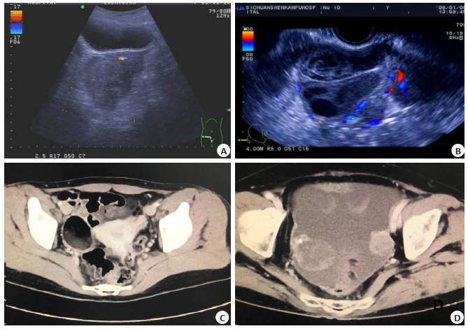
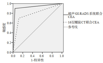
Abstract:
ObjectiveTo explore value of ultrasound gynecological image report and data system (GI-RADS) classification and 16- slice spiral CT in differential diagnosis of benign and malignant ovarian tumors. MethodsA total of 85 patients with ovarian tumors treated in the hospital from January 2015 to August 2019 were enrolled as study objects. All underwent ultrasound and 16-slice spiral CT examination. GI-RADS was applied to evaluate performances of ultrasonic images, and to detect the CEA level. The pathological examination was performed on lesions. The consistency of ultrasound GI-RADS system and 16-slice spiral CT combined with CEA was compared with those of pathological examination. Using pathology test results as the gold standard, the sensitivity, specificity, positive predictive value, negative predictive value and diagnostic accuracy of ultrasonic GI-RADS system, 16 slice spiral CT combined with CEA were compared in the diagnosis of identification of benign and malignant ovarian tumors. ROC curve was used to analyze the diagnostic value of ultrasound gi-rads system, 16 slice spiral CT combined with CEA in the diagnosis of benign and malignant ovarian tumors. ResultsThe consistency of ultrasound GI-RADS combined CEA examination with pathological examination was higher than that of 16-slice spiral CT combined CEA (Kappa: 0.791 vs 0.487). There were no significant differences in sensitivity, specificity, malignant predictive value and benign predictive value of the two methods for diagnosing benign and malignant ovarian tumors (P>0.05). The accuracy of ultrasound GI-RADS combined with CEA in the diagnosis of benign and malignant ovarian tumors was higher than that of 16-slice spiral CT combined with CEA (P < 0.05). ROC curve analysis showed that AUC of ultrasound GI-RADS combined with CEA was greater than that of 16-slice spiral CT combined with CEA for diagnosing benign and malignant ovarian tumors (P < 0.05). ConclusionThe ultrasound GI-RADS with CEA is of relatively high diagnostic value for benign and malignant ovarian tumors, and its diagnostic accuracy is relatively high.
2020, 43(4): 664-667.
doi: 10.12122/j.issn.1674-4500.2020.04.24
Abstract:
ObjectiveTo explore the diagnostic value of color Doppler flow imaging (CDFI) and CT for acute pancreatitis. Methods81 patients with acute pancreatitis admitted in the hospital from January 2013 to June 2020 were retrospectively analyzed. The examination results of CDFI and CT were compared among patients. The imaging features of CDFI and CT were explored in the diagnosis of acute pancreatitis patents. The diagnostic accuracy rate of acute pancreatitis, and diagnostic valu by the two methods were compared in oedematous and necrotic hemorrhagic pancreatitis. ResultsBy summarizing the imaging features of CT and CDFI in 81 patients with acute pancreatitis, The results showed that the expansion rate of pancreatic duct was 25.93% higher than that of CT 12.35%. The rates of small intrapancreatic liquefaction and extrapancreatic high density detected by CDFI were 12.35%, which were lower than 30.86% and 27.16% of CT examination respectively, and the difference was statistically significant (P < 0.05). There were no significant differences in the detection rates of extrapancreatic abscess, common bile duct stones (CBDS), fat layer blur and parenchyma unevenness between the two examination methods (P>0.05). Taking the clinical comprehensive diagnosis results as the gold standard, CDFI missed 16 cases diagnosis and CT missed 9 cases. The diagnostic coincidence rates of CDFI and CT for acute pancreatitis were 80.25% (65/81) and 88.89% (72/81), respectively, with no significant difference (P>0.05). The diagnostic coincidence rates of CDFI for oedematous pancreatitis and necrotic hemorrhagic pancreatitis were 78.79% and 86.67%, without significant difference compared with those of CT (87.88%, 93.33%) (P>0.05). ConclusionAbdominal CDFI can provide more accurate judgment for patients with acute pancreatitis. It has natural advantages in the judgment of pancreatic duct dilatation, and can be applied as one of the first choice for clinical diagnosis and treatment.
2020, 43(4): 668-671.
doi: 10.12122/j.issn.1674-4500.2020.04.25
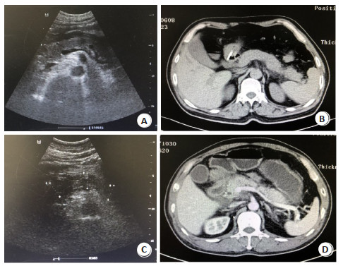
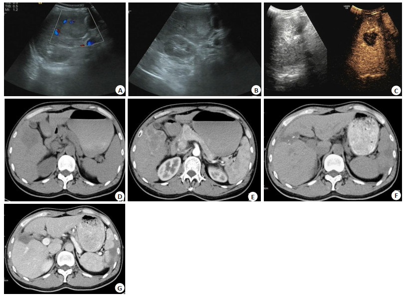
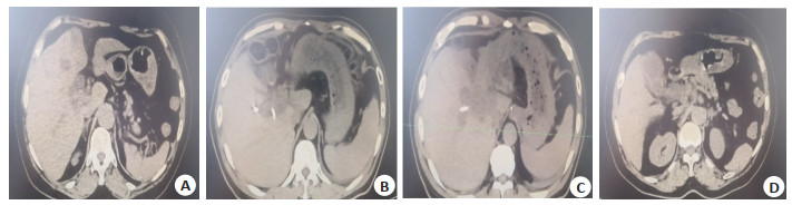
Abstract:
ObjectiveTo observe the application effect of contrast-enhanced ultrasound (CEUS) and multi-slice spiral computed tomography (MSCT) in the diagnosis and surgical assessment of primary clear carcinoma cell of the liver (PCCCL). Methods100 patients with PCCCL admitted in the hospital and 42 patients with focal nodular hyperplasia of the liver from February 2016 to March 2019 were enrolled in this study. CEUS and MSCT were performed respectively.The diagnostic effects of CEUS and MSCT on different diameter lesions, peritoneal effusion, retroperitoneal lymph node enlargement and unclear margin in PCCCL patients were compared with surgical pathological results as the gold standard; Sensitivities, specificities, missed diagnosis rates and misdiagnosis rates of CEUS and MSCT were statistically analyzed. The results of CEUS and MSCT were used to evaluate the surgical efficacy (tumor recurrence and metastasis) of PCCCL patients at 1 year after operation. ResultsMSCT was superior to CEUS in detecting lesions smaller than 1 cm in diameter, hydrops abdominis, posterior peritoneal lymphadenopathy and unclear margins and the difference was statistically significant (P < 0.05). MSCT had higher sensitivity and specificity than CEUS in the diagnosis of PCCCL (P < 0.05), but the misdiagnosis rate and missed diagnosis rate were lower than those of CEUS (P < 0.05). The detection rates of postoperative tumor recurrence and metastasis in patients with PCCCL by MSCT was higher than those by CEUS (P < 0.05). ConclusionBoth CEUS and MSCT can be used for diagnosis of PCCCL, but MSCT is better for diagnosing small lesions and abnormal imaging findings, with higher sensitivity and specificity. Besides, it is better for surgical effect evaluation.
2020, 43(4): 672-675.
doi: 10.12122/j.issn.1674-4500.2020.04.26
Abstract:
ObjectiveTo analyze the value of transvaginal sonography (TVS) combined with MRI in the diagnosis of residual myometrial thickness in cesarean scar pregnancy (CSP). MethodsEighty CSP cases with residual myometrium less than 3 mm were collected from January 2019 to June 2020. All patients were requested for termination of pregnancy and received TVS and MRI after admission. Besides, the examination results were compared with surgical and pathological findings. Imaging features of the two diagnostic methods were analyzed, and their clinical diagnostic value was discussed. ResultsIn this study, surgery and pathology confirmed 71 cases with residual myometrial thickness smaller than 3mm at the incision of CSP. TVS combined with MRI diagnostic results were positive in 72 cases and negative in 8 cases, and the diagnostic accuracy, specificity, sensitivity, positive predictive value, negative predictive value and Kappa value were 96.25%, 77.78%, 98.59%, 97.22%, 87.50%, and 0.803, respectively. TVS found 40 cases with yolk sac structure, 25 cases with germ structure, 15 cases with pulsating primitive heart tube. MRI was far inferior to ultrasound in observing internal structure of the gestational sac but it was better than ultrasound in observing adjacent structure around the gestational sac. ConclusionBoth TVS and MRI have high accurac in the diagnosis of residual myometrial thickness of CSP, and combination of these two methods greatly improves the diagnostic accuracy. Ultrasound has more advantages in observing blood flow signals at incision muscular layer, resistance index, yolk sac inside the pregnancy sac, blastema and fetal heartbeat, which is also real-time, cheap and highly repeatable. MRI can be used to better assess the thickness of residual muscle layer at the incision and its relationship with surrounding tissues. The joint detection of these two methods can better display the residual myometrial thickness at the incision, thereby providing a strong basis for patients and surgeons to choose the operation method.
2020, 43(4): 676-679.
doi: 10.12122/j.issn.1674-4500.2020.04.27
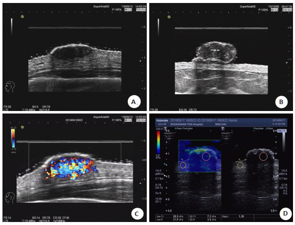
Abstract:
ObjectiveTo summarize the high-frequency ultrasound and shear wave elastograhpy findings of irritated seborrheic keratosis. Methods32 cases of ISK confirmed by surgery and pathology in our hospital were collected. All of them were single lesions, including 18 males and 14 females, aged from 47 to 81 years old, with an average age of 59.7 ± 9.4 years old. The position, involvement level, shape, size, boundary and internal echo of the tumor were observed with a high-frequency probe, and the mode of shear wave elastography was switched to, the mean young's modulus of the tumor and the Ratio of elastic strain between the tumor and adjacent normal dermal tissue were measured. ResultsAll the 32 patients were single lesions, and the lesions were most common in the head and face (30 cases, accounting for 93.8%). In 29 cases (accounting for 90.6%), the tumor increased significantly recently. 21 cases (accounting for 65.6%) had pain and itching at the tumor site and 9 cases (accounting for 28.1%) had superficial bleeding. The 25 cases' tumor was detected to involve dermis. The lesions were mainly elliptic (23 cases, accounting for 71.9%), and the increase in the thickness of the mass was mainly reflected by the increase in the thickness of the epidermis. The interior of the tumor was mainly hypoechoic, with point-like strong echoes in some cases (12 cases, accounting for 37.5%), and the internal blood flow in 32 cases was grade Ⅱ and Ⅲ. The mean young's modulus was 21.1±3.6 kPa, with a mean elastic strain ratio of 1.73±0.64 to the surrounding normal dermis. ConclusionIrritated seborrheic keratosis is mostly located in the head and face. The ultrasonic features are exophytic oval hypoechoic nodules, which are not clearly demarcated with dermis. In some lesions, there are point-like strong echoes. The internal blood flow is mainly grade Ⅱ and Ⅲ, combined with the value of 30.5 kPa of young's modulus, and the elastic strain ratio of the tumor to the surrounding normal dermis of 4.00 as the threshold, which could be differentiated from common malignant tumors of the skin.
2020, 43(4): 680-683.
doi: 10.12122/j.issn.1674-4500.2020.04.28
Abstract:
ObjectiveTo explore the application value of 64-slice spiral CT combined with multi-mode image reconstruction in preoperative resectability evaluation of hilar cholangiocarcinoma (HCCA). MethodsThe clinical data of 180 patients with HCCA who were admitted and treated in the hospital from May 2017 to March 2019 were retrospectively analyzed. All patients completed 64-slice spiral CT scanning before surgery. Differential diagnosis, TNM staging, Bismuth-corlette typing and resectability evaluation were carried out, and the results were compared with clinical pathological diagnosis results. The accuracy rates in diagnosis, classification, staging and resectability evaluation of HCCA were analyzed. ResultsThe accuracy of 64-slice spiral CT combined with multi-mode image reconstruction in detecting HCCA was 100%. CT examination results of all patients showed intrahepatic bile duct dilatation. Most patients had bile duct dilatation at the proximal end of the tumor. 14 patients had different degrees of hepatic lobe atrophy, 38 patients had hilar lymph node enlargement, and 16 patients had liver metastases. There were no significant differences between CT scanning results and surgical pathological results (P>0.05). Differences in results of clinical Bismuth-Corlett classification, TNM staging and resectability evaluation between the two methods were not statistically significant (P>0.05). Conclusion64-slice spiral CT combined with multi-mode image reconstruction has a high accuracy rate in the diagnosis, classification and clinical staging of HCCA. It can more intuitively and accurately evaluate the resectability of HCCA, which can be used in the development of personalized surgical plans and improvement of surgical safety.
2020, 43(4): 684-687.
doi: 10.12122/j.issn.1674-4500.2020.04.29
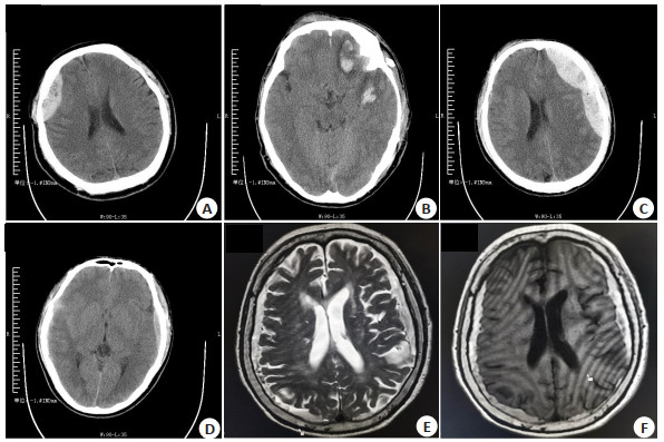
Abstract:
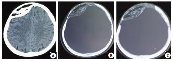
ObjectiveTo analyze the value of CT and MRI multiple sequences in evaluating the condition of patients with traumatic brain injury. MethodsThe clinical data of 70 patients with traumatic brain injury admitted and treated in the hospital were collected from February 2018 to April 2020. All patients were undergone with CT, magnetic resonance T1WI, T2WI, fluid attenuated inversion recovery (FLAIR), diffusion weighted imaging (DWI) and enhanced gradient echo T2- star weighted angiography (ESWAN) sequence scanning. Location, number and shape of the lesion were recorded. Compared with the results of surgical pathology, the evaluation value of the two diagnostic methods was analyzed. The patients were followed up 3 months after injury, and the Glasgow Outcome Score (GOS) was recorded. Spearman correlation was used for comparative analysis. ResultsAmong the 60 patients with traumatic brain injury, 42 cases were diagnosed as subdural hematoma, 21 cases as subarachnoid hemorrhage, 17 cases as epidural hematoma and 23 cases as brain contusion. The diagnostic rates of subdural hematoma, brain contusion and subarachnoid hemorrhage by MRI were higher than those by CT (P < 0.05), but there was no significant difference in the accuracy between the two methods in diagnosis of epidural hematoma (P>0.05). For microbleeds in the 60 patients detected by MRI multiple sequences, the number detected by ESWAN was the largest, followed by FLAIR sequence, DWI sequence, T2WI sequence and T1WI sequence. There was no significant difference in the number between CT and T2WI sequence (P>0.05). Microbleeds were mainly distributed in the frontal lobe, temporooccipital parietal lobe, corpus callosum, basal ganglia, thalamus and brainstem. The total volume of bleeding detected by ESWAN sequence was 288 557 mm3 with the largest volume of 63153 mm3 in the white matter of temporal occipito parietal lobe. Spearman correlation test showed that there was a significant negative correlation between the number and volume of hemorrhagic foci found by ESWAN sequence and the GCS score at admission (r=-0.753, P < 0.01; r=-0.736, P < 0.01). The GOS score at 3 months after injury was negatively correlated with the number and volume of hemorrhagic foci found by ESWAN sequence (r=-0.648, P < 0.01; r=-0.612, P < 0.01). ConclusionCompared with CT, MRI multiple sequences combined examination is more accurate in the diagnosis of subdural hematoma, brain contusion and subarachnoid hemorrhage in patients with traumatic brain injury. Besides, ESWAN sequence has more advantages in detecting the number and volume of hemorrhagic foci, which has important reference value for patients' condition and long-term prognosis evaluation.
2020, 43(4): 688-691.
doi: 10.12122/j.issn.1674-4500.2020.04.30
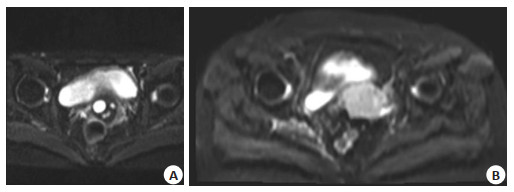
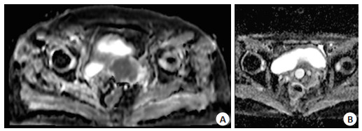
Abstract:
ObjectiveTo explore the value of 1.5 T MRI signs in determining the benign and malignant tumor of female pelvic cystic solid mass lesions. MethodsA total of 110 pelvic space-occupying lesions female patients admitted to our hospital for 1.5 T MRI examination from August 2017 to August 2018 were selected as the study objects. Their MRI imaging manifestations were analyzed, and the data of Fast ADC, Slow ADC, Standard ADC and Fraction of Fast ADC for benign and malignant tumors were recorded and analyzed. ResultsAmong 110 patients, 65 were benign pelviclesions and 45 were malignant tumor. The values of Standard ADC, Fast ADC and Slow ADC in the benign tumor group were significantly lower than those in the m alignant tumor group, and the differences were statistically significant(P < 0.05). Conclusion1.5 T MRI is of great significance for the determination of benign and malignant tumors and is worth promoting in clinical practice.

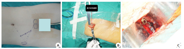
2020, 43(4): 692-696.
doi: 10.12122/j.issn.1674-4500.2020.04.31
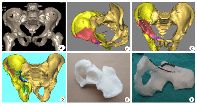
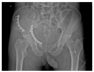
Abstract:
ObjectiveTo determine the effect of pararectus abdominis approach combined with 3D printing in the treatment of pelvic fracture. MethodsFifty patients with pelvic fracture undergoing operation performed by the researchers from September 2013 to June 2015 were selected as the study objects. In the control group, routine X-ray, CT plain scan and threedimensional reconstruction were performed before operation; In the observation group, virtual fracture reduction, internal fixation design, simulated internal fixation and steel plate pre- bending were used on the basis of X-ray, CT plain scan and three-dimensional reconstruction. Then we used the steel plate and screw which had been pre bent and implanted into the body during the simulated internal fixation under the minimally invasive pararectus abdominis approach. Both groups were treated with the incision beside the replicator. The average operation time, intraoperative blood loss and incision length were observed. In both groups, pelvic fractures were treated by a pararectus abdominis approach. The average operation time, intraoperative blood loss and incision length were observed. The therapeutic effect was evaluated by Matta and majeed scores, and the recovery of internal fixation and postoperative pelvic fracture was evaluated by imaging methods. ResultsCompared with the group of pararectus abdominis approach, the length of incision was significantly shortened, the amount of bleeding was significantly reduced and the time of operation were significantly shortened (P < 0.05); The excellent rate of fracture reduction was 96% by the evaluation with Matta standard. The excellent rate of fracture reduction was 96% by the evaluation with Majeed standard. The postoperative image evaluation of internal fixation is basically the same as that of preoperative simulated internal fixation. ConclusionCompared with the traditional operation, the pararectus abdominis approach combined with 3D printing technology in the treatment of pelvic fracture has the advantages of small trauma, small incision, low blood loss, light postoperative pain, fast recovery and high satisfaction of fracture reduction. It can make the patients return to work as soon as possible, improve the rehabilitation effect, reduce the medical cost and save the blood source for the society, which is worth popularizing widely.
2020, 43(4): 697-700.
doi: 10.12122/j.issn.1674-4500.2020.04.32
Abstract:
ObjectiveTo explore the application value of multi-slice spiral CT (MSCT) and MRI in the differential diagnosis of thyroid nodules. Methods94 thyroid nodules patients (94 nodules) admitted to the hospital from March 2018 to October 2019 and obtained pathological diagnosis results after surgery were enrolled as the research objects. All patients underwent MSCT and MRI examination. Taking histopathological examination as the golden standard, the diagnosis results of the two examination methods were compared. The sensitivity, specificity, accuracy, positive predictive value, negative predictive value and kappa value of the two examination methods were calculated. ResultsIn 94 cases of thyroid nodules, 73 cases (77.66%) were diagnosed as benign nodules, 21 cases (22.34%) as malignant nodules. There were no significant differences in sensitivity, specificity, accuracy, positive predictive value and negative predictive value between MSCT and MRI for diagnosis of benign and malignant thyroid nodules (P>0.05). However, the kappa value of MSCT diagnosis and pathological diagnosis was 0.746, which was higher than that of MRI diagnosis and pathological diagnosis (0.737). ConclusionThe sensitivity, specificity and accuracy of both MSCT and MRI are high in the diagnosis of benign and malignant thyroid nodules. They are highly consistent with the pathological diagnosis results and have high clinical application value. However, MSCT is more advantageous in diagnosis of malignant thyroid nodules calcification.
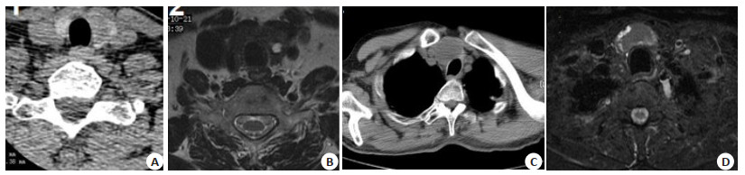
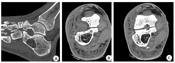
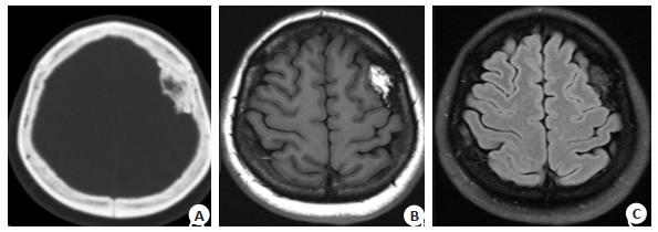
2020, 43(4): 701-704.
doi: 10.12122/j.issn.1674-4500.2020.04.33
Abstract:
ObjectiveTo investigate the CT and MRI manifestations findings of bone Intraosseous lipoma and angiolipoma and review of literature to improve the understanding of the disease. MethodsThe clinical and imaging data of 9 cases of Intraosseous lipoma and angiolipoma confirmed by surgical and pathology were collected, 8 cases received CT scan before operation, and 3 case received MRI examination. ResultsAmong the 9 cases, 6 cases were osteolipoma and 3 cases were bone angiolipoma. 6 cases of bone lipoma occurred in 4 cases in the intraosseous lipoma and 2 cases parosteal lipoma. Anong the 6 case osteolipoma, occurred in 1 case of calcaneus, 1 case of talus, 1 case of orbital bone, and 1 case of humerus. Among the 2 cases parosteal lipoma, there was 1 case of cranial and 1 case of femur. Angiolipoma of the bone occurred in 1 case of ribs, 1 case of skull, and 1 case of vertebrae.CT of the intraosseous lipoma showed mild swelling changes in the bone mass, mild sclerosis rings at the edges, fat density and bar- shaped, nodular calcifications in the lesions, MRI showed that the lesions showed high signal intensity on T1WI, PDWI and T2WI suppression Lipid sequence showed low signal. The CT scan of the parosteal lipoma showed uneven high- density masses. the lesions showed high signal intensity on T1WI, and the lipid suppression sequence on PDWI and T2WI showed low signal intensity. 3 cases of Intraosseous angiolipoma showed significant expansive changes. CT showed multiple crest and large bone trabecular at the edges and inside of bone destruction. ConclusionThe CT and MRI featrues of intraosseous lipomas and angiolipomas are mainly characterized by fat components, CT of parosteal lipomas are characterized by ossifying masses. Intraosseous angiolipoma has a more pronounced change in bone swelling than intraosseous lipoma. The edges and center of the lesion were characterized by multiple bone crests and large bone trabecular.
2020, 43(4): 705-708.
doi: 10.12122/j.issn.1674-4500.2020.04.34
Abstract:
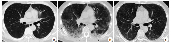
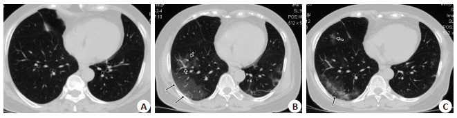
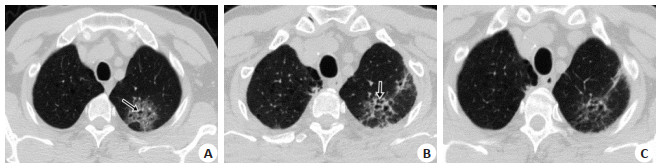
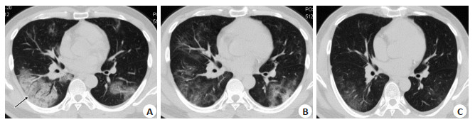
ObjectiveTo investigate the absorptive characteristics and the CT imaging features of COVID-19 in different stages and the changes in lung lesions with positive for nucleic acid test after discharge. MethodsThe CT imaging and clinical manifestations of 54 patients with COVID-19 were restrospectively analyzed. Through more than 3 times of chest CT examination to dynamically observe the changes of lesions, the changes of chest CT in patients with positive of nucleic acid test after discharge were compared with the last chest CT examination during hospitalization. Results42 patients (78%) come from Wuhan or Hubei to Huizhou in recent 14 days and 22% of the cases had no history of going to Wuhan or Hubei Province. Among them, 51 common type case (94%), 3 critical type case (6%) were divided by clinical symptoms and CT findings. Single lesion was located in the single pulmonary lobe in 2 cases; multiple lesions were located in the single pulmonary lobe in 3 cases; multiple lesions were located in the multiple pulmonary lobe in 49 cases; most lesions were located on the subpleural area in 46 cases; 6 cases were extended along the bronchovascular bundle; 2 cases were distributed along the pulmonary lobe. Ground glass opacities (GGO) were found in 54 patients; Consolidation was found in 28 cases; Interlobular septal thickening was appeared in GGO in 34 cases; Halo sign with GGO was found in 23 cases; Air sacs was occurred in the lesions of GGO or consolidation in 3 cases, and honeycomb sign was appeared in 1 case. Vascular bundle thickening in 14 cases, Bronchial thickening or dilatation in 18 cases, bilateral small amount of pleural effusion in 1 case. In early stage, patchy GGO was found on the subpleural area in 54 patients. In advanced stage, the number, density and range of lesions were more, denser and larger than those in the early stage. Segment or lobe consolidation in the background of diffuse or multiple ground glass opacities with double lungs in critical stage. In absorption stage, the lesions were significantly smaller and lighter in density, most of which disappeared, and a small amount of fibrosis were formed. The lesions in 47 cases began to be absorbed within 7-14 days, and the lesions in 7 cases began to be absorbed after 14 days. Positive for nucleic acid test occurred in 9 patients during 7-15 days after discharge, and 2 patients were occurred after 15 days, but the lesions were reduced compared with the previous CT (before discharge), and no new lesion was found. When 46 patients met the clinical discharge criteria, multiple ground glass lesions were still found on chest CT before discharge. ConclusionThe imaging manifestations of COVID-19 are different in different periods, and its evolution process has certain characteristics and rules. It is of great clinical value to correctly understand evolution process of COVID-19 for the early diagnosis, screening and efficacy evaluation.
2020, 43(4): 709-712.
doi: 10.12122/j.issn.1674-4500.2020.04.35
Abstract:
The lesion size, pathological type, stage, tissue differentiation and lymph node metastasis of cervical cancer can affect the prognosis of patients. Imaging examination can evaluate the pathological characteristics of cervical cancer and have great clinical value in guiding treatment and monitoring prognosis. DWI sequence of magnetic resonance can reflect the movement of water molecules, which is better than conventional sequence of magnetic resonance for the detection and examination of cervical cancer lesions, which is no harm to human body. ADC value is a quantitative indicator of diffusionweighted imaging. And on this basis, the histogram of ADC values obtained from high b value can describe the overall heterogeneity of the tumor at the cellular and molecular levels in the body. According to literatures, multiple parameters of ADC value histogram can better evaluate the pathological characteristics (pathological type, tissue differentiation degree, and stage) of cervical cancer, and which is also can predict its lymph node metastasis, therapeutic effect, effect of concurrent chemoradiotherapy, and recurrence. And it has clinical evaluation significance for other tumors in the body. This article reviews the selection of b value for diffusion-weighted imaging, the main parameters of histogram of ADC value, and the pathological type, stage, differentiation degree, lymph node metastasis and prognosis of cervical cancer.
The lesion size, pathological type, stage, tissue differentiation and lymph node metastasis of cervical cancer can affect the prognosis of patients. Imaging examination can evaluate the pathological characteristics of cervical cancer and have great clinical value in guiding treatment and monitoring prognosis. DWI sequence of magnetic resonance can reflect the movement of water molecules, which is better than conventional sequence of magnetic resonance for the detection and examination of cervical cancer lesions, which is no harm to human body. ADC value is a quantitative indicator of diffusionweighted imaging. And on this basis, the histogram of ADC values obtained from high b value can describe the overall heterogeneity of the tumor at the cellular and molecular levels in the body. According to literatures, multiple parameters of ADC value histogram can better evaluate the pathological characteristics (pathological type, tissue differentiation degree, and stage) of cervical cancer, and which is also can predict its lymph node metastasis, therapeutic effect, effect of concurrent chemoradiotherapy, and recurrence. And it has clinical evaluation significance for other tumors in the body. This article reviews the selection of b value for diffusion-weighted imaging, the main parameters of histogram of ADC value, and the pathological type, stage, differentiation degree, lymph node metastasis and prognosis of cervical cancer.
2020, 43(4): 713-716.
doi: 10.12122/j.issn.1674-4500.2020.04.36
Abstract:
ObjectiveTo investigate the application value of endoscopic ultrasonography in the depth of invasion and treatment of early gastric cancer. MethodsEighty patients with early gastric cancer who underwent endoscopic ultrasonography in our hospital from January 2019 to August 2020 were selected as the observation group, and 60 patients with early gastric cancer who did not receive endoscopic ultrasonography were selected as control group. The results of endoscopic ultrasonography and pathology were compared, the value of endoscopic ultrasonography in the diagnosis of tis/T1a and T1b were analyzed. And the difference of curative incision were compared between the observation group and the control group. ResultsThe ratio of tis/T1a and T1b in the observation group was 86.23% and 13.75%, respectively. Comparing with the control group, the difference was not statistically significant (P>0.05). The Kappa value of endoscopic ultrasonography consistent with the pathologic results was 0.634 (P < 0.05); The diagnostic accuracy of endoscopic ultrasonography was 90.00%, among which 2 cases of T1b were misdiagnosed as Tis/T1a and 6 cases of Tis/T1a were misdiagnosed as T1b. The sensitivity, specificity, positive predictive value and negative predictive value of endoscopic ultrasonography in Tis/T1a diagnosis were 91.30%, 81.82%, 96.92% and 60.00%, respectively. The sensitivity, specificity, positive predictive value and negative predictive value of endoscopic ultrasonography in T1b diagnosis were 81.82%, 91.30%, 60.00% and 96.92%, respectively. The curative resection ratio in the observation group was significantly higher than that in the control group (P < 0.05). ConclusionEndoscopic ultrasonography is of great value in the diagnosis of invasion depth of early gastric cancer, which is helpful to guide doctors to understand the scope of resection and improve the therapeutic effect.
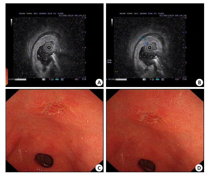
2020, 43(4): 717-720.
doi: 10.12122/j.issn.1674-4500.2020.04.37
Abstract:
ObjectiveTo explore the relationship between vaginal ultrasound measurement of ET, RI and β-HCG and clinical symptoms of patients with EP and its application value in the differential diagnosis of EP. MethodsSixty patients with EP who were admitted between January 2017 and March 2019 were selected as the EP group, 50 pregnant women with artificial abortion and 50 pregnant women with spontaneous abortion were selected as the artificial abortion group and spontaneous abortion group; vaginal ultrasound was used to measure ET and RI Level, and detect the β-HCG level in patients' blood; ROC curve was used to analyze the application value of combined detection of ET, RI and β-HCG in differential diagnosis of EP. Logistic multiple regression model was used to analyze the relationship between each index and clinical symptoms of EP patients. ResultsThere were significant differences in the levels of ET, RI and β-HCG in the EP group, spontaneous abortion group and artificial abortion group, and the differences were statistically significant (P < 0.05). Among the patients in the EP group, ET and β-HCG levels were the lowest, and RI was the highest. ET, RI and β-HCG are independent factors influencing the clinical symptoms of EP patients, which are statistically significant (P < 0.05). The sensitivity, specificity, and AUC of differential diagnosis in EP combined with ET, RI, and β-HCG are obviously higher than each index alone, and the difference was statistically significant (P < 0.05). ConclusionVaginal ultrasound measurement of ET, RI, and blood β-HCG levels are independent factors affecting the clinical symptoms of EP patients, and the combined application of each index can effectively increase the value of clinical application of single application.
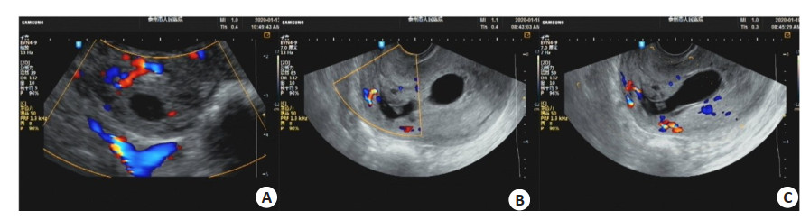
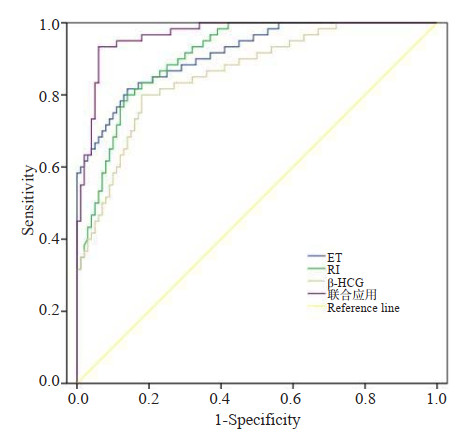
2020, 43(4): 721-725.
doi: 10.12122/j.issn.1674-4500.2020.04.38
Abstract:
ObjectiveTo retrospectively analyze the value of magnetic resonance dynamic contrast enhancement (DCE-MRI) and diffusion-weighted imaging (DWI) in predicting sentinel lymph node (SLN) metastasis in breast cancer. MethodsA total of 216 breast cancer patients in our hospital were collected, including 39 cases with SLN metastasis and 177 cases without SLN metastasis. The difference of image features and clinicopathological features were compared between metastatic and nonmetastatic groups. The risk factors of SLN metastasis were analyzed by multiple logistic regression. And by receiver operating characteristic (ROC), the predictive power of imaging and clinicopathological features were compared on SLN. ResultsThe imaging and clinicopathological characteristics of the two groups were statistically significant. The age, lesion size, internal enhancement method, and the ratio of peritumoral/tumor apparent diffusion coefficient (apparent diffusion coefficient, ADC) were statistically significant (P=0.003, 0.001, 0.020, 0.001). Logistic regression analysis showed that tumor size, edge enhancement and uneven enhancement, peritumoral/tumor ADC value ratio were independently related to SLN metastasis (OR= 1.15, P < 0.001; OR=15.54, P=0.007; OR=5.24, P=0.006; OR=70.62, P < 0.001); age, CD31 and SLN metastasis were independently related in the clinicopathological data (OR=0.96, P=0.012; OR=2.90, P=0.049). The predictive power of MRI features was significantly higher than clinicopathological variables (P=0.048). ConclusionThe preoperative DCE-MRI lesion size, internal enhancement pattern, and peritumoral/tumor ADC ratio may help predict SLN metastasis in breast cancer patients.
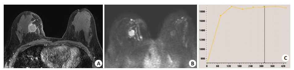
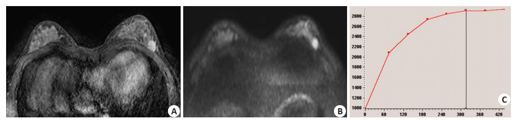
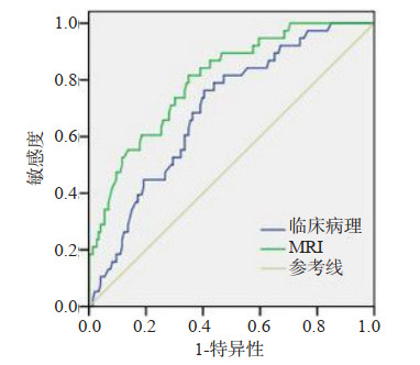
2020, 43(4): 726-730.
doi: 10.12122/j.issn.1674-4500.2020.04.39
Abstract:
ObjectiveTo explore the correlation between dietary inflammatory index (DII) and enhancement characteristics of carotid plaque by contrast-enhanced ultrasound. MethodsFrom March 2015 to October 2019, 77 patients with carotid plaque reexamined in our hospital were selected as the study objects. Food frequency questionnaire was used to collect the data of patients' daily diet, The DiI index of dietary components/nutrients of food was calculated by dietary nutrition software. According to the DiI index of patients, the positive DiI index (35 cases) was included in the anti-inflammatory diet group, and the negative DiI index (42 cases) was included in the anti-inflammatory diet group. According to the DiI index of patients, 5 grades were divided, That is ≤-1.05, -1.05~-0.33, -0.33~0.38, 0.38~ < 1.22 and ≥1.22. Carotid plaque was detected by color Doppler ultrasound. The plaque thickness, echo type and the distribution of plaque contrast agent were analyzed. Kendall correlation coefficient method was used to test the bivariate correlation analysis. ResultsThere were 41 plaques in 35 patients in the pro-inflammatory diet group and 47 plaques in 42 patients in the anti-inflammatory diet group. Inflammation promoting diet group of plaque thickness relatively anti-inflammatory diet group of thickening. The difference was statistically significant (P < 0.05). Inflammation promoting diet group contrast enhanced ultrasound enhance many to antiinflammatory diet group. The difference was statistically significant (P < 0.05). There was a significant positive correlation between DiI index and carotid plaque intima enhancement (Kendall correlation coefficient 0.659, P < 0.001). ConclusionsCarotid plaques are quite thick in patients with pro-inflammatory diet. The higher the DiI index, the more obvious the enhancement characteristics of carotid plaque.
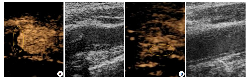
2020, 43(4): 731-734.
doi: 10.12122/j.issn.1674-4500.2020.04.40
Abstract:
ObjectiveTo explore the clinical diagnostic value of HR-VW-MRI in evaluating cerebrovascular stenosis compared with DSA. MethodsA total of 25 patients with recent ischemic encephalopathy admitted to our hospital from January 2016 to December 2019 were collected and confirmed with CTA and MRA examinations. Within one week, DSA examination and HRVW-MRI were performed to measure and calculate the stenosis rates of DSA and HR-VW-MRI in the stenosis segment. Highresolution magnetic resonance vascular wall reconstruction and T1WI enhancement were performed on the vessels in the responsible area. Linear regression analysis and Pearson correlation analysis were used to evaluate the correlation between cerebral vascular stenosis rate measured by HR-VW-MRI and DSA stenosis rate, and to explore the causes of cerebral artery stenosis. ResultsA total of 20 of the 25 patients completed HR-VW-MRI and DSA data measurement. The mean stenosis rate of HR-VW-MRI was 62.66±13.79, and that of DSA was 63.78±13.10. According to Pearson correlation analysis (P < 0.05), there was positive correlation between the two groups. The linear regression analysis showed that the r-squared value was 0.880, and the P < 0.05, and the correlation between the two groups was good. HR-VW-MRI found the responsible plaque of cerebral artery stenosis. ConclusionHR-VW-MRI has a good correlation with DSA in the measurement of cerebrovascular stenosis. Because of its non-invasive and good resolution of tissue structure, it can better display the structure of cerebrovascular wall and the size of vascular cavity from a three-dimensional Angle, analyze the etiology and make diagnosis, which has certain advantages in replacing DSA.
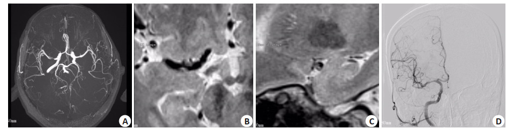
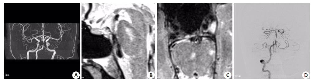
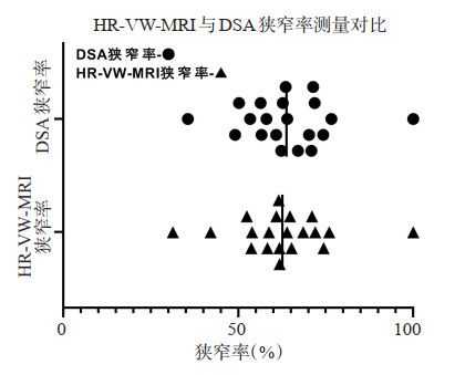
2020, 43(4): 735-739.
doi: 10.12122/j.issn.1674-4500.2020.04.41
Abstract:
ObjectiveTo observe the correlation between the thickness of the fetal heart wall in the third trimester and the control status of HbA1c levels in pregnant women with diabetes in gestation and discuss its clinical significance. MethodsA total of 122 single pregnant women suffering from gestational diabetes and their fetuses in our hospital from January 2015 to June 2019 were randomly selected as the research objects. The maternal HbA1c information in the first, second and third trimester of pregnancy and the fetal ventricular wall thickness information in the third trimester of pregnancy were collected by ultrasound detection. The correlation between fetal heart wall thickness and maternal HbA1c level control state were analyzed during the pregnancy. Use SPSS software to complete statistical analysis. ResultsThe HbA1c level of the subjects in the first trimester in the study was (5.80±1.60)%, the HbA1c% level in the second trimester was (5.91±1.61)%, and the third trimester was (5.97±1.86)%. There was no significant difference between the three periods (P>0.05). No statistically significant differences were found in the distribution characteristics of HbA1c control during different pregnancy periods. Among pregnant women with different HbA1c control levels in the first, second and third trimesters of pregnancy, the fetal ventricular wall thickness and interventricular septal thickness were measured by ultrasound during the third trimester. It can be seen that the fetuses with different HbA1c control levels in the first and second trimesters have late diastolic and contractile. There is no statistically significant difference in the thickness of left and right ventricles and interventricular septum. But the absolute figures show that the fetal ventricular wall and interventricular septal thickness of pregnant women with poorly controlled blood glucose levels are relatively higher. Thickness has a relatively large impact. The results of this study show that the mothers with poor control of HbA1c in the third trimester can see that the left ventricular wall thickness in the late fetal contraction is greater (P < 0.05); on the basis of the foregoing analysis, this study uses related analysis, HbA1c level control in the second and third trimesters of pregnancy and fetal ventricular wall thickness measured in the third trimester were correlated, and the results suggest that poorly controlled HbA1c levels in the third trimester and the late diastole measured in the third trimester fetus Ventricular thickness, left ventricular thickness in late systole, and late diastolic septum thickness were positively correlated (r=0.404, 0.361, 0.332, P < 0.05). ConclusionPoor blood glucose control in pregnant women suffering from gestational diabetes can cause thickening of the fetal ventricular wall and septum, which may adversely affect the fetal heart development. Therefore, it is suggested that the blood glucose level of pregnant women should be controlled in a good range as far as possible, especially in the third trimester of pregnancy, so as to reduce the heart damage of fetus and newborn.
2020, 43(4): 740-746.
doi: 10.12122/j.issn.1674-4500.2020.04.42
Abstract:
ObjectiveTo explore the correlation of m6A RNA methylation regulators and THCA. MethodsWe analyzed RNAseq data and related clinical information in the Cancer Genome Atlas (TCGA) database. The Wilcoxon test was used to analyze the different expressions of m6A RNA methylation-regulated genes between tumors and normal samples. A lasso Cox regression analysis was used to construct a risk prognosis model of m6A RNA methylation-regulated genes and THCA. The median risk score was used to divide THCA patients into high-risk group and low-risk group. Kaplan-Meier was performed for survival analysis. The ROC curve wa used to assess the sensitivity and feasibility of the prediction model. ResultsDifferent expression of m6A RNA methylation-regulated genes between tumors and normal samples showed 3 up-regulated genes and 14 down-regulated genes. Lasso Cox regression analysis screened four m6A RNA methylation regulating genes IGF2BP2, FTO, IGF2BP1 and RBM15. OS in the high-risk group was significantly lower than that in the low-risk group (P= 4.278e-02). The AUC value in the ROC curve was 0.783. The univariate and multivariate Cox regression were used to test the effectiveness of the model. The HR value in the univariate Cox regression analysis was 2.083, (P < 0.001). The HR value in the multivariate Cox regression analysis was 2.653 (P < 0.001). ConclusionThe m6A RNA methylation regulatory genes IGF2BP2, FTO, IGF2BP1 and RBM15 can construct predictive models to predict OS in patients with different clinical subgroups.