


CT imaging features and clinical analysis of superior mesenteric artery calcification in elderly patients
-
摘要:
目的探讨老年患者肠系膜上动脉(SMA)钙化性病变CT影像学特征,分析其临床特征。 方法回顾性分析2016年6月~ 2020年4月在我院进行SMA CT检查的186例患者,分析患者的一般临床资料,根据是否存在缺血性肠病(IBD)分为IBD组(n=26)与非IBD组(n=160),分析比较两组患者SMA钙化的检出率以及钙化斑块的部位、形态、数目及狭窄程度,探讨SMA钙化的CT影像学特征及与IBD的相关性。 结果IBD组SMA钙化斑块总数高于非IBD组(P < 0.05);IBD组SMA钙化斑块主要以SMA远段为主,其次是SMA近段,而非IBD组钙化斑块主要以SMA近段、SMA中段,位置分布情况差异有统计学意义(P < 0.05),IBD组SMA斑块的血管夹角低于非IBD组(P < 0.05);两组开口处狭窄程度比较差异有统计学意义(P < 0.05);IBD组患者SMA斑块钙化程度与其开口狭窄程度呈正相关(P < 0.05)。 结论老年患者SMA钙化性病变多以远段为主,且患者若同时存在开口处狭窄程度≥25%,提示可能存在IBD。 -
关键词:
- 肠系膜上动脉钙化性病变 /
- CT影像学特征 /
- 临床分析
Abstract:ObjectiveTo explore CT imaging features of superior mesenteric artery (SMA) calcification in elderly patients, and further analyze its clinical features. MethodsA total of 186 patients who had completed SMA CT examination in the hospital between June 2016 and April 2020 were selected as the research subjects, and their general clinical data were retrospectively analyzed. According to the presence or absence of ischemic bowel disease (IBD), the patients were divided into IBD group (n=26) and non-IBD group (n=160). The detection rate of SMA calcification, location, morphology, number and stenosis of calcified plaques were compared between the two groups. CT imaging features of SMA calcification and its correlation with IBD were discussed. ResultsThe number of SMA calcified plaques in IBD group was significantly larger than that in non-IBD group (P < 0.05). SMA calcified plaques in IBD group were mainly located in the distal segment of SMA, followed by the proximal segment, while those in non-IBD group were mainly located in proximal and middle segments of SMA. Differences in location distribution were significant (P < 0.05). Vascular angles of SMA plaques in IBD group were significantly smaller than those in non-IBD group (P < 0.05), and difference between the two groups in opening stenosis was also statistically significant (P < 0.05). The degree of SMA plaque calcification was positively correlated with the degree of opening stenosis in IBD group (P < 0.05). ConclusionSMA calcified lesions in elderly patients are mostly located in distal segment, and opening stenosis ≥25% may indicate the presence of IBD. -

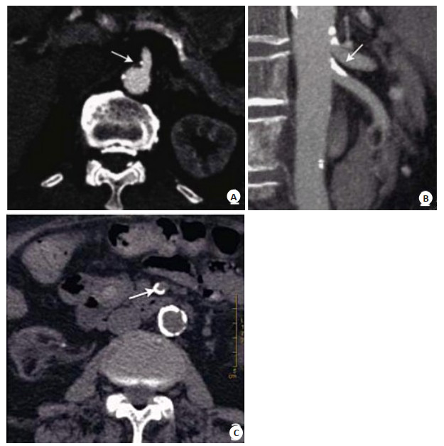
图 1 SMA钙化斑块形态CT表现
A:女性患者, 68岁, CT检查发现SMA近段处存在点状钙化斑块; B:男性患者, 62岁, CT检查发现SMA近段存在条状钙化斑块; C:女性患者, 65岁, CT检查发现SMA中段存在环状钙化斑块.
Figure 1. CT findings of the shape of SMA calcified plaque.
表 1 两组一般资料比较
Table 1. Comparison of general information of two groups (n)
组别 年龄(岁, Mean±SD) 性别(男/女) 冠心病 高血压 高血脂症 IBD组(n=26) 75.41±5.29 14/12 10 15 8 非IBD组(n=160) 75.49±6.22 92/68 61 90 50 t/χ2 0.062 0.122 0.001 0.019 0.002 P 0.951 0.727 0.974 0.891 0.961  下载: 导出CSV
表 2 两组CA钙化斑块及SMA钙化斑块形态比较
Table 2. Comparison of shape between CA calcified plaque and SMA calcified plaque of two groups (n)
组别 CA钙化斑块 SMA钙化斑块 总数[n (%)] 条形 环状 总数[n (%)] 条形 环状 点状 IBD组(n=26) 22 (84.62) 14 8 16 (61.54) 5 8 3 非IBD组(n=160) 112 (70.00) 72 40 40 (25.00) 24 8 8 χ2 2.372 14.189 P 0.124 0.000
下载: 导出CSV
表 3 SMA钙化斑块的狭窄情况及斑块部位比较
Table 3. Comparison of the stenosis and position of the SMA calcified plaque[n (%)]
组别 部位 血管夹角(°, Mean±SD) SMA近段 SMA中段 SMA远段 SMA末段 IBD组(n=26) 8 (30.77) 2 (7.69) 16 (61.54) 0 (0) 52.69±5.33 非IBD组(n=160) 78 (48.75) 72 (45.00) 10 (6.25) 0 (0) 61.47±6.29 t/χ2 16.310 6.732 P 0.000 0.000
下载: 导出CSV
表 4 两组SMA开口处狭窄程度比较
Table 4. Comparison of stenosis degree of SMA opening of two groups (n)
组别 SMA开口狭窄程度 1级 2级 3级 4级 IBD组(n=26) 6 9 8 3 非IBD组(n=160) 135 19 6 0 Z 51.01 P 0.000
下载: 导出CSV
表 5 SMA斑块钙化程度与其开口狭窄程度的相关性分析
Table 5. Correlation analysis between calcification degree and stenosis of opening of the SMA plaque
SMA斑块钙化程度 SMA开口狭窄程度 1级 2级 3级 4级 1级 3 1 1 0 2级 1 8 1 0 3级 2 0 1 0 4级 0 0 5 3
下载: 导出CSV
-
[1] 陈娟, 刘玥, 赵丹丹, 等.颅内动脉钙化与腔隙性脑梗死患者影像学特征的关系[J].中国老年学杂志, 2016, 36(22): 5604-5. http://d.old.wanfangdata.com.cn/Periodical/zglnxzz201622049 [2] Yu YM, Guo ML, Han XY. Comparison of multi- slice computed tomographic angiography and dual- source computed tomographic angiography in resectability evaluation of pancreatic carcinoma[J]. Cell Biochem Biophys, 2014, 70(2): 1351-6. http://cn.bing.com/academic/profile?id=181e1e599ca84fe537fcaaa3154d9f2a&encoded=0&v=paper_preview&mkt=zh-cn [3] 夏立栋, 胡少波. 16排CT与DSA在肠系膜上动脉狭窄诊断中临床价值分析[J].医学影像学杂志, 2015, 25(9): 1693-5. http://d.old.wanfangdata.com.cn/Periodical/yxyxxzz201509057 [4] Verde F, Bleich KB, Oshmyansky A, et al. Isolated celiac and superior mesenteric artery dissection identified with MDCT: imaging findings and clinical course[J]. J Comput Assist Tomogr, 2012, 36(5): 539-45. http://cn.bing.com/academic/profile?id=9440945d1d850ccd5145e4f25a82126d&encoded=0&v=paper_preview&mkt=zh-cn [5] Liu DN, Lv A, Tian ZH, et al. Superior mesenterie artery margin in pancre aticoduodenee tomy for pansereatie adenoeane inoma[J]. Oncotanget, 2017, 8(5): 7766-76. [6] 李海飞, 衣高峰. 64层螺旋CT血管重建诊断肠系膜上动脉及相关病变[J].医学影像学杂志, 2016, 26(6): 1048-51. http://d.old.wanfangdata.com.cn/Periodical/yxyxxzz201606027 [7] 邹循锋, 顾腾辉, 顾建华, 等.老年患者肠系膜上动脉钙化性病变的CT影像学分析[J].天津医药, 2017, 45(10): 1049-52. http://d.old.wanfangdata.com.cn/Periodical/tjyy201710011 [8] Ohnuki T, Takahashi W, Ohnuki Y, et al. Response letter regarding the article"significance of the presence of metabolic syndrome in patients with asymptomatic arteriosclerosis affecting the aorta and the cerebral, Extra- cranial carotid and coronary arteries"[J]. Intern Med, 2013, 52(20): 2391-7. [9] Schaefer PJ, Pfarr J, Trentmann J, et al. Comparison of noninvasive imaging modalities for Stenosis grading in mesenteric arteries[J]. Rofo, 2013, 185(7): 628-34. http://cn.bing.com/academic/profile?id=8212996df905ea9a6b688758852fb40c&encoded=0&v=paper_preview&mkt=zh-cn [10] 吴晓东, 赵衡.多层螺旋CT肠系膜血管成像对肠系膜上动脉疾病的诊断[J].中南医学科学杂志, 2015, 43(2): 204-5, 209. http://d.old.wanfangdata.com.cn/Periodical/zgwsbzgl201607133 [11] 马跃虎, 吴刚, 刘浩, 等.多层螺旋CT血管成像在肠系膜上动脉缺血性疾病诊断中的应用[J].山东医药, 2014, 54(43): 48-50. http://d.old.wanfangdata.com.cn/Periodical/shandyy201443019 [12] Luan JY, Li X. Computed tomography imaging features and classification of isolated dissection of the superior mesenteric artery[J]. Eur J Vasc Endovasc Surg, 2013, 46(2): 232-5. http://cn.bing.com/academic/profile?id=33aee70c57da8d394d2ceaca587f2c39&encoded=0&v=paper_preview&mkt=zh-cn [13] 贾永, 赵斐, 高亮, 等.肠系膜上动脉急性闭塞性疾病的CT诊断效果分析[J].中国急救医学, 2017, 37(z2): 54-5. http://d.old.wanfangdata.com.cn/Periodical/zgjjyx2017z2045 [14] 廖冠义, 高青.经手术及血管影像学检查证实的66例急性肠系膜上动脉栓塞的临床分析[J].中华消化杂志, 2019, 39(2): 111-4. http://d.old.wanfangdata.com.cn/Periodical/zhxhzz201902007 [15] 齐瑞楠, 曾书娥, 查莉, 等.孤立性肠系膜上动脉夹层的影像学表现1例[J].中国超声医学杂志, 2017, 33(9): 849-57. http://d.old.wanfangdata.com.cn/Periodical/zgcsyxzz201709029 [16] Aimi M, Amano C, Yoshida R, et al. Isolated superior mesenteric artery dissection with small intestine ischemia[J]. Case Rep Gastroenterol, 2015, 9(3): 341-6. http://cn.bing.com/academic/profile?id=052cfefdc2300b12d7501fe36f08912a&encoded=0&v=paper_preview&mkt=zh-cn [17] 张辉, 于天池, 汪弢, 等. 50例孤立性肠系膜上动脉夹层患者CTA特征及治疗效果分析[J].山东医药, 2019, 59(23): 61-3. http://d.old.wanfangdata.com.cn/Periodical/shandyy201923016 [18] 陈佳莉, 步军, 吴禹, 等. CTA观察右半结肠缺血性结肠炎与肠系膜上动脉钙化斑块的关系[J].中国医学影像技术, 2019, 35(3): 395-9. http://d.old.wanfangdata.com.cn/Periodical/zgyxyxjs201903020 [19] 靳明旭, 张娣, 苏浩波, 等. MSCTA评估孤立性肠系膜上动脉夹层累及分支血管特征[J].实用放射学杂志, 2016, 32(8): 1308-11. http://d.old.wanfangdata.com.cn/Periodical/syfsxzz201608040 [20] Kim YW. Current understandings of spontaneous isolated superior mesenteric artery dissection[J]. Vasc Specialist Int, 2016, 32(2): 37-43. http://cn.bing.com/academic/profile?id=2cafca90d5e1c84c3cd96d7e62439495&encoded=0&v=paper_preview&mkt=zh-cn [21] Guo K, Ma QY, Li JH, et al. Interaction of the sympathetic nerve with pancreatic cancer cells promotes perineural invasion through the activation of STAT3 signaling[J]. Mol Cancer Ther, 2013, 12 (3): 264-73. http://cn.bing.com/academic/profile?id=6853247a7841b3f4d259de6c19aaa890&encoded=0&v=paper_preview&mkt=zh-cn [22] Hirono S, Kawai M, Okada KL, et al. Treatment strategy for borderline resectable pancreatie eancer with nadiographie artery involvement[J]. Panereas, 2016, 45(10): 1438-46. https://www.researchgate.net/publication/301533163_Treatment_Strategy_for_Borderline_Resectable_Pancreatic_Cancer_With_Radiographic_Artery_Involvement [23] 刘文徽, 边祥兵, 杨立, 等.急性肠系膜上动脉栓塞患者血管基础病变的CT影像分析[J].胃肠病学和肝病学杂志, 2015, 24(5): 586-9. http://d.old.wanfangdata.com.cn/Periodical/wcbxhgbxzz201505029 [24] 李好鹏, 叶贤旺, 孙勤学. 320排CT对自发性孤立性肠系膜上动脉夹层的诊断价值[J].医学影像学杂志, 2015, 25(1): 88-91. http://d.old.wanfangdata.com.cn/Periodical/yxyxxzz201501026 [25] 梁腾飞, 李保卫. 55例肠系膜血管病变患者64排128层螺旋CT血管造影表现分析[J].实用心脑肺血管病杂志, 2018, 26(4): 106-8. http://d.old.wanfangdata.com.cn/Periodical/syxnfxgbzz201804028 [26] 高云, 郑晓林, 杨沛钦, 等.肠系膜动脉供血障碍性肠缺血肠管可逆性CT表现探讨[J].影像诊断与介入放射学, 2016, 25(4): 311-5. http://d.old.wanfangdata.com.cn/Periodical/yxzdyjrfsx201604010 [27] 宁武, 单前进, 祁小江, 等.多层螺旋CT血管成像对肠系膜上动脉的影像学结构研究[J].中国临床研究, 2014, 27(1): 87-8. http://d.old.wanfangdata.com.cn/Periodical/zgckyx201401042 -

 点击查看大图
计量
- 文章访问数: 863
- HTML全文浏览量: 310
- PDF下载量: 5
- 被引次数: 0