


Diagnostic value of magnetic resonance imaging in patients with IgG4-related ophthalmic disease
-
摘要:
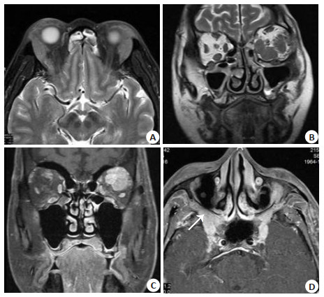
目的分析IgG4相关性疾病眼眶受累(IgG4-ROD)的磁共振表现,探讨其诊断价值。 方法回顾性分析18例IgG4-ROD患者MR图像,分析MR图像病变部位、双侧还是单侧、形状、边缘、眶内结构、T1WI及T2WI信号特点及强化方式。 结果18例IgG4-ROD病例全部经手术病理检查证实,根据MR表现分为4类:泪腺型、眼外肌型、肿块型、弥漫型。6例(33.3%)表现为泪腺增大,其中4例双侧对称增大,2例单侧增大;7例(38.9%)表现为肌锥内外软组织肿块,其中2单发,5例多发;1例(5.56%)眼外肌增粗;4例(25%)表现为沿邻近颅底孔道、生理腔隙及三叉神经走行区弥漫病变。18例中8例可见看到眶下神经增粗。14例行动态增强检查,时间-信号曲线均表现为快速上升缓慢流出的Ⅱ型曲线。 结论IgG4-ROD会累及泪腺、眼外肌、内眦、肌锥外间隙等,眶下神经增粗和沿三叉神经分支浸润性生长是该病特征性表现,认识了解IgG4-ROD典型影像学表现,对诊断提高诊断率有帮助。 Abstract:ObjectiveTo evaluate MRI findings of IgG4-related ophthalmic disease. MethodsWe included 18 patients with histopathologically proven IgG4-ROD. MR images were retrospectively evaluated for location, laterality, shape, margin, T1 and T2 signal intensity on precontrast MRI, internal architecture, ocular adnexal lesion enhancement patterns. ResultsThe lesions involved the lacrimal gland (n=6), focal mass (n=7), extraocular muscles (n=1) and multiple areas (n=4). All lacrimal gland lesions presented as diffuse enlargements. 4 patients had lesions involving multiple areas which extended along the trigeminal nerve, accompanied by expansion of neural foramina along their courses, with no signs of bone destruction. Infraorbital nerve enlargement was present in 8 cases. All ocular adnexal lesions showed isointensity on T1- and hypointensity on T2-weighted images, homogenous enhancement patterns and bone remodelling without destruction. TIC exhibited a rapidly enhancing and slow washout pattern in 14 patients. ConclusionIgG4-ROD can involve the lacrimal gland, extraocular muscles, medial canthus, extraconal space, and infraorbital nerves, and pterygopalatine fossa. Recognition of the typical radiological features of IgG4-ROD may help in the diagnosis of this benign clinical entity. -
Key words:
- magnetic resonance imaging /
- IgG4-related ophthalmic disease /
- orbit
-
[1] Johnston J, Allen JE. IgG4-related disease in the head and neck[J]. Curr Opin Otolaryngol Head Neck Surg, 2018, 26(6): 403-8. doi: 10.1097/MOO.0000000000000487 [2] Flores Balverdi J, Baenas DF, Riscanevo NC, et al. IgG4-related orbital disease[J]. Arch Soc Esp Oftalmol, 2018, 93(10): 494-6. doi: 10.1016/j.oftal.2018.04.003 [3] Chidambaram VA, Anita CSY, Robert C, et al. IgG4 related orbit disease-An unusual cause of an orbital mass[J]. Med J Malaysia, 2018, 73(6): 415-7. http://www.ncbi.nlm.nih.gov/pubmed/30647218 [4] Goto H, Takahira M, Takahira M, et al. Diagnostic criteria for IgG4-related ophthalmic disease[J]. Jpn J Ophthalmol, 2015, 59 (1): 1-7. doi: 10.1007/s10384-014-0352-2 [5] Ferry JA, Klepeis V, Sohani AR, et al. IgG4-related orbital disease and its mimics in a western population[J]. Am J Surg Pathol, 2015, 39(12): 1688-700. doi: 10.1097/PAS.0000000000000497 [6] Wang L, Lu F. Clinical analysis of IgG4-related disease[J]. J Central South Univ (Med Ed), 2019, 44(10): 1151-6. http://d.old.wanfangdata.com.cn/Periodical/zhyx201842014 [7] Sogabe Y, Ohshima K, Azumi A, et al. Location and frequency of lesions in patients with IgG4-related ophthalmic diseases[J]. Albrecht Von Graefes Arch Fur Klinische Und Exp Ophthalmol, 2014, 252(3): 531-8. doi: 10.1007/s00417-013-2548-4 [8] Hiwatashi A, Togao O, Yamashita K, et al. High resolution diffusion-weighted imaging for solitary orbital tumors: 3D Turbo field Echo with diffusion-sensitized driven-equilibrium (DSDE-TFE) preparation technique[J]. Clin Neuroradiol, 2018, 28(2): 261-6. doi: 10.1007/s00062-016-0556-6 [9] Song YS, Choung HK, Park SW, et al. Ocular adnexal IgG4-related disease: CT and MRI findings[J]. Br J Ophthalmol, 2013, 97(4): 412-8. http://d.old.wanfangdata.com.cn/NSTLQK/NSTL_QKJJ0229295188/ [10] Chen LY, Mattman A, Seidman MA, et al. IgG4-related disease: what a hematologist need to know[J]. Haematologica, 2019, 104 (3): 444-55. [11] Tiegs-Heiden CA, Eckel LJ, Hunt CH, et al. Immunoglobulin G4-related disease of the orbit: imaging features in 27 patients[J]. AJNR Am J Neuroradiol, 2014, 35(7): 1393-7. doi: 10.3174/ajnr.A3865 [12] Umehara H, Okazaki K, Kawano M, et al. How to diagnose IgG4-related disease[J]Ann Rheum Dis, 2017, 76(11): e46-52. doi: 10.1136/annrheumdis-2017-211330 [13] Mulay K, Aggarwal E, Jariwala M, et al. Orbital immunoglobulin-G4-related disease: case series and literature review[J]. Clin Experiment Ophthalmol, 2014, 42(7): 682-7. doi: 10.1111/ceo.12284 [14] Lecler A, Zmuda M, Deschamps R. Infraorbital nerve involvement on magnetic resonance imaging in Igg4-related ophthalmic disease: a highly suggestive sign[J]. Ophthalmology, 2018, 125(4): 577-85. http://www.sciencedirect.com/science/article/pii/S0161642017337168 [15] Soussan JB, Deschamps R, Sadik JC, et al. Infraorbital nerve involvement on magnetic resonance imaging in European patients with IgG4-related ophthalmic disease: a specific sign[J]. Eur Radiol, 2017, 27(4): 1335-43. http://cn.bing.com/academic/profile?id=2aa1853bc2458e29936bf25d848cc2bf&encoded=0&v=paper_preview&mkt=zh-cn [16] Detiger SE, Karim AF, Verdijk RM, et al. The treatment outment in IgG4-related orbital disease[J] Acta Ophthalmol, 2019, 97(5): 451-9. doi: 10.1111/aos.14048 [17] Rammeh S, Ajouli W, Landolsi A, et al. Unilateral orbital mass as an unusual presentation of IgG4-related disease[J]. Neurochirurgie, 2016, 62(1): 64-6. http://www.sciencedirect.com/science/article/pii/S0028377015002751 [18] Andron A, Hostovsky A, Nair AG, et al. The impact of IgG-4-ROD on the diagnosis of orbital tumors: a retrospective analysis[J]. Orbit, 2017, 36(6): 359-64. doi: 10.1080/01676830.2017.1337192 [19] Ferreira TA, Saraiva P, Genders SW, et al. CT and MR imaging of orbital inflammation[J]. Neuroradiology, 2018, 60(12): 1253-66. doi: 10.1007/s00234-018-2103-4 [20] Andrew NH, Sladden N, Kearney DJ, et al. An analysis of IgG4-related disease (IgG4-RD) among idiopathic orbital inflammations and benign lymphoid hyperplasias using two consensus-based diagnostic criteria for IgG4-RD[J]. Br J Ophthalmol, 2015, 99(3): 376-81. http://bjo.bmj.com/cgi/content/short/bjophthalmol-2014-305545v1?rss=1 [21] Tooley AA, Salomao DR, Bradley EA, et al. Distinguishing IgG4-related ophthalmic disease from Graves orbitopathy[J]. Ophthalmic Plast Reconstr Surg, 2019, 35(2): 170-6. doi: 10.1097/IOP.0000000000001201 -

 下载:
下载:
 点击查看大图
图(1)
计量
- 文章访问数: 705
- HTML全文浏览量: 368
- PDF下载量: 3
- 被引次数: 0