


Typical and atypical HRCT manifestations of COVID-19
-
摘要:
目的分析COVID-19合并早期心脏损害的临床表现及心脏损害早期识别的指标。 方法收集2020年1月20日~2月20日我院收治的12例COVID-19患者的临床表现、实验室检查、心脏彩超等资料,心脏彩超主要收集左心室射血分数(LVEF)、左室总体纵向应变(GLS)。分析COVID-19早期心脏损害的发生情况,比较窦性心动过速、心电图新发改变、GLS作为COVID-19合并早期心脏损害表现的可能识别指标,并进一步分析COVID-19早期心脏损害的相关危险因素。 结果12例COVID-19患者包括2例重型、8例普通型和2例轻型,所有患者肌钙蛋白Ⅰ、LVEF均无明显异常。但窦性心动过速、心电图新发改变及GLS的异常率均为33%,肌酸激酶同工酶与B型钠尿肽的异常率均为8%;COVID-19早期心脏损害可能指标GLS的绝对值与年龄、中性粒细胞比值、C-反应蛋白存在负相关(r=-0.795,-0.816,-0.917,P < 0.05),与血钾存在正相关性(r=0.73,P < 0.01)。 结论窦性心动过速、心电图新发改变及GLS在COVID-19患者中存在一定的异常比例,可能作为早期心脏损害识别指标。在肌钙蛋白Ⅰ、LVEF未见异常的COVID-19,通过观察患者的心电图的是否存在窦速、新发异常改变、GLS的异常,有助于对COVID-19心脏损害的早期识别。 Abstract:ObjectiveTo analyze the clinical manifestations of new coronavirus pneumonia (COVID-19) combined with early heart damage, and explore indicators that can identify COVID-19 heart damage early. MethodsThe clinical manifestations, laboratory examinations, and cardiac color Doppler ultrasound of 12 COVID-19 patients admitted to our hospital from January 20, 2020 to February 20, 2020 were collected. Among the patients, left ventricular ejection fraction (LVEF) and general left ventricular longitudinal strain (GLS) were collected by cardiac color Doppler ultrasound. We used the data above to analyze the occurrence of COVID-19 early heart damage. The sinus tachycardia, new ECG changes and GLS were compared as possible identification indicator of COVID-19 combined early heart damage performance. The risk factor of COVID-19 early heart damage was analyzed. ResultsThe 12 COVID-19 patients included 2 severe cases, 8 normal cases, and 2 mild cases. All patients had no significant abnormalities in troponin Ⅰ and left ventricular ejection fraction. However, abnormal rates of sinus tachycardia, new changes in ECG and GLS were all 33.3%. The abnormal rates of CKMB and BNP were both 8.3%. The absolute value of GLS, the possible index of COVID-19 with early heart damage, were negatively correlated with age, neutrophil ratio, and C-reactive protein (r=-0.795, -0.816, -0.917, P < 0.05). They were positively correlated with hypokalemia (r= 0.73, P < 0.05). ConclusionsSinus tachycardia, new ECG changes and GLS have a certain abnormal proportion in COVID-19 patients, which may be used as indicators of early heart damage identification. In COVID-19 patients with normal aTNI and LVEF, observing whether they appear sinus tachycardia, new abnormal ECG changes, and abnormal GLS will help to identify COVID-19 with early heart damage. -
Key words:
- COVID-19 /
- early heart damage /
- global longitudinal strain /
- electrocardiogram
-

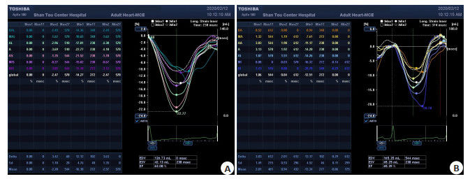
图 1 2例重症患者(病例5、6)的GLS图像
A:病例5, GLS明显减低,最低-11, 绝对值11; B:病例6, GLS明显减低,最低-8, 绝对值8;注: GLS正常值为≤-20(绝对值≥20)
Figure 1. GLS images of the two severe patients.

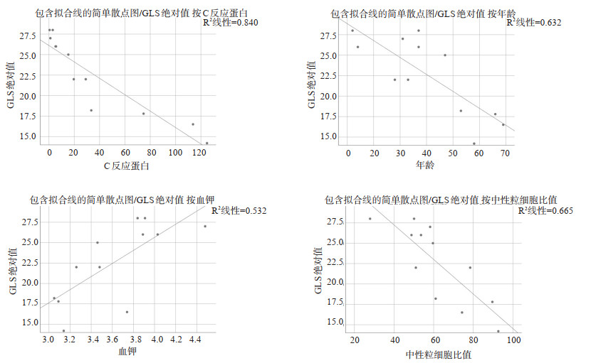
图 2 GLS与COVID-19患者年龄、中性粒细胞比值、C反应蛋白、血钾浓度的Spearman相关性分析
Figure 2. Significant linear correlation between GLS and age, neutrophil ratio, C-reactive protein, serum potassium.
表 1 12例COVID-19患者的实验室检验特征
Table 1. Laboratory test results of the COVID-19 patients
检验指标 参考区间 病例1 病例2 病例3 病例4 病例5 病例6 病例7 病例8 病例9 病例10 病例11 病例12 异常率(%) 血常规 白细胞计数(109/L) 3.5~9.5 11.4 12.6 6.3 4.8 7.4 16.2 3.7 4.5 5.7 6 5.5 4.1 25 中性粒细胞比值(%) 50~70 89.6 78.4 51 48.8 74.3 92.6 61 27.9 59.6 58.2 53.6 50.1 50 淋巴细胞比值(%) 20~40 8.3 15.1 36 38.4 17.1 1.3 24 60 27.5 30.3 27.4 39.2 33.3 单核细胞比值(%) 3~8 2 4.9 11.6 10.7 7.1 6 14.2 8.6 11.7 8.6 16.1 8.8 75 血小板计数(109/L) 125~350 310 246 367 254 268 133 117 239 273 288 247 188 16.7 血红蛋白浓度(g/L) 130~175 (男) 107 132 161 156 129 124 162 122 101 143 153 127 33.3 115~150 (女) 血生化(代谢、心、肝、肾功能指标) 血钾(mmol/L) 3.5~5.3 3.09 3.48 3.26 4.03 3.74 3.14 3.05 3.84 3.46 4.48 3.89 3.91 50 血钠(mmol/L) 137~147 135 131 138.7 139 131 136 136 138 132 139 139 137 50 葡萄糖(mmol/L) 3.89~6.11 5.04 10.78 5.01 4.43 6.15 9.51 5.16 4.69 5.13 4.93 5.34 5.13 25 尿素(mmol/L) 2.5~7.14 4.29 3.36 3.22 5.11 4.09 6.94 7.07 4.91 2.55 4.25 4.15 3.05 0 肌酐(μmol/L) 40~133 63.8 75.3 119.5 106.6 91.3 111.2 105.5 50.8 53.8 89.1 93.1 53.5 0 尿酸(μmol/L) 150~430 220 206 465 356 305 266 468 331 202 284 446 194 25 白蛋白(g/L) 40~55 33.7 34.6 42.8 46.2 29.4 36 34.3 36.7 39.4 43.6 39.6 35 75 甘油三酯(mmol/L) 0.23~1.71 1.05 0.95 2.43 2.22 1.7 1.07 2.58 1.3 1.37 0.97 3.3 2.15 41.7 总胆固醇(mmol/L) 2.9~5.72 5.2 3.61 4.23 4.55 5.1 4.43 4.05 5.65 4.86 4.67 4.34 4.44 0 总胆红素(μmol/L) 2~20 16.1 25.4 30.6 16.9 19.3 13.7 13 19 13.6 14.7 15.7 14.1 16.7 谷丙转氨酶(U/L) 9~50 (男) 22 10 34 12 19 21 19 16 12 23 31 16 0 7~40 (女) 谷草转氨酶(U/L) 15~40 (男) 32 14 23 18 21 33 17 28 23 22 21 15 0 13~35 (女) 肌酸激酶(U/L) 0~174 79 69 71 76 34 373 26 97 58 68 69 35 8.3 乳酸脱氢酶(U/L) 100~300 332 194 226 193 171 270 199 175 220 149 181 202 8.3 凝血功能 纤维蛋白原(g/L) 1.8~4.88 5.42 2.3 2.5 3.2 8.4 5.88 4.85 2.5 3.49 1.53 2.9 2.64 25 D-二聚体(μg/L) 0~550 310 150 164 220 1920 1470 290 153 2110 750 320 160 33.3 感染指标 C反应蛋白(mg/L) 0~8 74.7 29.06 19.6 5.2 113.76 124.78 33.43 < 0.499 15.3 1.14 5.61 2.99 83.3 降钙素原(ng/mL) 0~0.5 0.05 0.034 < 0.1 < 0.1 0.112 0.116 < 0.1 < 0.02 < 0.1 < 0.1 0.13 < 0.1 16.7  下载: 导出CSV
表 2 12例COVID-19心脏损伤评估指标
Table 2. Cardiac injury assessment indexes of the COVID-19 patients
心脏损伤
评估指标正常范围 病例1 病例2 病例3 病例4 病例5 病例6 病例7 病例8 病例9 病例10 病例11 病例12 异常率(%) 心率(次/min) 60~100 86 102 65 92 112 118 106 84 74 68 84 78 25.0 CKMB (U/L) 0~24 15 14 16 10 11 74 8 19 12 8 12 7 8.3 aTnI (ng/mL) 0~0.04 0.001 0 0 0 0 0.03 0 0 0 0 0 0 0 BNP (pg/mL) 0~300 220 60 120 30 205.3 556.2 280 20 125 224 60.7 < 18 8.3 心电图新
发改变- 早搏 无 无 无 早搏 早搏
ST段压低无 无 无 无 无 早搏 33.3 LVEF (%) ≥55 65 71 75 74 63 65 68 75 67 72 70 71 0 GLS ≤-20 -17.8 -22 -22 -26 -16.5 -14.2 -18.2 28 -25 -27 -26 -28 25 CKMB: Creatine Kinase Isoenzyme; BNP: B-type natriuretic peptide; LVEF: Left Ventricular Ejection Fraction; GLS: Global Longitudinal Strain.
下载: 导出CSV
-
[1] de Groot RJ, Baker SC, Baric R, et al. Coronaviridae.In: virus taxonomy: ninth report of the international committee on taxonomy of viruses [M].San Diego: Elsevier Academic Press, 2012. [2] Zumla A, Chan JF, Azhar EI, et al. Coronaviruses-drug discovery and therapeutic options[J]. Nat Rev Drug Discov, 2016, 15(5):327-47. http://www.wanfangdata.com.cn/details/detail.do?_type=perio&id=693220aecda5d4878ae44df69eb4ed7c [3] Wang C, Horby PW, Hayden FG, et al. A novel coronavirus outbreak of global health concern[J]. Lancet, 2020, 395(10223):470-3. http://d.old.wanfangdata.com.cn/Periodical/yrdyyzz-e202004007 [4] Zhu N, Zhang DY, Wang WL, et al. A novel coronavirus from patients with pneumonia in China, 2019[J]. N Engl J Med, 2020, 382(8): 727-33. http://www.researchgate.net/publication/338816497_A_Novel_Coronavirus_from_Patients_with_Pneumonia_in_China_2019 [5] Munster VJ, Koopmans M, van Doremalen N, et al. A novel coronavirus emerging in China -key questions for impact assessment [J]. N Engl J Med, 2020, 382(8): 692-4. http://www.researchgate.net/publication/338806807_A_Novel_Coronavirus_Emerging_in_China_-_Key_Questions_for_Impact_Assessment [6] Perlman S. Another decade, another coronavirus[J]. N Engl J Med, 2020, 382(8): 760-2. doi: 10.1056/NEJMe2001126 [7] Heymann DL, Shindo N, WHO Scientific and Technical Advisory Group for Infectious Hazards. COVID-19: what is next for public health?[J]. Lancet, 2020, 395(10224): 542-5. http://d.old.wanfangdata.com.cn/Periodical/dzkjdxxb202003007 [8] 国家卫生健康委办公厅.新型冠状病毒肺炎诊疗方案(试行第七版)[J].传染病信息, 2020, 33(1): 1-6, 26. http://d.old.wanfangdata.com.cn/Periodical/zhongguoyy202006002 [9] 赵双全, 周永生, 殷亮, 等. COVID-19的临床特征和CT表现[J].分子影像学杂志, 2020, 43(1): 7-11. doi: 10.12122/j.issn.1674-4500.2020.01.13 [10] Huang CL, Wang YM, Li XW, et al. Clinical features of patients infected with 2019 novel coronavirus in Wuhan, China[J]. Lancet, 2020, 395(10223): 497-506. https://www.sciencedirect.com/science/article/pii/S0140673620301835 [11] Chen NS, Zhou M, Dong X, et al. Epidemiological and clinical characteristics of 99 cases of 2019 novel coronavirus pneumonia in Wuhan, China: a descriptive study[J]. Lancet, 2020, 395(10223):507-13. http://www.sciencedirect.com/science/article/pii/S0140673620302117 [12] Xu XT, Chen P, Wang JF, et al. Evolution of the novel coronavirus from the ongoing Wuhan outbreak and modeling of its spike protein for risk of human transmission[J]. Sci China Life Sci, 2020, 63(3):457-60. http://www.wanfangdata.com.cn/details/detail.do?_type=perio&id=zgkx-ec202003015 [13] Wang DW, Hu B, Hu C, et al. Clinical characteristics of 138 hospitalized patients with 2019 novel coronavirus-infected pneumonia in Wuhan, China[J]. J Am Med Assco, 2020, 323(11):1061-9. http://www.researchgate.net/publication/339112553_Clinical_Characteristics_of_138_Hospitalized_Patients_With_2019_Novel_Coronavirus-Infected_Pneumonia_in_Wuhan_China [14] 中华医学会超声医学分会超声心动图学组, 中华医学会心血管病学分会心血管病影像学组, 中国医药教育协会超声医学专业委员会.新型冠状病毒肺炎(NCP)床旁超声心动图检查及远程超声检查实施方案(第一版)[EB/OL].[2020.02.07]. https://mp.weixin.qq.com/s/6xTLgJqBTImq6EYtTUQFVg. [15] 侯涛.武汉金银潭医生前线手记: 新型冠状病毒肺炎临床特征初探[EB/OL]. [2020.01.29]. http://news.medlive.cn/all/info-news/show-165664_97.html. [16] Xu Z, Shi L, Wang Y, et al. Pathological findings of COVID-19 associated with acute respiratory distress syndrome[J]. Lancet Respir Med, 2020, 8(4): 420-2. https://pubmed.ncbi.nlm.nih.gov/32085846/ [17] Zou X, Chen K, Zou JW, et al. The single-cell RNA-seq data analysis on the receptor ACE2 expression reveals the potential risk of different human organs vulnerable to Wuhan 2019-nCoV infection[J]. Front Med, 2020, 14(2): 185-92. http://www.researchgate.net/publication/338835763_Single-cell_RNA_expression_profiling_of_ACE2_the_putative_receptor_of_Wuhan_2019-nCov [18] 杨杰孚, 王华, 柴坷. 2018中国心力衰竭诊断和治疗指南亮点[J].中国心血管病研究, 2018, 42(12): 1057-60. http://d.old.wanfangdata.com.cn/Periodical/zgxxgbyj201812001 [19] Alexander LK, Keene BW, Yount BL, et al. ECG changes after rabbit coronavirus infection[J]. J Electrocardiol, 1999, 32(1): 21-32. http://d.old.wanfangdata.com.cn/NSTLQK/NSTL_QKJJ02872336/ -

 点击查看大图
计量
- 文章访问数: 922
- HTML全文浏览量: 266
- PDF下载量: 7
- 被引次数: 0