


Value of 16-slice spiral CT in clinical diagnosis and postoperative evaluation of comminuted calcaneal fractures
-
摘要:
目的观察16排螺旋CT在跟骨粉碎性骨折临床诊断及术后评价中的应用价值。 方法选取2019年3月1日~2020年3月1日我院154例跟骨粉碎性骨折患者,均经术中所见证实,术前行X线片、16排螺旋CT与64排螺旋CT检查,比较3种方式诊断准确性;术后予以16排螺旋CT检查,比较手术前后跟骨Bohler角以及Gissane角。 结果手术发现跟骨粉碎性骨折154例(174足),16排螺旋CT诊断准确性91.38%,64排螺旋CT诊断准确性92.53%,16排螺旋CT与64排螺旋CT诊断准确性高于X线片82.18%(P < 0.05),16排螺旋CT与64排螺旋CT诊断准确性比较差异无统计学意义(P>0.05);跟骨粉碎性骨折患者术后跟骨Bohler角、Gissane角较术前升高(P < 0.05)。 结论与X线片相比,16排螺旋CT可有效提高跟骨粉碎性骨折临床诊断准确性,并且能为术后骨折恢复评估提供可靠依据,具有重要应用价值 Abstract:ObjectiveTo observe the application value of 16-slice spiral CT in clinical diagnosis and postoperative evaluation of comminuted calcaneal fractures. MethodsWe selected 154 patients with suspected comminuted calcaneal fractures who were admitted to the hospital between March 1st, 2019 and March 1st, 2020 and confirmed by surgical findings. All patients completed X-ray film, 16-slice spiral CT and 64-slice spiral CT examinations before surgery. The diagnostic accuracy rates of the 3 methods were compared. After surgery, 16-sice spiral CT examination was performed. The calcaneal Bohler angle and Gissane angle were compared before and after surgery. ResultsA total of 154 cases (174 feet) with comminuted calcaneal fractures were found by surgery. The diagnostic accuracy rates of 16-slice spiral CT and 64-slice spiral CT (91.38% and 92.53%) were significantly higher than that of X-ray film (82.18%, P < 0.05). The difference of diagnostic accuracy between 16-slice spiral CT and 64-slice spiral CT was not significant (P>0.05). The calcaneal Bohler angle and Gissane angle in patients with comminuted calcaneal fracture were significantly increased after surgery (P < 0.05). ConclusionCompared with X-ray film, 16-slice spiral CT can effectively improve the clinical diagnostic accuracy of comminuted calcaneal fractures, and provide a reliable basis for postoperative fracture recovery evaluation. -
Key words:
- 16-slice spiral CT /
- comminuted calcaneal fracture /
- diagnosis /
- postoperative evaluation
-

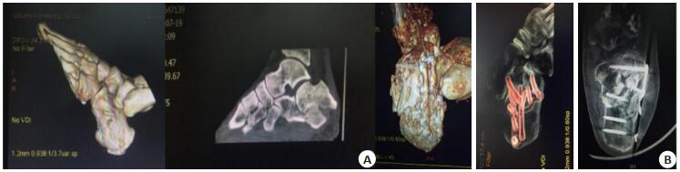
图 1 跟骨粉碎性骨折手术前后16排螺旋CT图片
患者诊断为跟骨粉碎性骨折; A:术前16排螺旋CT扫描其跟骨骨折部位所得后处理图像,发现跟骨横径增加, 碎骨块对合不良, 并且跟骨结节角减小; B:术后16排螺旋CT扫描所得后处理图像, 跟骨见金属内固定, 碎骨块集中, 骨位与跟骨结节角恢复情况良好.
Figure 1. 16-slice spiral CT images of comminuted calcaneal fracture before and after operation.
表 1 X线片与16排螺旋CT诊断准确性比较
Table 1. Comparison of diagnostic accuracy between X-ray film and 16-slice spiral CT[n=174, n(%)]
检查方式 准确性 X线片 143 (82.18) 16排螺旋CT 159 (91.38)# 64排螺旋CT 161 (92.53)# χ2 11.16 P 0.004 #P < 0.05 vs X线片  下载: 导出CSV
表 2 手术前后跟骨Bohler角以及Gissane角比较
Table 2. Comparison of calcaneal Bohler angle and Gissane angle before and after operation (°, n=174, Mean±SD)
时间点 Bohler角 Gissane角 术前 7.86±1.24 87.53±12.15 术后 30.72±4.09 127.36±14.31 t 70.556 27.988 P < 0.001 < 0.001
下载: 导出CSV
-
[1] Zhang W, Lin F, Chen EM, et al. Operative versus nonoperative treatment of displaced intra-articular calcaneal fractures: a meta-analysis of randomized controlled trials[J]. J Orthop Trauma, 2016, 30(3): e75-81. doi: 10.1097/BOT.0000000000000446 [2] Williams JR, Little MT, Kramer PA, et al. Incidence of Preoperative Deep Venous Thrombosis in Isolated Calcaneal Fractures[J]. J Orthop Trauma, 2016, 30(7): e242-5. doi: 10.1097/BOT.0000000000000568 [3] 孙辉, 臧学慧, 高立华, 等.可吸收人工骨粉修复跟骨粉碎性骨折[J].中国组织工程研究, 2014, (39): 6375-80. doi: 10.3969/j.issn.2095-4344.2014.39.025 [4] 孙昌俊, 李力更, 毕若杰, 等". L"入路与"八字"微创入路治疗跟骨骨折的比较[J].中国医学科学院学报, 2015, 37(6): 733-6. doi: 10.3881/j.issn.1000-503X.2015.06.017 [5] 么贵军, 尚剑.跟骨骨折的生物力学研究进展[J].中华创伤骨科杂志, 2014, 16(9): 803-5. doi: 10.3760/cma.j.issn.1671-7600.2014.09.015 [6] Konda SR, Goch AM, Haglin J, et al. Ultralow-dose CT (REDUCTION protocol) for extremity fracture evaluation is as safe and effective as conventional CT: an evaluation of quality outcomes[J]. J Orthop Trauma, 2018, 32(5): 216-22. doi: 10.1097/BOT.0000000000001137 [7] Tresley J, Subhawong TK, Singer AD, et al. Incidence of tendon entrapment and dislocation with calcaneus and pilon fractures on CT examination[J]. Skeletal Radiol, 2016, 45(7): 977-88. doi: 10.1007/s00256-016-2380-0 [8] Meng QT, Wang QX, Wu XR, et al. Clinical application of the sinus tarsi approach in the treatment of intra-articular calcaneal fracture [J]. Medicine (Baltimore), 2018, 97(13): e0175-82. doi: 10.1097/MD.0000000000010175 [9] Yue ZS, Tang YH, Hu ZQ, et al. Sanders type IIIAB calcaneal fracture without broken lateral wall [J]. Medicine, 2018, 97(7): e9926-33. doi: 10.1097/MD.0000000000009926 [10] Chen CH, Hung C, Hsu YC, et al. Biomechanical evaluation of reconstruction plates with locking, nonlocking, and hybrid screws configurations in calcaneal fracture: a finite element model study [J]. Med Biol Eng Comput, 2017, 55(10): 1799-807. doi: 10.1007/s11517-017-1623-0 [11] Zwipp H, Paša L, Žilka L, et al. Introduction of a new locking nail for treatment of intraarticular calcaneal fractures[J]. J Orthop Trauma, 2016, 30(3): e88-92. doi: 10.1097/BOT.0000000000000482 [12] Rammelt S. An update on the treatment of calcaneal fractures[J]. J Orthop Trauma, 2014, 28(10): 549-50. doi: 10.1097/BOT.0000000000000227 [13] Durand D, Wu X, Kalra VB, et al. Predictors of vertebral artery injury in isolated C2 fractures based on fracture morphology using CT angiography[J]. Spine, 2015, 40(12): E713-8. doi: 10.1097/BRS.0000000000000893 [14] 强敏菲, 陈雁西, 贾小阳, 等. CT三维重建在髋臼骨折术后评估中的应用价值[J].中华创伤杂志, 2016, 32(11): 974-9. doi: 10.3760/cma.j.issn.1001-8050.2016.11.004 [15] 王郑浩, 李开南, 兰海.基于三维CT的股骨转子间骨折后内侧壁骨折地图的研究[J].中华创伤骨科杂志, 2019, 21(9): 745-51. doi: 10.3760/cma.j.issn.1671-7600.2019.09.003 [16] 李胜, 邹文远, 侯明伟, 等. CR与CT对桡骨小头骨折Mason分类法的应用价值比较[J].临床放射学杂志, 2014, 33(5): 756-60. http://d.old.wanfangdata.com.cn/Periodical/lcfsxzz201405028 [17] 梅刚, 吴卫东, 欧阳钧, 等.跟骨骨折畸形愈合的数字化虚拟重建及在矫形手术设计中的初步应用[J].中国临床解剖学杂志, 2014, 42(3): 243-7. http://d.old.wanfangdata.com.cn/Periodical/zglcjpxzz201403002 [18] 陈木养, 黄伟坚, 朱裕明, 等.股骨颈骨折的多层螺旋CT与临床分析[J].分子影像学杂志, 2017, 40(2): 144-7. doi: 10.3969/j.issn.1674-4500.2017.02.06 [19] 杨力, 蒲红, 朱缨. MSCT扫描及三维重建技术在降低隐匿性骨折漏诊率中的临床应用[J].中国CT和MRI杂志, 2017, 15(7): 137-40. doi: 10.3969/j.issn.1672-5131.2017.07.043 [20] 马卓, 张世民, 胡孙君, 等. Schatzker Ⅳ型胫骨平台双髁骨折的CT亚型分类及临床意义[J].中华创伤骨科杂志, 2016, 18(10): 832-9. doi: 10.3760/cma.j.issn.1671-7600.2016.10.002 -

 点击查看大图
计量
- 文章访问数: 759
- HTML全文浏览量: 362
- PDF下载量: 4
- 被引次数: 0