


Image of breast phyllodes tumor misdiagnosed as fibroadenoma in MRI: a case report and literature review
-
摘要:
目的探讨乳腺叶状肿瘤与纤维腺瘤的MRI影像差异。 方法通过1例乳腺叶状肿瘤在MRI上误诊为纤维腺瘤的病例,分析二者在MRI上的差异及误诊原因。 结果乳腺叶状肿瘤的MRI表现与纤维腺瘤类似,极易混淆,乳腺叶状肿瘤中可见裂隙状T2WI更高信号区,乳腺纤维腺瘤的特征性表现则是内部不强化的T1WI、T2WI低信号的间隔,另外ADC值也有助于区分乳腺纤维腺瘤和乳腺叶状肿瘤,本病例为乳腺良性叶状肿瘤,MRI影像上未出现典型的裂隙状T2WI更高信号区,总体的影像表现为良性肿瘤,诊断医师首先考虑诊断为临床上更常见的乳腺纤维腺瘤,导致误诊。 结论乳腺叶状肿瘤与乳腺纤维腺瘤的MRI影像表现存在一定的差异,但二者的MRI影像表现较为相近时,需密切结合临床病史以提高诊断准确性。 Abstract:ObjectiveTo explore the differences between the breast phyllodes tumor and the fibroadenoma in MRI. MethodsA case of breast phyllodes tumor misdiagnosed as fibroadenoma in MRI was analyzed. The differences in MRI and the cause of misdiagnosis were analyzed. ResultsThe MRI performance of breast phyllodes tumor was easily mistaken for fibroadenoma. The higher signal area of fissured T2WI was seen in the breast phyllodes tumor. The characteristic expression of breast fibroadenoma was the low signal interval of T1WI and T2WI which were not strengthened internally. In addition, ADC value was also helpful to distinguish breast fibroadenoma from breast phyllodes tumor. This case was benign phyllodes tumor of breast. There was no typical fissured T2WI higher signal area on MRI image. The overall image showed benign tumor. The diagnosis doctor had considered the diagnosis of breast fibroadenoma firstly, which was more common in clinic and leading to misdiagnosis easily. ConclusionThe diagnosis of breast phyllodes tumor in MRI should be made combined with clinical. -
Key words:
- breast phyllodes tumor /
- breast fibroadenoma /
- MRI /
- diffusion weithted image /
- misdiagnosis
-

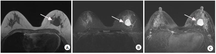
图 1 左侧乳腺良性叶状肿瘤患者MRI图像
A: MRI平扫T1WI图, 左侧乳腺可见类圆形均匀低信号灶, 边界清; B: MRI平扫T2WI图, 左侧乳腺可见类圆形均匀高信号灶, 边界清; C: MRI增强最大密度投影(MIP)图, 左侧乳腺肿物呈明显均匀强化灶, 边界清, 周围可见粗大血管影, 双侧乳腺可见背景实质强化.
Figure 1. MRI image of a patient with benign phyllodes tumor in left breast.
-
[1] Lakhani SR, Ellis IO, Schnitt SJ, et al. WHO classification of tumours of the breast. 4 ed[M]. Lyon: IARC Press, 2012. [2] Zafrakas M, Papasozomenou P, Hatzibougias D. Mixed fibroadenoma and Phyllodes breast tumor: Limitations of core needle biopsy[J]. Hippokratia, 2017, 21(2): 111-8. https://pubmed.ncbi.nlm.nih.gov/30455567/ [3] 芮春朵, 单海荣. MRI鉴别诊断乳腺叶状肿瘤与纤维腺瘤价值分析[J].中外医疗, 2018, 37(21): 160-2. http://d.old.wanfangdata.com.cn/Periodical/hgzy201821057 [4] Kawashima H, Miyati T, Ohno N, et al. Differentiation between Phyllodes tumours and fibroadenomas using intravoxel incoherent motion magnetic resonance imaging: comparison with conventional diffusion-weighted imaging[J]. Br J Radiol, 2018, 91(1084): 20170687-95. https://reference.medscape.com/medline/abstract/29231040 [5] Mai H, Mao YF, Dong TF, et al. The utility of texture analysis based on breast magnetic resonance imaging in differentiating Phyllodes tumors from fibroadenomas [J]. Front Oncol, 2019, 9: 1021-30. doi: 10.3389/fonc.2019.01021 [6] Duman, Gezer NS, Balcı P, et al. Differentiation between Phyllodes tumors and fibroadenomas based on mammographic sonographic and MRI features [J]. Breast Care (Basel), 2016, 11(2): 123-7. doi: 10.1159/000444377 [7] Yii N, Read T, Tan CC, et al. Diagnosing Phyllodes tumours of the breast: how successful are our current preoperative assessment modalities [J]. ANZ J Surg, 2018, 88(10): 988-92. doi: 10.1111/ans.14815 [8] 许晶晶, 李晓冬, 王增奎.乳腺叶状肿瘤的3.0T MRI表现及病理分析[J].实用癌症杂志, 2019, 34(7): 1090-2. doi: 10.3969/j.issn.1001-5930.2019.07.013 [9] Zhou ZR, Wang CC, Sun XJ, et al. Prognostic factors in breast Phyllodes tumors: a nomogram based on a retrospective cohort study of 404 patients [J]. Cancer Med, 2018, 7(4): 1030-42. doi: 10.1002/cam4.1327 [10] 钱跃军, 刘宏军, 王殊, 等.乳腺巨大恶性叶状肿瘤一例[J].中华乳腺病杂志:电子版, 2017, 11(4): 252-4. http://d.old.wanfangdata.com.cn/Periodical/zhrxbzz201704013 [11] Stoffel E, Becker AS, Wurnig MC, et al. Distinction between Phyllodes tumor and fibroadenoma in breast ultrasound using deep learning image analysis [J]. Eur J Radiol Open, 2018, 5: 165-70. doi: 10.1016/j.ejro.2018.09.002 [12] Mitus JW, Blecharz P, Jakubowicz J, et al. Phyllodes tumors of the breast. The treatment results for 340 patients from a single cancer centre[J]. Breast, 2019, 43: 85-90. doi: 10.1016/j.breast.2018.11.009 [13] Iimori N, Kashiwagi S, Ishikawa T, et al. Mammary Phyllodes tumor with six episodes of a relapse: a case report[J]. J Med Case Rep, 2017, 11(1): 261-8. doi: 10.1186/s13256-017-1432-y [14] Moo TA, Alabdulkareem H, Tam A, et al. Association between recurrence and Re-excision for close and positive margins versus observation in patients with benign Phyllodes tumors[J]. Ann Surg Oncol, 2017, 24(10): 3088-92. doi: 10.1245/s10434-017-5955-7 [15] Spanheimer PM, Murray MP, Zabor EC, et al. Long-term outcomes after surgical treatment of malignant/borderline Phyllodes tumors of the breast [J]. Ann Surg Oncol, 2019, 26(7): 2136-43. doi: 10.1245/s10434-019-07210-4 [16] Moon SH, Jung JH, Lee J, et al. Complete remission of giant malignant Phyllodes tumor with lung metastasis: a case report[J]. Medicine (Baltimore), 2019, 98(22): e15762-9. doi: 10.1097/MD.0000000000015762 [17] Yoshiba S, Saotome T, Mikogami T, et al. Metastasis of mammary gland malignant Phyllodes tumor to the mandibular region: a case report and review of the literature[J]. J Oral Maxillofac Surg, 2017, 75(2): 440-8. https://www.sciencedirect.com/science/article/pii/S0278239116308849 [18] 王岸飞, 胡瑛, 王晓燕, 等.乳腺叶状肿瘤的3.0TMRI表现[J].放射学实践, 2017, 32(8): 847-50. http://www.cqvip.com/QK/94342X/201708/673020365.html [19] Kalambo M, Adrada BE, Adeyefa MM, et al. Phyllodes tumor of the breast: ultrasound-pathology correlation[J]. AJR Am J Roentgenol, 2018, 210(4): W173-9. doi: 10.2214/AJR.17.18554 [20] Yabuuchi H, Soeda H, Matsuo Y, et al. Phyllodes tumor of the breast: correlation between MR findings and histologic grade[J]. Radiology, 2006, 241(3): 702-9. doi: 10.1148/radiol.2413051470 [21] 刘洋, 王攀鸽, 谭红娜, 等.乳腺叶状肿瘤的影像学表现与病理对照分析[J].实用放射学杂志, 2017, 33(8): 1191-5. doi: 10.3969/j.issn.1002-1671.2017.08.009 [22] 谷红玉, 罗松, 邓小毅, 等.不同病理级别的乳腺叶状肿瘤MRI成像分析[J].临床放射学杂志, 2019, 38(7): 1194-7. http://www.cnki.com.cn/Article/CJFDTotal-LCFS201907010.htm [23] 徐茂林, 谢东, 李芳, 等.乳腺叶状肿瘤的MRI表现与病理学分级的相关性研究[J].临床放射学杂志, 2019, 38(7): 1203-7. http://www.cnki.com.cn/Article/CJFDTotal-LCFS201907012.htm [24] 李雪梅, 陈颖, 崔春玲.乳腺叶状肿瘤的MRI表现[J].哈尔滨医科大学学报, 2018, 52(3): 289-91, 296. doi: 10.3969/j.issn.1000-1905.2018.03.021 [25] Guo Y, Tang WJ, Kong QC, et al. Can whole-tumor apparent diffusion coefficient histogram analysis be helpful to evaluate breast phyllode tumor grades?[J]. Eur J Radiol, 2019, 114: 25-31. doi: 10.1016/j.ejrad.2019.02.035 -

 下载:
下载:
 点击查看大图
图(2)
计量
- 文章访问数: 1318
- HTML全文浏览量: 417
- PDF下载量: 10
- 被引次数: 0