


Clinical significance of CDFI in the detection of carotid artery and retro-ocular blood flow parameters in patients with NAION
-
摘要:
目的探讨彩色多普勒超声(CDFI)在非动脉炎性前部缺血性视神经病变(NAION)患者颈动脉、眼球后血管血流参数检测中的价值。 方法选取我院收治的NAION患者80例作为NAION组,另选80例无眼部相关疾病的志愿者作为对照组;采用CDFI检测两组研究对象的颈内动脉血流参数、颈内动脉血管弹性参数、眼球后动脉血流参数,采用相干光层析血管成像术检测两组的视杯视盘直径比、黄斑神经节细胞复合体厚度、视盘视网膜神经纤维层厚度;采用简单线性相关Pearson相关分析法分析颈内动脉血流参数、颈内动脉血管弹性参数、眼球后动脉血流参数与NAION病变的关系。 结果NAION组颈内动脉舒张期峰值流速值低于对照组(P < 0.05),NAION组颈内动脉RI、IMT值高于对照组(P < 0.05);NAION组颈内动脉动脉扩张性、动脉紧张度、压力-应变性系数值低于对照组(P < 0.05);NAION组眼动脉动脉的收缩期峰值流速、舒张期峰值流速测定值低于对照组(P < 0.05);NAION组眼动脉的RI测定值高于对照组(P < 0.05);NAION组的水平及垂直视杯视盘直径比、黄斑神经节细胞复合体厚度均低于对照组(P < 0.05)。EDV值与水平及垂直视杯视盘直径比、黄斑神经节细胞复合体厚度呈正相关(P < 0.05),RI值与水平及垂直视杯视盘直径比、黄斑神经节细胞复合体厚度呈负相关(P < 0.05)。 结论NAION患者颈动脉、眼球后血管血流参数常发生显著改变,并且与NAION患者眼部病变程度有一定的关系。 -
关键词:
- 彩色多普勒超声 /
- 非动脉炎 /
- 前部缺血性视神经病变 /
- 颈动脉 /
- 球后动脉
Abstract:ObjectiveTo explore the value of color Doppler ultrasound (CDFI) in the detection of blood flow parameters of carotid artery and retro-ocular vascular in patients with non-arteritic anterior ischemic optic neuropathy (NAION). MethodsEighty NAION patients admitted to our hospital were selected as the NAION group and 80 volunteers without eye-related diseases as the control group. CDFI was used to detect the internal carotid artery blood flow parameters, internal carotid artery vascular elasticity parameters, and eyeballs of the two groups of subjects. The posterior arterial blood flow parameters were measured by coherent optical tomography to detect the optic disc diameter ratio, macular ganglion cell complex thickness, and optic disc retinal nerve fiber layer thickness in the two groups. Simple linear correlation Pearson correlation analysis method was used Analyze the relationship between internal carotid artery blood flow parameters, internal carotid artery vascular elastic parameters, posterior arterial blood flow parameters and NAION lesions. ResultsThe Vd value of the internal carotid artery in the NAION group was lower than that of the control group (P < 0.05). The RI and IMT values of the internal carotid artery in the NAION group were higher than those in the control group (P < 0.05). The dilatation, arterial tension, and pressure of the internal carotid artery in the NAION group-The strain coefficient value was lower than the control group (P < 0.05). The measured values of Vs and Vd of the OA artery in the NAION group were lower than the control group (P < 0.05). The measured value of arterial RI was higher than the control group (P < 0.05). The horizontal and vertical optic disc diameter ratio and the thickness of the macular ganglion cell complex in the NAION group were lower than those of the control group (P < 0.05). EDV value was significantly positively correlated with the ratio of the optic disc diameter of the horizontal and vertical optic cups, the thickness of the macular ganglion cell complex (P < 0.05). The RI value was significantly correlated with the ratio of the optic disc diameter of the horizontal and vertical optic cups, and the macular nerve The thickness of the ganglion cell complex was significantly negatively correlated (P < 0.05). ConclusionThe blood flow parameters of the carotid artery and posterior blood vessels of NAION patients often have significant changes. There is a certain relationship with the degree of ocular lesions in NAION patients. -

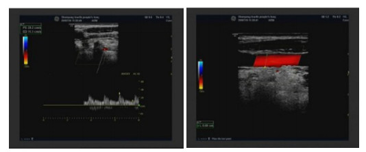
图 1 某女性患者,69岁,眼部Ⅲ浅表器官+球后血管彩色多普勒超声图片
左右眼玻璃体轻度混浊(左眼明显), 左眼玻璃体后脱离, 右眼PCACRA血流峰值流速略减低, 左眼CRA血流峰值流速轻度减低.
Figure 1. A69-year-old female of a patient with ocular surface Ⅲ superficial organ

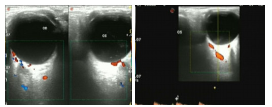
图 2 某男性患者,62岁双侧颈部血管常规超声检测图片,双侧颈总及颈内动脉内-中膜回声欠规整
Figure 2. Amale patient, CDFI shows the routine ultrasound of bilateral cervical vessels at the age of 62, and the echo of bilateral common neck and internal carotid artery is irregular.
表 1 NAION组与对照组一般资料比较
Table 1. Comparison of baseline data between NAION group and control group (n=80)
一般资料 NAION组 对照组 t/χ2 P 年龄(岁, Mean±SD) 61.5±6.5 60.5±7.2 0.922 0.358 BMI(kg/m2,
Mean±SD)23.1±2.4 23.3±2.2 -0.549 0.583 眼压(mmHg, Mean±SD) 15.3±2.0 14.9±2.4 1.145 0.254 眼轴长度(mm, Mean±SD) 22.5±2.4 22.9±2.1 -1.122 0.264 性别[n(%)] 0.637 0.425 男 43
(53.75)38
(47.50)女 37
(46.25)42
(52.50)高血压[n(%)] 1.185 0.276 是 15
(18.75)10
(12.50)否 65
(81.25)70
(87.50)糖尿病[n(%)] 1.765 0.184 是 9
(11.25)15
(18.75)否 71
(88.75)65
(81.25)NAION: 非动脉炎性前部缺血性视神经病变.  下载: 导出CSV
表 2 NAION组与对照组颈内动脉血流参数及IMT值比较
Table 2. Comparison of internal carotid artery blood flow parameters and IMT values between NAION group and control group (n=80, Mean±SD)
组别 Vs(cm/s) Vd(cm/s) RI IMT(mm) NAION组 11.56±1.32 3.38±0.73 0.69±0.08 1.18±0.23 对照组 11.42±1.18 3.70±0.70 0.64±0.09 1.11±0.18 t 0.707 -2.830 3.714 2.144 P 0.480 0.005 0.000 0.034 Vs: 收缩期峰值流速; Vd: 舒张期峰值流速; RI: 阻力指数; IMT: 颈内动脉内-中膜厚度.
下载: 导出CSV
表 3 NAION组与对照组的颈内动脉血管弹性参数比较
Table 3. Comparison of vascular elastic parameters of internal carotid artery between NAION group and control group (n=80, Mean±SD)
组别 动脉扩张性(mmHg) 动脉紧张度 动脉顺应性(mm2/mmHg) 压力-应变性系数(mmHg) NAION组 2.40±0.61 7.68±1.43 0.36±0.08 116.7±16.3 对照组 2.58±0.56 8.74±1.38 0.38±0.10 107.6±14.8 t -1.944 -4.771 -1.397 3.697 P 0.054 0.000 0.164 0.000
下载: 导出CSV
表 4 NAION组与对照组的球后动脉血流参数比较
Table 4. Comparison of retrobulbar arterial blood flow parameters between NAION group and control group(n=80, Mean±SD)
组别 眼动脉 视网膜中央动脉 Vs(cm/s) Vd(cm/s) RI Vs(cm/s) Vd(cm/s) RI NAION组 31.82±2.68 7.63±1.04 0.68±0.09 11.63±1.10 4.81±0.86 0.65±0.08 对照组 33.05±2.80 8.38±1.17 0.63±0.08 11.38±0.96 4.97±0.80 0.63±0.09 t -2.838 -4.285 3.714 1.532 -1.218 1.486 P 0.005 0.000 0.000 0.128 0.225 0.139
下载: 导出CSV
表 5 NAION组与对照组的OCTA检测结果比较
Table 5. Comparison of OCTA test results between NAION group and control group (n=80, Mean±SD)
组别 视杯视盘直径比 黄斑神经节细胞复合体厚度(μm) 视盘视网膜神经纤维层厚度(μm) 水平 垂直 NAION组 0.410±0.118 0.382±0.104 89.8±13.1 114.2±17.6 对照组 0.612±0.130 0.508±0.122 102.0±16.7 110.5±15.8 t -10.291 -7.030 -5.141 1.399 P 0.000 0.000 0.000 0.164
下载: 导出CSV
表 6 Pearson线性分析结果
Table 6. Pearson linear analysis results
指标 颈内动脉 EDV RI 垂直视杯视盘直径比 r 0.441 -0.337 P 0.000 0.010 水平视杯视盘直径比 r 0.398 -0.418 P 0.001 0.000 黄斑神经节细胞复合体厚度 r 0.427 -0.296 P 0.000 0.014
下载: 导出CSV
-
[1] 郝宇, 王岩, 熊丹丹, 等. 激素联合个性化药物治疗非动脉炎性前部缺血性视神经病变的疗效[J]. 武警医学, 2020, 31(9): 771-3, 777. doi: 10.3969/j.issn.1004-3594.2020.09.009 [2] Huang HM, Wu PC, Kuo HK, et al. Natural history and visual outcome of nonarteritic anterior ischemic optic neuropathy in Southern Taiwan: a pilot study[J]. Int Ophthalmol, 2020, 40(10): 2667-76. doi: 10.1007/s10792-020-01448-8 [3] Kumar MK, Daswani M, Shah VM. Prothrombin G20210A mutation causing nonarteritic anterior ischemic optic neuropathy in a young patient[J]. J Neuro-Ophthalmol, 2020, 40(3): 442-3. http://journals.lww.com/jneuro-ophthalmology/Fulltext/2020/09000/Prothrombin_G20210A_Mutation_Causing_Nonarteritic.27.aspx [4] 付智勇, 李红阳, 王薇, 等. 非动脉炎性前部缺血性视神经病变的影像学分析[J]. 临床眼科杂志, 2020, 28(5): 408-12. doi: 10.3969/j.issn.1006-8422.2020.05.006 [5] Foster RC, Bhatti MT, Crum OM, et al. Stroke rate, subtype, and cardiovascular risk factors in nonarteritic anterior ischemic optic neuropathy: a population-based study[J]. J Neuroophthalmol, 2020, 40(3): 328-32. doi: 10.1097/WNO.0000000000000923 [6] Chen J, Zhu J, Chen L, et al. Steroids in the treatment of nonarteritic anterior ischemic optic neuropathy: a PRISMA-compliant metaanalysis[J]. Medicine, 2019, 98(46): e17861. doi: 10.1097/MD.0000000000017861 [7] Egan RA, Arnold AC, Lee AG, et al. Should aspirin be prescribed to prevent recurrence in nonarteritic anterior ischemic optic neuropathy?[J]. J Neuro-Ophthalmol, 2020, 40(3): 428-33. doi: 10.1097/WNO.0000000000000930 [8] 中华医学会眼科学分会神经眼科学组. 我国非动脉炎性前部缺血性视神经病变诊断和治疗专家共识(2015年[) J]. 中华眼科杂志, 2015, 51(5): 323-6. doi: 10.3760/cma.j.issn.0412-4081.2015.05.002 [9] 张瑞君, 尹晓强, 王丽丽, 等. 颈动脉病变和高血压病与非动脉炎性前部缺血性视神经病变的相关性[J]. 临床心身疾病杂志, 2018, 24(2): 61-3. doi: 10.3969/j.issn.1672-187X.2018.02.017 [10] Rueløkke LL, Malmqvist L, Wegener M, et al. Optic disc drusen associated anterior ischemic optic neuropathy: prevalence of comorbidities and vascular risk factors[J]. J Neuro-Ophthalmol, 2020, 40(3): 1. http://journals.lww.com/jneuro-ophthalmology/Abstract/2020/09000/Optic_Disc_Drusen_Associated_Anterior_Ischemic.11.aspx [11] Kim JA, Lee EJ, Kim TW, et al. Differentiation of nonarteritic anterior ischemic optic neuropathy from normal tension Glaucoma by comparison of the Lamina cribrosa[J]. Investig Ophthalmol Vis Sci, 2020, 61(8): 21. http://www.researchgate.net/publication/342988441_Differentiation_of_Nonarteritic_Anterior_Ischemic_Optic_Neuropathy_from_Normal_Tension_Glaucoma_by_Comparison_of_the_Lamina_Cribrosa [12] 周洁, 周希瑗. 超声造影对非动脉炎性前部缺血性视神经病变患者视网膜中央动脉血流状态的初步研究[J]. 临床超声医学杂志, 2018, 20(3): 157-60. https://www.cnki.com.cn/Article/CJFDTOTAL-LCCY201803005.htm [13] Arnold AC. Aspirin should not be recommended to prevent second eye involvement in patients with nonarteritic anterior ischemic optic neuropathy[J]. J Neuro-Ophthalmol, 2020, 40(2): 271-3. doi: 10.1097/WNO.0000000000000931 [14] 郭金喜, 赵云, 路璐. 视野和图形视诱发电位对非动脉炎性前部缺血性视神经病变患者视功能的评估价值[J]. 临床与病理杂志, 2020, 40 (8): 2097-102. https://www.cnki.com.cn/Article/CJFDTOTAL-WYSB202008030.htm [15] Kuerten D, Plange N. Ocular hemodynamics in acute nonarteritic anterior ischemic optic neuropathy compared with normal tension Glaucoma[J]. J Glaucoma, 2019, 28(12): e180-1. doi: 10.1097/IJG.0000000000001404 [16] 安建斌, 周娜磊, 王一, 等. 非动脉炎性前部缺血性视神经病变患者眼部血流动力学观察[J]. 中华超声影像学杂志, 2018, 27(10): 887-90. doi: 10.3760/cma.j.issn.1004-4477.2018.10.013 [17] Francis CE, Patel VR. Should a hypercoagulable work-up be performed on young patients with nonarteritic anterior ischemic optic neuropathy?[J]. J Neuroophthalmol, 2019, 39(4): 523-8. doi: 10.1097/WNO.0000000000000840 [18] Rizzo JF. Unraveling the Enigma of nonarteritic anterior ischemic optic neuropathy[J]. J Neuroophthalmol, 2019, 39(4): 529-44. doi: 10.1097/WNO.0000000000000870 [19] 武炳慧, 陆慧琴, 吴惠琴. 全身疾病致非动脉炎性前部缺血性视神经病变42例临床研究[J]. 陕西医学杂志, 2018, 47(7): 830-2. doi: 10.3969/j.issn.1000-7377.2018.07.004 [20] Harvey JP, O'Sullivan E. Ocular hemodynamics in acute nonarteritic anterior ischemic optic neuropathy compared to normal tension Glaucoma[J]. J Glaucoma, 2019, 28(8): e146-7. doi: 10.1097/IJG.0000000000001299 -

 点击查看大图
计量
- 文章访问数: 650
- HTML全文浏览量: 271
- PDF下载量: 8
- 被引次数: 0