


ROC curve analysis of diagnostic value of ultrasound combined with serum HE4 and Ddimer in ovarian cancer
-
摘要:
目的研究超声联合血清HE4、D-二聚体(D-D)检测在诊断卵巢癌中的价值。 方法将我院2016年1月~2020年6月接收的70例经病理检查证实卵巢病变性质的卵巢占位患者及20例健康志愿者纳为研究对象,其中经病理检查证实卵巢癌44例(恶性组),卵巢良性病变26例(良性组),检测并统计恶性组、良性组及健康组血清HE4、D-D水平及超声成像特点,分析超声联合血清HE4、D-D在诊断卵巢癌中的价值。 结果对照组、良性组及恶性组间血清HE4及D-D水平差异具有统计学意义(P < 0.05),其中恶性组及良性组血清HE4水平高于对照组,且恶性组血清HE4水平高于良性组(P < 0.05),恶性组血清D-D水平高于良性组与对照组,而良性组与对照组间血清D-D水平差异无统计学意义(P>0.05);随着肿瘤分期的上升,恶性组患者血清HE4及DD水平均呈依次上升趋势,组间差异有统计学意义(P < 0.05);当HE4及D-D分别取临界值97.12 pmol/L与0.45 mg/L时,两物质在诊断卵巢癌中的效能最高,但两者联合可有效提高各物质单独诊断效能;以病理检查结果作为“金标准”,比较发现,卵巢良性病变组超声评分低于恶性组,差异有统计学意义(P < 0.05),良、恶性卵巢病变组成分、分隔、厚度RI及PI等超声征象差异均有统计学意义(P < 0.05);且超声联合血清HE4及D-D诊断卵巢癌的灵敏度、特异度及准确率最高,分别为92.31%、93.18%和92.86%。 结论超声、血清HE4及D-D在诊断恶性卵巢肿瘤中均具有一定的价值,而三者联合能有效提高诊断效果,为临床诊断提供更可靠的参考。 Abstract:ObjectiveTo analyze the diagnostic value of ultrasound combined with serum HE4 and D-dimer in ovarian cancer based on ROC curve. MethodsSeventy patients with ovarian lesions in the hospital from January 2016 to June 2020 were enrolled, including 44 cases of ovarian cancer (malignant group) and 26 cases of benign ovarian lesions (benign group), and 20 healthy individuals (control group). Serum HE4 and D-D levels and ultrasound imaging characteristics of three groups were compared, and the value of ultrasound combined with serum HE4 and D-D in the diagnosis of ovarian cancer was analyzed. ResultsSerum HE4 and D-D levels had significant difference among three groups (P<0.05). Serum HE4 level among three groups was the highest in malignant group, followed by benign group and control group (P<0.05). Serum D-D level among three groups was the highest in malignant group (P<0.05), while the D-D level showed no significant difference between benign group and control group (P>0.05). With the increase of tumor stage, there was an increasing trend in the levels of serum HE4 and D-D in malignant group, with statistic difference (P<0.05). When the critical values of HE4 and D-D were 97.12 pmol/L and 0.45 mg/L respectively, the two substances had the highest efficiency in the diagnosis of ovarian cancer, while the diagnostic efficacy of combined detection of HE4 and D-D was higher than that of single detection. Taking pathological examination results as "gold standard", the ultrasound score of benign group was significantly lower than that of malignant group (P<0.05). And there were significant differences in ultrasound signs such as the composition, separation, and thickness RI and PI between benign group and malignant group (P<0.05). The sensitivity, specificity and accuracy of ultrasound combined with serum HE4 and D-D in diagnosing ovarian cancer were the highest, being 92.31%, 93.18% and 92.86%, respectively. ConclusionUltrasound, serum HE4 and D-D are of certain value in diagnosis of malignant ovarian cancer, and the combined detection of the three can effectively improve the diagnosis efficacy and provide a more reliable reference for clinical diagnosis. -
Key words:
- ultrasound /
- serum HE4 /
- D-D /
- ovarian cancer /
- diagnostic value analysis
-

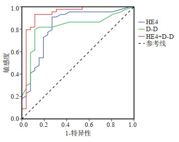
图 1 血清HE4及D-D诊断卵巢癌的价值分析
Figure 1. Value of serum HE4 and D-D in the diagnosis of ovarian cancer.

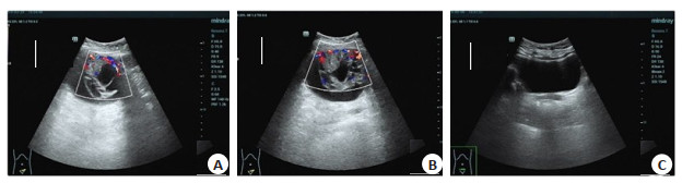
图 2 典型个例手术前后超声图像
A: 术前超声图像提示子宫左旁可见一液实性回声团,大小约125 mm×77 mm, 边界尚清, 内回声欠均匀, 内可见多条厚薄不均的分隔; B: CDFI显示团块实性部位见丰富彩流信号, 术前拟诊断为囊腺癌; C: 术后超声图像子宫及双侧卵巢未探及(自述已切除), 盆腔内未见明显异常团块及液性区, 术后超声提示盆腔内未见明显异常, 手术病理证实为左侧卵巢粘液性囊腺癌.
Figure 2. Ultrasound images of typical cases before and after operation.
表 1 Lerner评分标准
Table 1. Lerner score
项目 0分 1分 2分 3分 形态 规则 不规则 - - 包膜 完整 不完整 - - 壁厚 < 3 mm ≥3 mm - ≥3 mm且内见乳头 回声 无回声或低回声 - - 混合回声或强回声 周边血流情况 Ⅰ级 Ⅱ级 - Ⅲ级 RI - > 0.5 ≤0.5 - PI - > 1.0 ≤1.0 - 腹腔积液 无 - 有 - RI: 阻力指数; PI: 搏动指数.  下载: 导出CSV
表 2 三组血清HE4及D-D水平比较
Table 2. Comparison of serum HE4 and D-D levels among three groups (Mean±SD)
组别 HE4(pmol/L) D-D(mg/L) 恶性组(n=44) 410.15±56.68* 2.13±0.52*# 良性组(n=26) 26.78±13.25* 0.39±0.07 对照组(n=20) 20.56±11.25 0.34±0.04 F 1261.324 334.049 P 0.000 0.000 *P < 0.05 vs对照组; #P < 0.05 vs良性组.
下载: 导出CSV
表 3 恶性组不同分期患者血清HE4及D-D水平比较
Table 3. Comparison of serum HE4 and D-D levels among patients in malignant group at different stages (Mean±SD)
组别 HE4(pmol/L) D-D(mg/L) Ⅰ期(n=11) 133.68±35.45 1.36±0.26 Ⅱ期(n=14) 161.54±33.21* 1.41±0.31 Ⅲ期(n=13) 589.15±113.21*# 2.54±0.54*# Ⅳ期(n=6) 591.45±134.54*# 3.48±0.75*#△ F 86.545 44.436 P 0.000 0.000 *P < 0.05 vs Ⅰ期; #P < 0.05 vs Ⅱ期; P < 0.05 vs Ⅲ期.
下载: 导出CSV
表 4 血清HE4及D-D诊断卵巢癌的价值分析
Table 4. Value of serum HE4 and D-D in the diagnosis of ovarian cancer
指标 临界值 AUC 95%CI P 敏感度 特异性 HE4 97.12 pmol/L 0.829 0.722~0.936 0.000 65.9 80.8 D-D 0.45 mg/L 0.821 0.716~0.926 0.000 79.5 88.5 HE4+D-D - 0.933 0.859~1.000 0.000 93.2 88.5
下载: 导出CSV
表 5 不同性质卵巢癌声像图及血流特点
Table 5. Ultrasound signs and blood flow characteristics of benign and malignant ovarian cancer (n=70, Mean±SD)
征象 病理检查结果 合计 恶性率(%) χ2 P 良性(n=26) 恶性(n=44) 大小 0.082 0.775 > 5 mm 18 29 47 61.70 ≤5 mm 8 15 23 65.22 成分 38.661 0.000 囊实性 2 37 39 94.87 囊性 24 7 31 22.58 分隔 14.726 0.000 有 8 34 42 80.95 无 18 10 28 35.71 厚度 15.664 0.000 > 3 mm 5 30 35 85.71 ≤3 mm 21 14 35 40.00 RI 20.886 0.000 > 0.5 24 16 40 40.00 ≤0.5 2 28 30 93.33 PI 7.593 0.006 > 1.0 23 25 48 52.08 ≤1.0 3 19 22 86.36
下载: 导出CSV
表 6 超声、血清HE4及D-D在诊断卵巢癌中的价值研究
Table 6. Value of ultrasound, serum HE4 and D-D in the diagnosis of ovarian cancer (n=70)
方式 病理诊断 合计 灵敏度(%) 特异度(%) 准确率(%) 良性 恶性 超声 73.08 63.64 67.14 良性 19 16 35 - - - 恶性 7 28 35 - - - 合计 26 44 - - - 血清HE 65.38 81.82 75.71 良性 17 8 25 - - - 恶性 9 36 45 - - - 合计 26 44 - - - 血清D-D 80.77 72.73 75.71 良性 21 12 33 - - - 恶性 5 32 37 - - - 合计 26 44 - - - 联合诊断 92.31 93.18 92.86 良性 24 3 27 - - - 恶性 2 41 43 - - - 合计 26 44 - - -
下载: 导出CSV
-
[1] 邓宇傲, 陈阿, 李秀云, 等. 白杨素调控miR-320和Twist1表达抑制卵巢癌干细胞样细胞体外致瘤功能[J]. 湖南师范大学学报: 医学版, 2018, 15(4): 1-4. https://www.cnki.com.cn/Article/CJFDTOTAL-HNYG201804001.htm [2] 欧阳璟雯, 唐荣, 郑佳. 卵巢癌CT及超声影像特征与术后病理检查结果的比较分析[J]. 中国CT和MRI杂志, 2019, 17(7): 110-2. https://www.cnki.com.cn/Article/CJFDTOTAL-CTMR201907033.htm [3] 李玮珊, 王丹波. 肿瘤标志物HE4在卵巢癌中的应用研究进展[J]. 现代肿瘤医学, 2019, 27(6): 1095-8. https://www.cnki.com.cn/Article/CJFDTOTAL-SXZL201906048.htm [4] 刘建军, 崔林, 刘兴祥, 等. 血浆D-二聚体水平与卵巢癌患者临床特征及化疗预后的关系研究[J]. 癌症进展, 2018, 16(6): 766-9. https://www.cnki.com.cn/Article/CJFDTOTAL-AZJZ201806029.htm [5] 胡蝶, 马玲. 术前NLR与卵巢癌预后的相关性研究[J]. 现代肿瘤医学, 2018, 26(17): 2790-3. https://www.cnki.com.cn/Article/CJFDTOTAL-SXZL201817038.htm [6] 李杰, 赵卫东, 王慧妍, 等. 错配修复基因缺陷对卵巢癌预后影响的系统评价[J]. 中国临床保健杂志, 2018, 21(6): 771-6. https://www.cnki.com.cn/Article/CJFDTOTAL-LZBJ201806015.htm [7] 余红星, 张春玲. 阴道超声检查与腹部超声检查在妇产科疾病诊断中的应用价值[J]. 陕西医学杂志, 2019, 48(4): 445-8. https://www.cnki.com.cn/Article/CJFDTOTAL-SXYZ201904010.htm [8] 李克婷, 姜中慧, 田家玮, 等. Mainz超声评分系统结合三维能量多普勒超声鉴别盆腔肿瘤良恶性的价值[J]. 中华超声影像学杂志, 2018, 27(1): 53-7. [9] 曾瑾, 祝子范, 邓宗科, 等. 超声和血清RBP4及CA125对卵巢肿瘤的诊断价值分析[J]. 西南国防医药, 2019, 29(11): 1125-7. https://www.cnki.com.cn/Article/CJFDTOTAL-XNGF201911013.htm [10] 韩碧波, 魏海东, 郑磊, 等. 经阴道彩色多普勒超声联合血清糖链抗原125和人附睾蛋白4水平检测对卵巢癌的诊断价值[J]. 新乡医学院学报, 2019, 36(10): 971-4. https://www.cnki.com.cn/Article/CJFDTOTAL-XXYX201910016.htm [11] 杨阿芳. 卵巢癌超声造影参数与血管新生、癌细胞生长的相关性研究[J]. 海南医学院学报, 2018, 24(2): 273-6, 279. https://www.cnki.com.cn/Article/CJFDTOTAL-HNYY201802035.htm [12] Dehdashti SM, Acharjee S, Nomani A, et al. Production of pharmaceutical active recombinant globular adiponectin as a secretory protein in Withania Somnifera hairy root culture[J]. J Biotechnol, 2020, 323: 302-12. http://www.sciencedirect.com/science/article/pii/S0168165620301930 [13] 韦金虎. CA125、HE4联合超声检查在卵巢肿瘤中的应用价值[J]. 检验医学与临床, 2019, 16(12): 1715-7. https://www.cnki.com.cn/Article/CJFDTOTAL-JYYL201912025.htm [14] 伍紫娟. D-二聚体与卵巢癌的诊断、治疗及预后关系的研究进展[J]. 检验医学与临床, 2018, 15(1): 135-8. https://www.cnki.com.cn/Article/CJFDTOTAL-JYYL201801044.htm [15] 许洁, 韩洪超, 崇庆国, 等. 血浆D-二聚体及血小板水平检测在以VTE为早期表现的恶性肿瘤中的意义[J]. 中国医师杂志, 2019(4): 596-8. [16] 张成大, 何君, 黄婷, 等. 结直肠癌患者血清CEA和D-二聚体检测临床意义[J]. 中华肿瘤防治杂志, 2018, 25(4): 282-6. https://www.cnki.com.cn/Article/CJFDTOTAL-QLZL201804011.htm [17] 梁巍, 胡雨薇, 应媛媛. 联合检测D-二聚体、糖类抗原-199、癌胚抗原在结直肠癌患者中的临床价值[J]. 中国实验诊断学, 2019, 23 (1): 83-5. https://www.cnki.com.cn/Article/CJFDTOTAL-ZSZD201901108.htm [18] 王玉婷, 古丽米拉木·艾热提, 刘春花, 等. D-二聚体、纤维蛋白原升高的影响因素及其与恶性实体肿瘤患者临床特征及预后的关系[J]. 现代肿瘤医学, 2018, 26(12): 1921-4. https://www.cnki.com.cn/Article/CJFDTOTAL-SXZL201812031.htm [19] 苏媛媛, 李静, 梁义娟. HE4、CA125和D-dimer检测在卵巢癌早期诊断筛查中的应用价值[J]. 陕西医学杂志, 2018, 47(3): 368-70. https://www.cnki.com.cn/Article/CJFDTOTAL-SXYZ201803030.htm -

 点击查看大图
计量
- 文章访问数: 368
- HTML全文浏览量: 231
- PDF下载量: 6
- 被引次数: 0