


Value of DCE-MRI in evaluation of concurrent chemoradiotherapy for locally advanced cervical cancer
-
摘要:
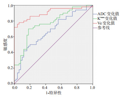
目的探讨动态对比增强磁共振成像(DCE-MRI)在局部晚期宫颈癌同步放化疗效果评估中的价值。 方法选取2019年1月~2020年1月在我院治疗的局部晚期宫颈癌患者200例,所有患者均给予同步放化疗疗效,观察治疗前后DCE-MRI参数变化情况,同时分析治疗有效和无效患者DCE-MRI参数差异。 结果200例患者经同步放化疗后,达到完全缓解患者21例,部分缓解患者67例,疾病稳定患者72例,疾病进展患者40例,治疗有效率为44.00%;治疗无效患者FIGO分期ⅣA期比例、卡氏评分高于治疗有效患者(P < 0.05);治疗后病灶表观扩散系数(ADC)值、容量转移常数(Ktrans)、血管外细胞外间隙容积(Ve)较治疗前升高(P < 0.05);治疗有效患者病灶ADC变化值、Ktrans变化值、Ve变化值高于治疗无效患者(P < 0.05);ADC变化值、Ktrans变化值、Ve变化值预测治疗有效的ROC曲线下面积分别为0.675、0.770和0.905(P < 0.05),截断值分别为0.35×10⁻³ mm2/s、0.54 min-1和0.22,灵敏性分别为66.60%、78.80%和85.50%,特异性分别为62.00%、75.50%和82.00%。 结论DCE-MRI在局部晚期宫颈癌同步放化疗效果评估中有一定应用价值,值得临床使用。 -
关键词:
- 动态对比增强磁共振成像 /
- 局部晚期 /
- 宫颈癌 /
- 同步放化疗 /
- 治疗效果
Abstract:ObjectiveTo investigate the value of dynamic contrast- enhanced magnetic resonance imaging (DCE-MRI) in the evaluation of concurrent chemoradiotherapy for locally advanced cervical cancer. MethodsA total of 200 patients with locally advanced cervical cancer treated in our hospital from January 2019 to January 2020 were selected. All patients were treated with concurrent chemoradiotherapy, the changes of DCE-MRI parameters before and after treatment were observed, and the differences of DCE-MRI parameters between effective and ineffective patients were analyzed. ResultsAll patients received concurrent chemoradiotherapy. There were 21 patients with complete remission, 67 patients with partial remission, 72 patients with stable disease and 40 patients with disease progression, the effective rate was 44.00%. The proportion of FIGO stage ⅣA and Karnofsky score of patients with ineffective treatment were significantly higher than those of patients with effective treatment (P < 0.05). The apparent diffusion coefficient (ADC), volume metastasis constant (Ktrans) and volume of extracellular space (Ve) were significantly higher than those before treatment (P < 0.05). The change values of ADC, Ktrans and Ve in the effective patients were significantly higher than those in the ineffective patients (P < 0.05). The area under the ROC curve of ADC change, Ktrans change and Ve change to predict the treatment effectiveness were 0.675, 0.770 and 0.905 (P < 0.05), and the cutoff values were 0.35×10⁻³ mm2/s, 0.54 min-1 and 0.22, the sensitivity were 66.60%, 78.80% and 85.50%, and the specificity were 62.00%, 75.50% and 82.00%, respectively. ConclusionDCE-MRI has certain application value in evaluating the effect of concurrent chemoradiotherapy for local advanced cervical cancer and is worthy of clinical use. -

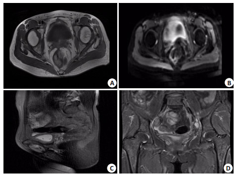
图 1 某患者DCE-MRI扫描图片
A: 斜轴位T1WI; B: 斜轴位T2WI; C: 矢状位T2WI; D: 冠状位压脂T2WI.
Figure 1. DCE-MRI scanning picture of a patient.
表 1 治疗有效和无效患者临床资料比较
Table 1. Comparison of clinical data between effective and ineffective patients
临床资料 治疗有效(n=88) 治疗无效(n=112) t/χ2 P 性别[n(%)] 0.514 0.473 男 49(55.68) 68(60.71) 女 39(44.32) 44(39.29) 年龄(岁, Mean±SD) 60.39±5.58 59.28±4.42 1.570 0.118 BMI(kg/m2, Mean±SD) 21.20±3.22 21.51±2.82 -0.725 0.469 婚姻状况[n(%)] 0.210 0.647 已婚 50(56.82) 60(53.57) 未婚 38(43.18) 52(46.43) FIGO分期[n(%)] 8.316 0.004 Ⅲ期 55(62.50) 47(41.96) ⅣA期 33(37.50) 65(58.04) 肿瘤直径(cm, Mean±SD) 5.50±1.10 5.67±1.03 -1.124 0.262 卡氏评分(分, Mean±SD) 85.50±5.54 79.90±6.10 6.708 0.000 FIGO: 国际妇产科联盟.  下载: 导出CSV
表 2 治疗前后DCE-MRI参数比较
Table 2. Comparison of DCE-MRI parameters before and after treatment (n=200, Mean±SD)
时期 ADC值(×10-3 mm2/s) Ktrans(min-1) Kep(min-1) Ve 治疗前 0.64±0.21 0.63±0.22 1.22±0.34 0.44±0.13 治疗后 0.98±0.18 1.15±0.28 1.23±0.31 0.64±0.20 t 17.385 20.652 0.307 11.857 P 0.000 0.000 0.759 0.000 Ktrans: 容量转移常数; Ve: 血管外细胞外间隙容积.
下载: 导出CSV
表 3 不同治疗疗效患者DCE-MRI参数变化值比较
Table 3. Comparison of DCE-MRI parameters in patients with different therapeutic effects
组别 ADC变化值(×10-3mm2/s) Ktrans变化值(min-1) Kep变化值(min-1) Ve变化值 治疗有效(n=88) 0.37±0.10 0.60±0.16 0.01±0.01 0.27±0.09 治疗无效(n=112) 0.32±0.12 0.46±0.13 0.01±0.00 0.15±0.05 t 3.144 6.827 0.000 11.960 P 0.002 0.000 1.000 0.000
下载: 导出CSV
-
[1] 冯煜森, 丁莹莹, 张娅, 等. 动态增强磁共振成像在宫颈癌新辅助化疗疗效评估中的价值[J]. 实用放射学杂志, 2018, 34(5): 725-9. doi: 10.3969/j.issn.1002-1671.2018.05.020 [2] Hillestad T, Hompland T, Fjeldbo CS, et al. MRI distinguishes tumor hypoxia levels of different prognostic and biological significance in cervical cancer[J]. Cancer Res, 2020, 80(18): 3993-4003. doi: 10.1158/0008-5472.CAN-20-0950 [3] Kim SH, Cho SH. Assessment of pelvic lymph node metastasis in FIGO IB and ⅡA cervical cancer using quantitative dynamic contrast-enhanced MRI parameters[J]. Diagn Interv Radiol, 2020, 26 (5): 382-9. doi: 10.5152/dir.2020.19365 [4] 李风莲, 周玲. 常规MRI联合DWI在宫颈癌术前分期诊断中的应用价值[J]. 中国CT和MRI杂志, 2019, 17(6): 101-3. https://www.cnki.com.cn/Article/CJFDTOTAL-CTMR201906031.htm [5] 米红兰, 程杰军, 殷霞, 等. 体素不相干运动与动态对比增强磁共振成像在评估早期宫颈癌淋巴脉管浸润状态中的应用[J]. 上海交通大学学报: 医学版, 2020, 40(2): 224-30. https://www.cnki.com.cn/Article/CJFDTOTAL-SHEY202002018.htm [6] Feng YS, Liu H, Ding YY, et al. Combined dynamic DCE-MRI and diffusion-weighted imaging to evaluate the effect of neoadjuvant chemotherapy in cervical cancer[J]. Tumori, 2020, 106(2): 155-64. doi: 10.1177/0300891619886656 [7] Lu Y, Peng W, Song J, et al. On the potential use of dynamic contrastenhanced (DCE) MRI parameters as radiomic features of cervical cancer[J]. Med Phys, 2019, 46(11): 5098-109. doi: 10.1002/mp.13821 [8] 张常青, 周星, 张文文, 等. 动态增强MRI及扩散加权成像对早期宫颈癌的诊断价值[J]. 兰州大学学报: 医学版, 2019, 45(1): 38-42. https://www.cnki.com.cn/Article/CJFDTOTAL-LZYX201901009.htm [9] Li X, Wu S, Li D, et al. Intravoxel incoherent motion combined with dynamic contrast-enhanced perfusion MRI of early cervical carcinoma: correlations between multimodal parameters and HIF-1α expression[J]. J Magn Reson Imaging, 2019, 50(3): 918-29. doi: 10.1002/jmri.26604 [10] 王月月, 夏春华. 扩散加权成像及动态增强磁共振成像在诊断子宫颈癌分期中的应用[J]. 临床与病理杂志, 2019, 39(4): 786-93. https://www.cnki.com.cn/Article/CJFDTOTAL-WYSB201904016.htm [11] Huang JW, Song JC, Chen T, et al. Making the invisible visible: improving detectability of MRI-invisible residual cervical cancer after conisation by DCE-MRI[ J]. Clin Radiol, 2019, 74(2): 166. e15-21. doi: 10.1016/j.crad.2018.10.013 [12] 何志兵, 陈首名, 罗鹰, 等. DCE-MRI和IVIM-DWI诊断宫颈癌病理分级和临床分期的价值分析[J]. 中国CT和MRI杂志, 2019, 17(8): 110-3. https://www.cnki.com.cn/Article/CJFDTOTAL-CTMR201908033.htm [13] Simonsen TG, Lund KV, Hompland T, et al. DCE-MRI-derived measures of tumor hypoxia and interstitial fluid pressure predict outcomes in cervical carcinoma[J]. Int J Radiat Oncol Biol Phys, 2018, 102(4): 1193-201. doi: 10.1016/j.ijrobp.2018.04.035 [14] 高歌. DWI和MR动态增强扫描诊断宫颈癌的临床价值分析[J]. 中国CT和MRI杂志, 2018, 16(6): 97-9, 107. https://www.cnki.com.cn/Article/CJFDTOTAL-CTMR201806031.htm [15] Wu Q, Shi D, Dou S, et al. Radiomics analysis of multiparametric MRI evaluates the pathological features of cervical squamous cell carcinoma[J]. J Magn Reson Imaging, 2019, 49(4): 1141-8. [16] 张禹, 张茜, 张雪健, 等. 3.0 T动态对比增强MRI定量参数在鉴别FIGOⅡ期宫颈癌宫旁浸润中的应用价值[J]. 临床放射学杂志, 2018, 37(7): 1163-7. https://www.cnki.com.cn/Article/CJFDTOTAL-LCFS201807026.htm [17] Hansen MB, Tietze A, Haack S, et al. Robust estimation of hemodynamic parameters in traditional DCE-MRI models[J]. PLoS One, 2019, 14(1): e0209891. [18] 赵晓艳, 赵鑫, 张小安, 等. IVIM-DWI与DCE-MRI对宫颈癌的诊断价值和相关性研究[J]. 实用放射学杂志, 2018, 34(5): 717-20. https://www.cnki.com.cn/Article/CJFDTOTAL-WZYX202010010.htm [19] Yang W, Qiang JW, Tian HP, et al. Multi-parametric MRI in cervical cancer: early prediction of response to concurrent chemoradiotherapy in combination with clinical prognostic factors[J]. Eur Radiol, 2018, 28(1): 437-45. [20] 李靖, 张孝先, 曲金荣, 等. MR扩散加权成像预测宫颈癌同步放化疗预后的价值[J]. 实用放射学杂志, 2019, 35(8): 1275-8. -

 点击查看大图
计量
- 文章访问数: 462
- HTML全文浏览量: 241
- PDF下载量: 5
- 被引次数: 0