


Value of DECT in diagnosis of bone marrow lesions in patients with knee osteoarthritis
-
摘要:
目的探讨双能CT(DECT)诊断膝关节骨关节炎(KOA)患者骨髓病变的价值。 方法回顾性分析2016年12月~2018年12月收治147例KOA患者的临床资料,所有患者入院后均行膝关节MRI和DECT平扫检查,将其膝关节根据解剖结构划分为12个区域,股骨下段及胫骨上段各6个区域,分别获得MRI图像、常规CT及虚拟去钙图像。以MRI结果作为标准,比较不同对比物质相对比(Rel.CM)值下DECT检查对患者骨髓病变的诊断价值,计算灵敏度、特异度、准确率、阴性预测值和阳性预测值。采用兴趣区定量测量法检测最佳Rel.CM值下虚拟去钙图像中的CT值,对比阳性区及阴性区在DECT图像上的骨髓CT值,经ROC曲线评估DECT图像上的骨髓CT值检测对KOA患者骨髓病变的诊断效能。 结果不同Rel.CM值下DECT诊断KOA患者骨髓病变的灵敏度、特异度、准确率和阳性阴性预测值对比差异无统计学意义(P>0.05),但当Rel.CM=1.45时其灵敏度、特异度、准确率和阳性阴性预测值等均最佳;最佳Rel.CM值下不同区域内骨髓病变阳性及阴性的骨髓CT值比较均有统计学意义(P < 0.05);ROC曲线显示,单独检测股骨下端CT及胫骨上端CT值均可作为诊断KOA患者骨髓病变的预测指标(P < 0.05)。 结论DECT技术在诊断KOA患者骨髓病变上具有较高准确率,且可通过CT值进行辅助诊断。 Abstract:ObjectiveTo explore the value of dual-energy CT (DECT) in diagnosis of bone marrow lesions in patients with Knee Osteoarthritis (KOA). MethodsThe clinical data of 147 patients with KOA admitted between December 2016 and December 2018 were retrospectively analyzed. All patients underwent knee MRI and DECT plain scan after admission, and their knee joints were divided into 12 regions according to the anatomical structure, including each 6 regions of the lower femur and upper tibia respectively. MRI images, conventional CT and virtual non-calcium images were obtained. Using MRI results as the standard, the diagnostic value of DECT examination on patients with bone marrow lesions under different contrast material relative ratio (Rel.CM) values, and the sensitivity, specificity, accuracy rate, negative predictive value and positive predictive value were calculated. The area of interest quantitative measurement method was used to detect the CT value of virtual noncalcium images under the best Rel.CM value, and the bone marrow CT values of the positive region and the negative region on the DECT images were compared, and the receiver operating characteristic (ROC) curve was used to evaluate the diagnostic efficacy of bone marrow CT value on the DECT images on bone marrow lesions in patients with KOA. ResultsThere were no significant differences in the sensitivity, specificity, accuracy rate and positive and negative predictive values of DECT under different Rel.CM values in diagnosing bone marrow lesions in patients with KOA (P>0.05), but when Rel.CM value=1.45, the sensitivity, specificity, accuracy rate and positive and negative predictive values were the best. There were statistically significant differences in the bone marrow CT values of positive and negative bone marrow lesions in different regions under the best Rel.CM value (P < 0.05). ROC curves showed the CT values of the lower end of the femur and the upper end of the tibia can be used as predictive indicators for the diagnosis of bone marrow lesions in patients with KOA (all P < 0.05). Conclusion DECT technology has a high accuracy rate in diagnosing bone marrow lesions in patients with KOA, and it can be used as an auxiliary diagnosis by CT value. -
Key words:
- dual-energy CT /
- magnetic resonance imaging /
- diagnosis /
- knee osteoarthritis /
- bone marrow lesions /
- value
-
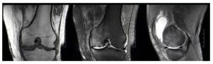
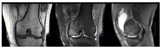
图 1 膝关节骨关节炎患者膝关节MRI影像学图像
Figure 1. MRI imaging features of knee joint in patients with knee osteoarthritis.

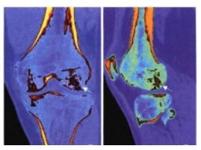
图 2 膝关节骨关节炎患者膝关节DECT影像学图像
患者男, 56岁, 左膝骨关节炎, DECT图像中箭头所示为水肿区域.
Figure 2. DECT imaging features of knee joint in patients with knee osteoarthritis.
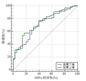
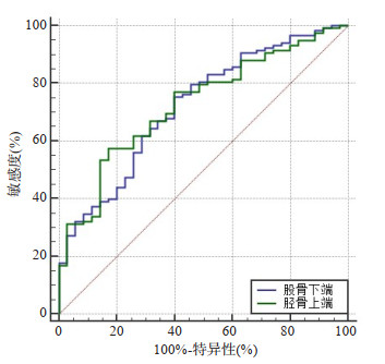
图 3 DECT检查骨髓CT值诊断KOA患者骨髓病变的ROC图像
Figure 3. ROC images of bone marrow lesions in patients with KOAdiagnosed by bone marrow CT values by DECT.
表 1 不同Rel.CM值下DECT检查诊断KOA患者骨髓病变结果比较
Table 1. Comparison of results of bone marrow lesions in patients with KOAdiagnosed by DECT under different Rel.CM values
检查方法 MRI 合计 阳性 阴性 DECT(Rel.CM=1.25) 阳性 281 166 447 阳性 33 1320 1353 DECT(Rel.CM=1.45) 阳性 301 80 381 阳性 13 1406 1419 DECT(Rel.CM=1.75) 阳性 286 157 443 阳性 28 1329 1357 DECT: 双能CT; Rel.CM: 不同对比物质相对比.  下载: 导出CSV
表 2 不同Rel.CM值下DECT检查的灵敏度、特异度、准确率和阳性阴性预测值
Table 2. Sensitivity, specificity, accuracy rate and positive and negative predictive values of DECT under different Rel.CM values
检查方法 灵敏度 特异度 准确率 阳性预测值 阴性预测值 Rel.CM=1.25 0.895 0.888 0.889 0.629 0.976 Rel.CM=1.45 0.959 0.946 0.948 0.790 0.991 Rel.CM=1.75 0.911 0.894 0.897 0.646 0.979
下载: 导出CSV
表 3 DECT检查诊断骨髓病变阳性及阴性区骨髓CT值比较
Table 3. Comparison of bone marrow CT values between positive and negative bone marrow lesions diagnosed by DECT (Hu, Mean±SD)
组别 股骨下端 胫骨上端 阳性组(n=35) -77.26±23.14 -85.42±16.47 阴性组(n=118) -96.47±24.08 -98.14±20.47 t 4.181 3.365 P 0.000 0.001
下载: 导出CSV
表 4 DECT检查骨髓CT值诊断KOA患者骨髓病变的预测效能
Table 4. Predictive efficacy of bone marrow CT values by DECT in the diagnosis of bone marrow lesions in patients with KOA
分组 最佳诊断临界点 AUC 95%CI 敏感度(%) 特异性(%) Z P 股骨下端CT值 81.74 0.721 0.643~0.791 75.42 60.00 4.589 < 0.0001 胫骨上端CT值 95.27 0.729 0.651~0.797 57.63 82.86 4.984 < 0.0001
下载: 导出CSV
-
[1] Moss P, Benson HAE, Will R, et al. Patients with knee osteoarthritis who score highly on the PainDETECT questionnaire present with multimodality hyperalgesia, increased pain, and impaired physical function[J]. Clin J Pain, 2018, 34(1): 15-21. doi: 10.1097/AJP.0000000000000504 [2] Yennan P, Suputtitada A, Yuktanandana P. Effects of aquatic exercise and land-based exercise on postural sway in elderly with knee osteoarthritis [J]. Asian Biomed, 2010, 4(5): 739-45. doi: 10.2478/abm-2010-0096 [3] Richardson C, Plaas A, Block JA. Intra-articular hyaluronan therapy for symptomatic knee osteoarthritis[J]. Rheum Dis Clin North Am, 2019, 45(3): 439-51. doi: 10.1016/j.rdc.2019.04.011 [4] Davatchi F, Abdollahi BS, Mohyeddin M, et al. Mesenchymal stem cell therapy for knee osteoarthritis. Preliminary report of four patients [J]. Int J Rheum Dis, 2011, 14(2): 211-5. doi: 10.1111/j.1756-185X.2011.01599.x [5] Bilbao A, García-Pérez L, Arenaza JC, et al. Psychometric properties of the EQ-5D-5L in patients with hip or knee osteoarthritis: reliability, validity and responsiveness[J]. Qual Life Res, 2018, 27(11): 2897-908. doi: 10.1007/s11136-018-1929-x [6] Panahi Y, Rahimnia AR, Sharafi M, et al. Curcuminoid treatment for knee osteoarthritis: a randomized double-blind placeb-controlled trial [J]. Phytother Res, 2014, 28(11): 1625-31. doi: 10.1002/ptr.5174 [7] Dong TH, Chen W, Zhang F, et al. Radiographic measures of settlement phenomenon in patients with medial compartment knee osteoarthritis [J]. Clin Rheumatol, 2016, 35(6): 1573-8. doi: 10.1007/s10067-015-3146-0 [8] 赵承勇, 罗松, 邓小毅, 等. 双能量CT虚拟去钙成像在鉴别急慢性椎体压缩性骨折中的研究[J]. 医学影像学杂志, 2018, 28(9): 1535-9. https://www.cnki.com.cn/Article/CJFDTOTAL-XYXZ201809034.htm [9] 中华医学会风湿病学分会. 骨关节炎诊断及治疗指南[J]. 中华风湿病学杂志, 2010, 14(6): 416-9. doi: 10.3760/cma.j.issn.1007-7480.2010.06.024 [10] Bongartz T, Glazebrook KN, Kavros SJ, et al. Dual-energy CT for the diagnosis of gout: an accuracy and diagnostic yield study[J]. Ann Rheum Dis, 2015, 74(6): 1072-7. doi: 10.1136/annrheumdis-2013-205095 [11] Shadab M, Zulkifle M, Ansari AH, et al. Correlation of predisposing factors and knee osteoarthritis: A cross-sectional study[J]. Noise Health, 2015, 2(2): 49-58. http://openurl.ebscohost.com/linksvc/linking.aspx?stitle=Noise%20and%20Health&volume=2&issue=2&spage=49 [12] Kean CO, Bennell KL, Wrigley TV, et al. Relationship between hip abductor strength and external hip and knee adduction moments in medial knee osteoarthritis [J]. Clin Biomech (Bristol, Avon), 2015, 30(3): 226-30. doi: 10.1016/j.clinbiomech.2015.01.008 [13] Nishioka H, Nakamura E, Hirose J, et al. MRI T1ρ and T2 mapping for the assessment of articular cartilage changes in patients with medial knee osteoarthritis after hemicallotasis osteotomy[J]. Bone Joint Res, 2016, 5(7): 294-300. doi: 10.1302/2046-3758.57.BJR-2016-0057.R1 [14] 王建辉, 曲智伟. 首次X线检查阴性, 复查中出现新病变19例外伤分析[J]. 检验医学与临床, 2017, 14(Z1): 319-20. doi: 10.3969/j.issn.1672-9455.2017.25.162 [15] Hart HF, Crossley KM, Ackland DC, et al. Effects of an unloader knee brace on knee-related symptoms and function in people with post-traumatic knee osteoarthritis after anterior cruciate ligament reconstruction[J]. Knee, 2016, 23(1): 85-90. doi: 10.1016/j.knee.2015.05.006 [16] 王昌盛, 杨海涛, 邓茗中, 等. 膝关节骨关节炎软骨下水肿与MRI及临床表现的相关性研究[J]. 实用放射学杂志, 2017, 33(8): 1236-40. doi: 10.3969/j.issn.1002-1671.2017.08.020 [17] Lu C, Shu J, Han Y, et al. The polymorphism of SMAD3 rs1065080 is associated with increased risk for knee osteoarthritis[J]. Mol Biol Rep, 2019, 46(4): 4501-5. doi: 10.1007/s11033-019-04905-5 [18] 王亮, 陈祁青, 童培建, 等. 膝关节骨性关节炎早期诊断的临床特征分析[J]. 中国骨与关节损伤杂志, 2015, 30(2): 161-3. https://www.cnki.com.cn/Article/CJFDTOTAL-GGJS201502018.htm [19] 董玉茹, 王宏, 梁莹, 等. 3.0T磁共振不同成像技术对膝骨关节炎骨髓病变SNR和CNR对比分析[J]. 中国CT和MRI杂志, 2015, 13(6): 92-4. https://www.cnki.com.cn/Article/CJFDTOTAL-CTMR201506029.htm [20] 马新荣, 苏萱, 郭荣洲, 等. 双源CT双能量成像技术在骨与关节疾病中的应用[J]. 中国医学装备, 2016, 13(1): 93-6. https://www.cnki.com.cn/Article/CJFDTOTAL-YXZB201601033.htm [21] 章辉庆, 刘海燕, 邱晓晖, 等. 双能量CT虚拟去钙图诊断椎体骨髓水肿[J]. 中国医学影像技术, 2019, 35(2): 260-3. https://www.cnki.com.cn/Article/CJFDTOTAL-ZYXX201902037.htm [22] 梁建超, 方义杰, 李文娟, 等. 双能量CT虚拟去骨图不同对比物质相对比值对膝关节创伤性骨髓水肿的诊断价值[J]. 中华放射学杂志, 2018, 52(1): 41-5. doi: 10.3760/cma.j.issn.1005?1201.2018.01.009 [23] Bratu VA, Häusermann P, Walker UA, et al. Do patients with skin psoriasis show subclinical axial inflammation on magnetic resonance imaging of the sacroiliac joints and entire spine?[J]. Arthritis Care Res (Hoboken), 2019, 71(8): 1109-18. doi: 10.1002/acr.23767 [24] 何晓清, 朱万寿, 梁汉欢, 等. 双源CT双能量成像在痛风性关节炎诊断中的价值[J]. CT理论与应用研究, 2018, 27(2): 171-7. https://www.cnki.com.cn/Article/CJFDTOTAL-CTLL201802008.htm [25] 冯媛媛, 罗亚萍, 师静, 等. 膝骨关节炎患者关节疼痛与软骨下骨髓水肿的相关性研究[J]. 现代生物医学进展, 2018, 18(6): 1155-8, 1196. https://www.cnki.com.cn/Article/CJFDTOTAL-SWCX201806034.htm -

 点击查看大图
计量
- 文章访问数: 550
- HTML全文浏览量: 295
- PDF下载量: 8
- 被引次数: 0