

 Find Duplicates
 Check Document
 Submission(new)
 Experts Office
 Editorial Office
2022 Vol. 45, No. 3
column
Display Method:
2022, 45(3): 313-316.
doi: 10.12122/j.issn.1674-4500.2022.03.01
Abstract:
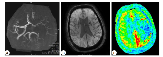
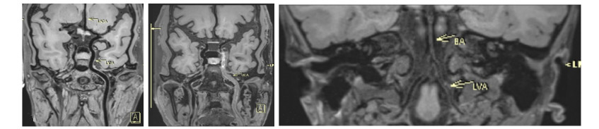
Objective To analyze the clinical value of T2* weighted angiography and three-dimensional arterial spin labeling in the evaluation of cerebral perfusion after unilateral internal carotid artery (ICA) chronic occlusion. Methods Forty-four patients with unilateral ICA occlusion diagnosed by three-dimensional time leap magnetic resonance angiography from January 2018 to March 2022 were retrospectively collected, and all underwent T2* weighted angiography and threedimensional arterial spin labeling sequence examination. The differences of cerebral blood flow (CBF) between the negative and positive groups of ICA supply area prominent vascular sign (PVS) on the occluded side were analyzed, and the CBF values of ICA occluded side and mirror area in the positive and negative groups were compared. Results The CBF values of frontal lobe, parietal lobe, temporal lobe and paraventricular white matter of ICA occlusion in PVS negative group were significantly higher than those in PVS positive group (P < 0.05). There was no significant difference between the CBF values of frontal lobe, parietal lobe, temporal lobe and paraventricular white matter area in ICA occlusion side in PVS negative group and those in mirror area (P>0.05). The CBF values of frontal lobe, parietal lobe, temporal lobe and paraventricular white matter area in ICA occlusion side in PVS positive group were significantly lower than those in mirror area (P < 0.05). Conclusion After chronic occlusion of unilateral ICA, T2* weighted angiography and three- dimensional arterial spin labeling can objectively reflect the establishment of collateral circulation and cerebral perfusion, which can provide image basis for the selection of clinical treatment.
2022, 45(3): 317-322.
doi: 10.12122/j.issn.1674-4500.2022.03.02
Abstract:
Objective To evaluate the accuracy of prenatal MRI for the diagnosis of placenta implantation. Methods MRI images of clinically suspected placenta implantation in our hospital from May 2018 to May 2021 were retrospectively analyzed. The images of 47 pregnant women were included in the study according to the criteria. The consistency of MRI signs of abnormal placenta implantation was analyzed. Combined with surgical pathological results, the diagnostic efficacy of MRI placenta implantation was analyzed. Two diagnostic methods were used to assess placenta implantation, with at least one MRI sign (method 1) and at least two MRI signs (method 2). Results Two physicians had high agreement with the following signs, hypointense band on T2WI sequence (Kappa=0.874), local interruption of placental tissue protrusion in the uterus (Kappa= 0.753), abnormal thickened tortuous vessels in placenta (Kappa=0.870). The overall accuracy of MRI in diagnosing placenta accreta was high: Youden index was 0.69, sensitivity was 90.91% and specificity was 78.57%. MRI diagnosis accuracy of adherent placenta is lower than that of implanted and penetrating placenta. The AUC area of method 1 was 0.714, and that of method 2 was 0.913. The accuracy of method 2 was higher than that of method 1. Conclusion MRI is an effective method to evaluate placenta implantation, and the diagnostic accuracy is highest when at least two abnormal MR signs occur.
2022, 45(3): 323-329.
doi: 10.12122/j.issn.1674-4500.2022.03.03
Abstract:
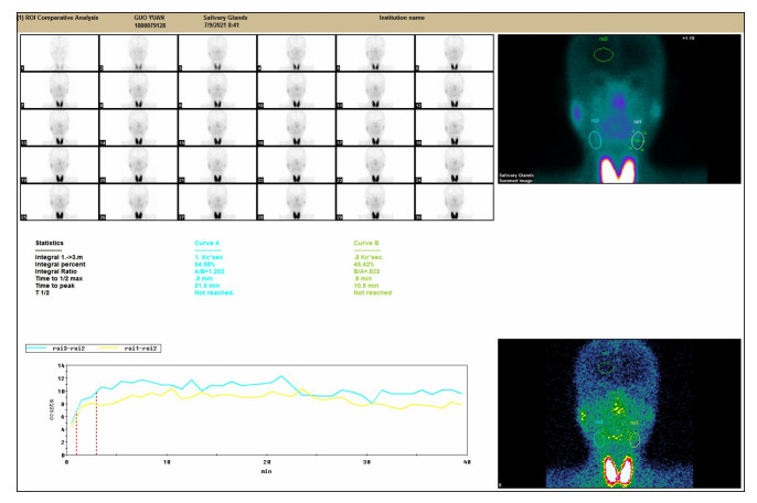
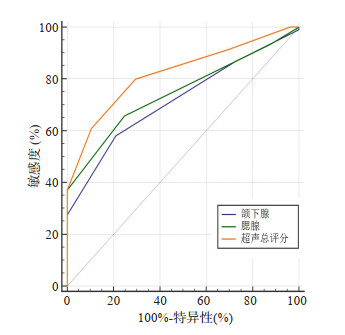
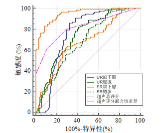
Objective To explore the diagnostic value of salivary gland ultrasound scoring combined with nuclide imaging parameters in sjogren's syndrome (SS). Methods A total of 105 SS patients admitted to our hospital from January 2018 to December 2021 were selected as the study group, and 105 non-SS patients with dry mouth and dry eye were selected as the control group. Salivary gland ultrasonography and radionuclide imaging were used to observe the ultrasonic image, radionuclide imaging and salivary gland function in two groups. ROC curve was used to analyze the diagnostic value of ultrasonic scoring combined with radionuclide imaging in SS patients. Results Ultrasound imaging showed diffuse lesions in parotid gland and one or more hypoechoic nodules in parotid gland or submandibular gland. Radionuclide imaging showed that the uptake function of bilateral parotid gland and submandibular gland was significantly reduced in SS patients, the uptake of 99TcmO4- in salivary gland was reduced, and the concentration of 99TcmO4- in oral cavity was less. No obvious concentration was observed after oral vitamin C. The bilateral parotid gland and submandibular gland were continuously increased. The submandibular gland score, parotid gland score and salivary gland ultrasound score in the study group were significantly higher than those in the control group (P < 0.05). ROC curve was made based on clinical diagnosis, among which the total salivary gland ultrasound score had the highest diagnostic value and had significant differences with the submandibular gland score and parotid gland score (P < 0.05). The levels of UR and SR in the submandibular gland and parotid gland in the study group were significantly lower than those in the control group (P < 0.05). ROC curve was made based on clinical diagnosis, in which the ultrasonic score of salivary gland combined with radionuclide imaging had the highest diagnostic value, and there were significant differences compared with the diagnosis alone (P < 0.05). Conclusion Salivary gland ultrasound scoring combined with radionuclide imaging has high diagnostic value in SS patients.

2022, 45(3): 330-334.
doi: 10.12122/j.issn.1674-4500.2022.03.04
Abstract:
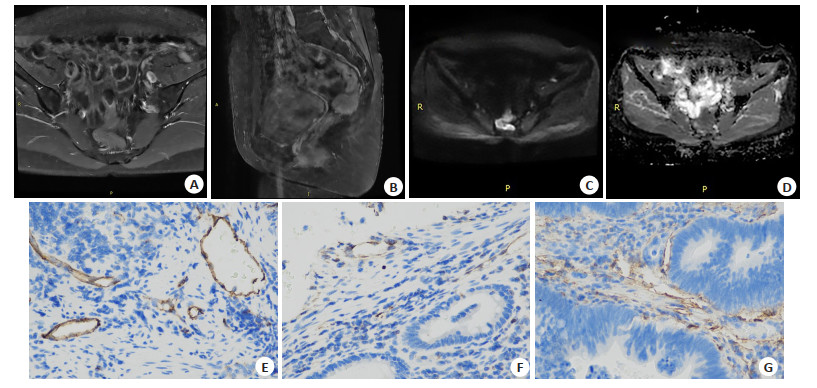
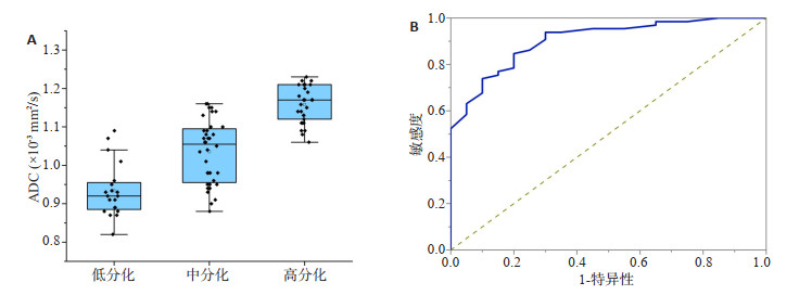
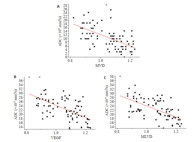
Objective To investigate the diagnostic value of the apparent diffusion coefficient (ADC) of MRI diffusion weighted imaging parameters in preoperative assessment of the differentiation degree and tumor microstructure of rectal cancer. Methods A total of 82 patients with rectal cancer confirmed pathologically from 2018 to 2021 were collected. The ADC values of tumors were measured in all patients after 3.0 T diffusion weighted imaging, and the difference in ADC values of rectal cancers with different levels of differentiation was analyzed. ROC curve was used to determine the efficacy of ADC value in differentiating the differentiation degree of rectal cancer. The correlation between ADC value and microvessel density, vascular endothelial growth factor and microvessel density was analyzed. Results The ADC value of high [(1.16±0.05)×10-3 mm2/s] and moderately [(1.04±0.08)×10-3 mm2/s] differentiated rectal cancer was significantly higher than that of poorly differentiated rectal cancer [(0.93±0.07)×10-3 mm2/s, P < 0.05]. ROC curve showed that the optimal critical value of ADC value to distinguish poorly differentiated from well-differentiated and moderately differentiated rectal cancer was 1.05×10⁻³ mm2/s, and the AUC was 0.902 (95%CI: 0.831-0.972). The ADC value and microvessel density, the percentage of vascular endothelial growth factor positive cells and microlymphatic vessel density of rectal cancer tumors were all negatively correlated (r=- 0.56, -0.61, - 0.59, P < 0.05). Conclusion The ADC value of rectal cancer tumor is helpful to evaluate tumor differentiation and microstructure characteristics. It indirectly reflects tumor angiogenesis and microlymphatic vessel density.
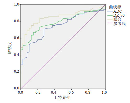
Application of 3.0 T MRI combined with serum DR-70 detection in early diagnosis of colorectal cancer
2022, 45(3): 335-338.
doi: 10.12122/j.issn.1674-4500.2022.03.05
Abstract:
Objective To investigate the value of 3.0 T MRI combined with serum fibrin- degrading complex (DR-70) in the diagnosis of early colorectal cancer. Methods Ninety-six patients with early colorectal cancer admitted to our hospital from August 2018 to August 2020 were selected as observation group, and forty patients with colorectal benign tumor were selected during the same period as control group. Implementation of 3.0T MRI dynamic enhancement and diffusion weighted imaging sequence scan and serum DR-70 test, using the Spearman correlation analysis the relationship between early colorectal cancer of 3.0T MRI parameters and serum DR-70 level. ROC was used to evaluate the diagnostic efficacy of the two detection indexes for early colorectal cancer. Results Among 96 patients, 94 cases of colorectal cancer were detected, 2 cases were missed, the detection rate was 97.92%. 3.0T MRI could better observe the location, morphology, invasion and other conditions of the tumor lesion. The tumor lesion showed medium low signal in T1 image, slightly high signal in T2 image, uneven enhancement in the early stage of dynamic enhancement scan, slightly low signal in the delay stage, significantly high signal in diffusion weighted imaging, and low signal in ADC image. The ADC value of MRI parameters in observation group was significantly lower than that in control group (P < 0.05), while the DR-70 level was significantly higher than that in control group (P < 0.05). Spearman correlation analysis showed that ADC value was significantly negatively correlated with colorectal cancer (r=-0.383, P < 0.05), while DR-70 level was significantly positively correlated with colorectal cancer (r=0.460, P < 0.05). ROC curve showed that the area under the curve of ADC and DR-70 combined diagnosis of colorectal cancer was 0.850, which was significantly higher than the area under the curve of each individual index (P < 0.05). Conclusion 3.0 T MRI and DR-70 combined detection can significantly improve the diagnostic and differential performance of colorectal cancer patients.
2022, 45(3): 339-343.
doi: 10.12122/j.issn.1674-4500.2022.03.06
Abstract:
Objective To investigate the predictive value of CT-based radiomics in lymph node metastasis in stage T1 lung adenocarcinoma. Methods A total of 140 patients with lung adenocarcinoma admitted to our hospital from June 2016 to May 2019 were retrospectively selected. With the results of pathological tissue samples in intraoperative examination as the gold standard, they were divided into the gold standard positive group (n=67) and the gold standard negative group (n=73). Preoperative enhanced CT and preoperative CT imaging were used to evaluate lymph node metastasis in the two groups. Delong test was used to evaluate the value of CT imaging and enhanced CT in predicting lymph node metastasis in both groups. Results In the two groups, the prediction ratioof positive lymph node metastasis by CT radiomics was 86.57% (58/67), which was higher than that by enhanced CT method [64.18% (43/67)] (P < 0.05). The prediction ratioof negative lymph node metastasis by CT radiomics was 100.00% (73/73), which was higher than that by enhanced CT [93.15% (68/73)] (P < 0.05). In the gold standard positive group, the risk score of patients with lymph node metastasis was significantly higher than that of patients without lymph node metastasisvia CT radiomics (P < 0.05). In the gold standard positive group, the AUC area of CTradiomics in prediction of lymph node metastasiswas significantly higher than that of enhanced CT (P < 0.05), and so was in the gold standard negative group (P < 0.05). Conclusions Compared with enhanced CT, CT radiomics has a higher positive predictive value and negative predictive value for predicting lymph node metastasis of lung adenocarcinoma before surgery with high diagnostic value.
2022, 45(3): 344-347.
doi: 10.12122/j.issn.1674-4500.2022.03.07
Abstract:
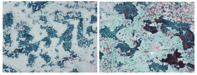
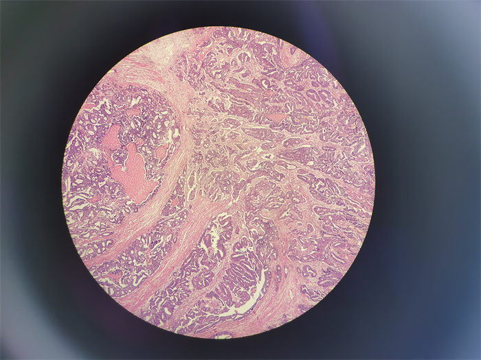
Objective To analyze the pathological characteristics of gastric polyps diagnosed by endoscopic forceps biopsy, and explore the difference between the pathological diagnosis of gastric polyp specimens diagnosed by endoscopic forceps biopsy and the pathological diagnosis of gastric polyp specimens. Methods Clinical data of 145 patients who underwent endoscopic forceps biopsy diagnosis and endoscopic gastric polypectomy in the department of Gastroenterology of our hospital from February 2021 to February 2022 were retrospectively selected. We analyzed the pathological characteristics of gastric polyps diagnosed by endoscopic forceps biopsy, and compared the consistency with the pathological results of endoscopic gastric polypectomy. The influencing factors affecting the difference between endoscopic clamp biopsy and pathological diagnosis of gastric polyp were evaluated. Results A total of 124 cases (85.52%) had single polyp and 21 cases (14.48%) had multiple polyp in 145 patients. A total of 171 polyps were detected, 119 polyps were consistent with the pathological diagnosis after endoscopic clamp biopsy, accounting for 69.59%, and 52 polyps were inconsistent, accounting for 30.41%. The pathological diagnosis result Kappa value of the two diagnoses was 0.182, the difference was statistically significant (P < 0.05). Logistic multivariate analysis showed that age≥60 years and polyp size≥2.0 cm were independent influencing factors influencing the histological difference between endoscopic clamp biopsy and electrosurgical diagnosis. Conclusion There is inconsistency between the nature of gastric polyp diagnosed by endoscopic clamp biopsy and the results of pathological diagnosis, especially for patients with age≥60 years and polyp size≥2.0 cm. Special attention should be paid to the coexistence of gastric polyps and cancerous lesions in such patients in clinical diagnosis.
2022, 45(3): 348-352.
doi: 10.12122/j.issn.1674-4500.2022.03.08
Abstract:
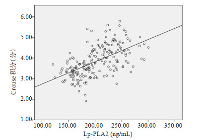
Objective To investigate the relationship between the imaging characteristics of carotid atherosclerotic plaque (CAS) and plasma lipoprotein- associated phospholipase A2 (Lp-PLA2) in patients with cerebral infarction by high resolution MRI scanning. Methods A total of 196 patients with cerebral infarction admitted to our hospital from June 2019 to June 2020 were selected as the cerebral infarction group. Forty patients without cerebral infarction admitted to our hospital wards during the same period were selected as the control group. The general information and plaque distribution of the two groups were compared. The plaque properties of all patients with cerebral infarction were compared. Patients with cerebral infarction were divided into stable plaque group (n=61), unstable plaque group (n=123) and no plaque group (n=12) according to whether the plaque was stable. Biochemical indexes (total cholesterol, triglyceride, high density lipoprotein cholesterol, low density protein cholesterol) and Lp-PLA2 levels in the three groups were compared. Crouse score and Lp-PLA2 level in patients with different plaque grades (grade 0, 1, 2 and 3) were compared. The correlation between Crouse score and Lp-PLA2 in patients with cerebral infarction was analyzed by Pearson analysis. Results Among 196 patients with cerebral infarction, 184 (93.88%) had CAS plaque. Of the 40 in control group, 9 (22.50%) had CAS plaques; The incidence of CAS plaque in cerebral infarction group was higher than that in control group (P < 0.05). The expression levels of total cholesterol, triglyceride, low density protein cholesterol and Lp- PLA2 in the stable plaque group, the unstable plaque group and the non-plaque group were lower than that in the stable plaque group and the unstable plaque group (P < 0.05), and the high density lipoprotein cholesterol expression levels in the three groups were lower than that in the stable plaque group and the non-plaque group (P < 0.05). There were statistically significant differences in Crouse score and Lp-PLA2 level in different plaque grades of cerebral infarction patients (P < 0.05). The Crouse score and Lp-PLA2 level were compared in grade 1 < grade 2 < grade 3 (P < 0.05). The Pearson correlation analysis showed that there was a positive correlation between plaque grade Crouse score and Lp-PLA2 level in patients with cerebral infarction (P < 0.05). Conclusion High resolution MRI scan of cerebral infarction patients can show significant abnormalities in the size and nature of CAS plaques. There is a certain correlation between CAS plaque grade and serum Lp-PLA2 level, and serum Lp-PLA2 level can reflect the status of CAS plaques in patients.
2022, 45(3): 353-357.
doi: 10.12122/j.issn.1674-4500.2022.03.09
Abstract:
Objective To explore the relationship between perfusion parameters of spiral CT and C-reactive protein, procalcitonin, serum amylase, hematocrit and clinical symptoms in patients with severe pancreatitis. Methods Clinical data of 102 patients with pancreatitis admitted to our hospital from January 2019 to January 2021 were retrospectively collected. The patients were divided into the mild group (n=64, score of Ranson < 3 points) and the severe group (n=38, score of Ranson 3-11 points) according to Ranson score. The inflammatory indicators, score of clinical symptom and CT concern parameters of the two groups were statistically analyzed, and the correlation between CT perfusion parameters and inflammatory indicators and clinical symptoms was analyzed by Pearson correlation. Results The levels of serum C-reactive protein, procalcitonin, serum amylase, peripheral blood hematocrit and scores of Ranson, acute physiology and chronic health score (APACHE- II) in the severe group were higher than those in the mild group (P < 0.05), while blood flow, blood flow volume were lower than those in the mild group (P < 0.05). Pearson correlation analysis showed that blood flow and blood flow volume were significantly negatively correlated with level of peripheral blood hematocrit, scores of Ranson and APACHE-II (r=-0.632, -0.673, -0.703 and -0.715, -0.662, -0.637, P < 0.05). Conclusion The levels of serum C-reactive protein, procalcitonin and serum amylase, peripheral blood hematocrit, scores of Ranson and APACHE- II are increased, while blood flow and blood flow volume decreased in patients with severe pancreatitis. Blood flow and blood flow volume are significantly negatively correlated with level of peripheral blood hematocrit, scores of Ranson and APACHE-II.
2022, 45(3): 358-362.
doi: 10.12122/j.issn.1674-4500.2022.03.10
Abstract:
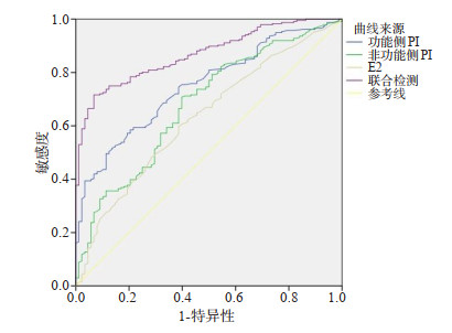
Objective To evaluate the efficacy of vaginal ultrasound measurement of bilateral pulsatility index (PI) in combination with serum estrogen and progesterone testing in the diagnosis of ectopic pregnancy. Methods A total of 107 women with ectopic pregnancy diagnosed and treated in Chongming Branch of Xinhua Hospital from February 2019 to February 2021 were selected as observation group, and another 107 women in early intrauterine pregnancy who underwent prenatal examination in our hospital during the same period were selected as control group. Parameters including resistance index (RI), bilateral PI, peak systolic velocity, end-diastolic velocity, follicle-stimulating hormone, luteinizing hormone, estradiol (E2) and progesterone were detected in both groups. Then the diagnostic efficacy of combined detection of PI, E2 and progesterone for ectopic pregnancy were evaluated. Results Compared with control group, observation group had a decrease in RI (t=51.534, P < 0.001), along with an increase in functional PI (t=5.884, P < 0.001), non functional PI (t=8.565, P < 0.001), peak systolic velocity (t=45.597, P < 0.001) and end-diastolic velocity (t=12.302, P < 0.001). Observation group had significantly lower levels of follicle-stimulating hormone (t=3.676, P < 0.001), luteinizing hormone (t=6.334, P < 0.001), E2 (t=28.259, P < 0.001) and progesterone (t=48.787, P < 0.001) compared with control group. The combined detection of bilateral PI, progesterone and E2 achieved higher diagnostic efficacy than the separate detection. ROC curve indicated that the area under the curve of combined detection of bilateral PI, progesterone and E2 in the diagnosis of ectopic pregnancy was larger than that of separate detection, and the cut- off values of functional PI, non functional PI, E2 and progesterone were 2.58, 2.76, 422.56 pmol/L and 15.13 ng/mL, respectively. Conclusion Application of vaginal ultrasound measurement of bilateral PI in combination with serum estrogen and testing is of great significance in the clinical diagnosis of ectopic pregnancy.
2022, 45(3): 363-368.
doi: 10.12122/j.issn.1674-4500.2022.03.11
Abstract:
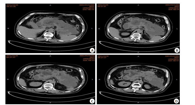
Objective To investigate the effect of laparoscopic pancreaticoduodenectomy in the treatment of pancreatic head malignant tumor, and analyze the imaging characteristics of pancreas before and after operation. Methods A total of 110 patients with pancreatic head malignant tumor treated in our hospital from January 2020 to June 2021 were selected. They were divided into 2 groups according to different surgical methods. Among them, 61 patients in the observation group were treated with laparoscopic duodenectomy, and 49 patients in the control group were treated with open duodenectomy. Perioperative indicators, surgical integrity and inflammatory response of patients in the two groups were observed. The incidence of complications, preoperative and postoperative pancreatic imaging characteristics were analyzed. Results There were no statistically significant differences in the operative time, the completeness of surgical resection and the incidence of complications between the two groups (P > 0.05). The amount of intraoperative blood loss and length of hospital stay were significantly shorter than those in the control group, with statistically significant differences (P < 0.05). The serum levels of C-reactive protein, IL-6 and TNF-α in observation group were significantly lower than those in control group (P < 0.05). Preoperative CT showed irregular cystic low-density shadow on the head of the pancreas, with lobed edges, internal separation, uneven density and clear enhanced boundary. After surgery, patients had encapsulated effusion in the operation area and slightly higher local density. Conclusion Laparoscopic pancreaticoduodenectomy in the treatment of pancreatic head malignant tumor can reduce inflammatory response, reduce intraoperative bleeding and shorten hospital stay. The integrity of surgical resection is similar to that of open surgery, and the preoperative CT images can effectively guide the surgical process and identify postoperative pancreatic fistula and other complications.
2022, 45(3): 369-373.
doi: 10.12122/j.issn.1674-4500.2022.03.12
Abstract:
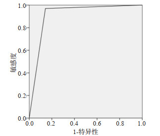
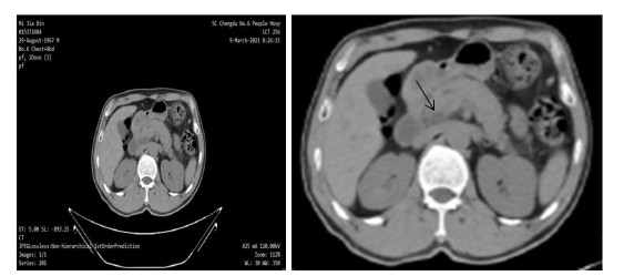
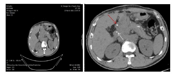
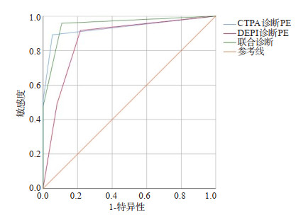
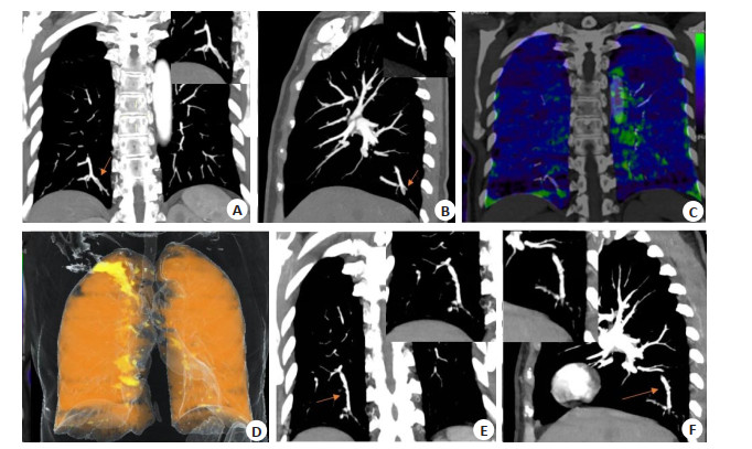
Objective To investigate the role of perfusion defects in dual-energy CT pulmonary perfusion in the diagnosis and risk stratification of pulmonary embolism. Methods A total of 157 patients diagnosed with suspected pulmonary embolism in our hospital from January 2018 to December 2020 were enrolled, and pulmonary embolism was finally confirmed in 120 patients. All patients were performed with dual- energy CT pulmonary perfusion imaging (DEPI) and pulmonary artery CT angiography (CTPA). The number of pulmonary embolisms, pulmonary artery loss area fraction, right/left ventricular short-axis maximum diameter ratio and cardiac biological markers were recorded and compared. All patients were followed up for 3 months with the outcomes recorded. Results Dual-energy CT was consistent with CTPA in the diagnosis of pulmonary embolism, with a diagnostic coincidence rate of 86.1%. For CTPA combined with DEPI, AUC value was 0.95, with the specificity, sensitivity, and Youden index of 89.20%, 95.80% and 0.85, respectively, better than CTPA and DEPI alone in the diagnosis of pulmonary embolism. There were significant differences in perfusion defect area, cardiac biological markers, and right/left ventricular short- axis maximum diameter ratio levels between the low-risk, medium-risk, and high- risk groups of pulmonary embolism (P < 0.05). Conclusion DEPI can be used as a supplement for the diagnosis of pulmonary embolism by CTPA. Through risk stratification for pulmonary artery perfusion loss area fraction, it is a new option for clinical diagnosis and treatment, suitable for clinical promotion.
2022, 45(3): 374-377.
doi: 10.12122/j.issn.1674-4500.2022.03.13
Abstract:
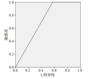
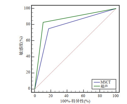
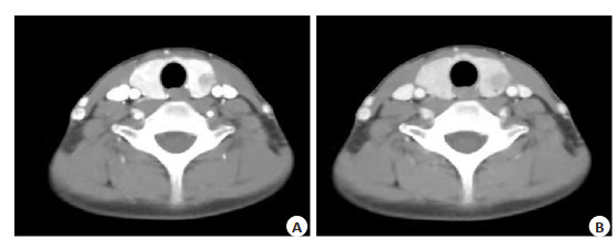
Objective To explore the characteristics and diagnostic value of ultrasound and multi-slice spiral CT (MSCT) for thyroid cancer. Methods A total of 349 cases of patients suspected of thyroid cancer who were admitted to the hospital between January 1, 2018 and October 8, 2021 were selected. According to the results of ultrasonography, 203 cases were divided into malignant group and 146 cases were divided into benign group. According to the results of MSCT, 198 cases were divided into malignant group and 151 cases were divided into benign group. Their ultrasound and MSCT characteristics were analyzed. The ROC curve was plotted to analyze the specificity and sensitivity of ultrasound and MSCT in the diagnosis of thyroid cancer. Results There were significant differences between the malignant group and benign group which were examined by ultrasonography in solid nodules, hypoecho, calcification, irregular morphology and fuzzy boundaries (P < 0.05). There were significant differences between the malignant group and benign group which were examined by MSCT in single lesions, fuzzy boundaries, irregular morphology, calcification, cystic degeneration, heterogeneous enhancement, and enlarged lymph nodes (P < 0.05). The area under the curve value, sensitivity and specificity of MSCT to diagnose thyroid cancer were 0.789, 75.18% and 82.69%, which of ultrasound were 0.862, 82.98% and 89.42%. Conclusion The imaging features of thyroid carcinoma diagnosed by ultrasound are solid nodules, hypoechoic, calcification, irregular morphology and fuzzy boundaries. The imaging features of MSCT in the diagnosis of thyroid cancer are single focus, fuzzy boundaries, irregular morphology, calcification, cystic change, uneven enhancement and enlarged lymph nodes. Ultrasound is more specific and sensitive than MSCT in the diagnosis of thyroid cancer.
2022, 45(3): 378-381.
doi: 10.12122/j.issn.1674-4500.2022.03.14
Abstract:
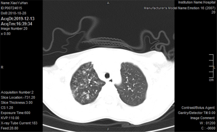
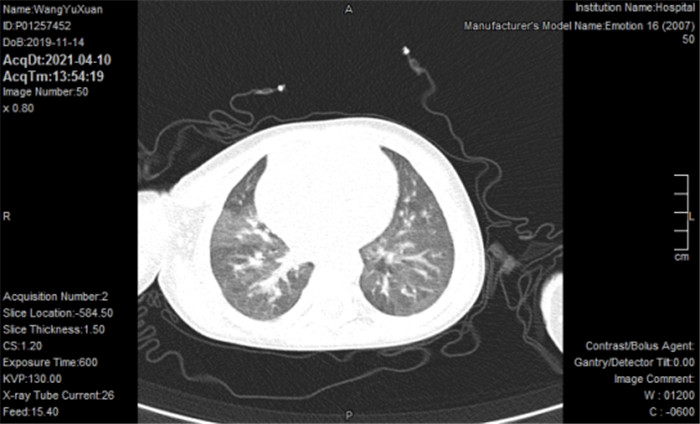
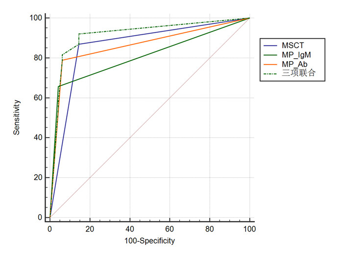
Objective To analyze the diagnostic efficacy of multi-slice spiral CT (MSCT) combined with serum mycoplasma pneumoniae immunoglobulin M (MP- IgM) and mycoplasma pneumoniae antibody (MP-Ab) in children with mycoplasma pneumonia. Methods A total of 86 children with suspected mycoplasma pneumonia admitted to our hospital from January 2019 to January 2021 were selected as the study subjects, and divided into the infection group (n=38) and non-infection group (n=48) according to the gold standard. MSCT was used to scan all children, and serum MP-IgM and MP-Ab levels were measured. ROC was used to analyze the diagnostic efficacy of MSCT combined with serum MP-igm and MP-Ab for mycoplasma pneumonia in children. Results MSCT images showed that mycoplasma infection pneumonia in children showed bronchial inflation phase, bronchial wall thickening, ground- glass opacities, reticular opacities and bronchovascular bundle thickening. In addition, there were obvious fan-shaped thin slices. Among 38 cases, 15 cases (39.47%) occurred in unilateral left lung, 17 cases (44.74%) in unilateral right lung, and 6 cases (15.79%) in bilateral lung. MSCT had good consistency with gold standard (Kappa=0.72). The positive rates of MP-IgM and MP-Ab in infected group were significantly higher than those in non-infected group (P < 0.05). ROC curve was made based on clinical diagnosis, among which three combinations had the highest diagnostic value, and there were significant differences compared with MSCT, MP-IgM and MP-Ab (P < 0.05). Conclusion MSCT combined with MP-IgM and MP-Ab has high diagnostic value in children with mycoplasma pneumonia.
2022, 45(3): 382-386.
doi: 10.12122/j.issn.1674-4500.2022.03.15
Abstract:
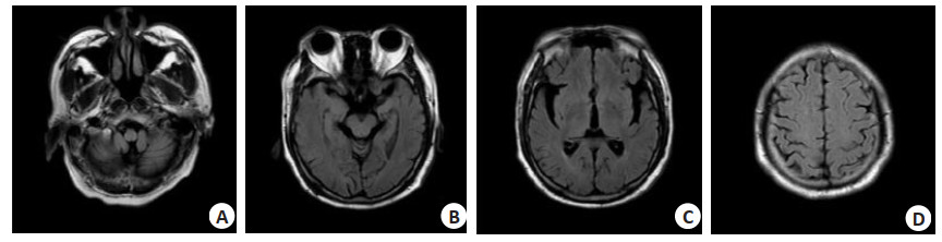
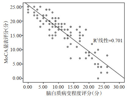
Objective To investigate the correlation between the intima-media thickness of the bilateral carotid arteries, the number of plaques and the degree of stenosis, white matter lesions (WHL) and cognitive function changes. Methods A total of 146 patients in our hospital from January 2020 to January 2021 were selected. Among the patients, 73 patients with WHL were treated as WHL group, and 73 non-WHL patients were treated as non-WHL group. For patients with WHL, a prospective observational cohort study method was performed. Cognitive function scores, cervical vascular color Doppler ultrasound, and head MRI WHL were scored for each case, and various indicators of carotid artery lesions and WHL scales were analyzed. The correlation between score and cognitive function scale score, to explore the relationship between carotid artery disease and WHL and cognitive dysfunction. Results The intima-media thickness, total number of plaque points and total stenosis degree points in WML group were higher than those in non-WML group (1.97±0.83 mm vs 0.87±0.12 mm, 6±3.28 vs 4±1.96, 4.83±1.17 vs 2.89±1.05, P < 0.05). The MoCA score of WML group was significantly higher than that of non-WML group (18±6.82 vs 10±5.67, P < 0.01). The correlation analysis of carotid artery color doppler ultrasound index and WML degree score showed that carotid intima-media thickness (r=0.828, P=0.020), stenosis degree (r=0.897, P=0.010) and WML degree score were correlated. The number of plaques and the WML degree showed no correlation (P > 0.05). The correlation analysis of carotid artery color doppler ultrasound index and MoCA score in the WML group showed that intima-media thickness (r=-0.793, P=0.020), stenosis degree (r=-0.873, P=0.010) and MoCA scale score were correlated in the WML group. There was no correlation between plaque number and MoCA score (P > 0.05). The correlation analysis of the MoCA scale score and the WHL degree score of the WHL group showed that the MoCA scale score and the WHL degree score of the WHL group were negatively correlated (r=-0.837, P < 0.01). Conclusion The carotid intima-media thickness and the degree of vascular stenosis of cervical vascular color Doppler ultrasound are related to the degree of WHL and cognitive dysfunction. The degree of WHL is negatively correlated with the degree of cognitive dysfunction. Carotid artery color Doppler ultrasound is useful for clinical diagnosis. And predict WML and cognitive dysfunction has a certain guiding role.
2022, 45(3): 387-393.
doi: 10.12122/j.issn.1674-4500.2022.03.16
Abstract:
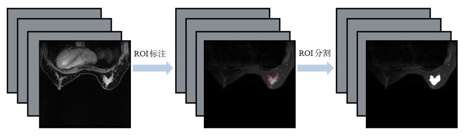
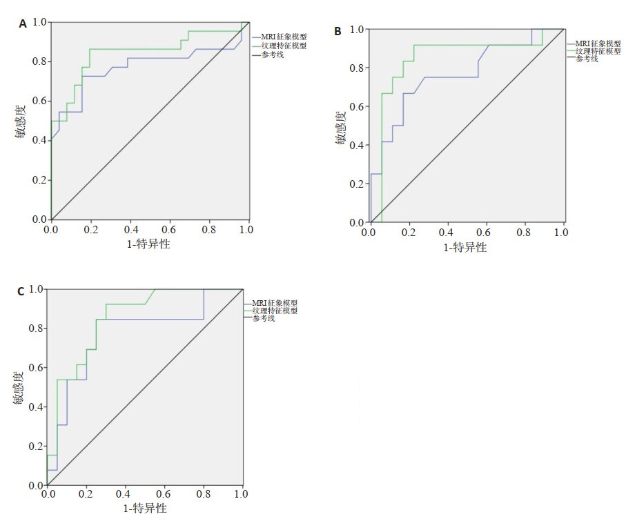
Objective To explore the value of MRI texture features in evaluating the vascular infiltration of invasive breast cancer, as well as the missing molecular typing and cytokeratin 5/6 gene expression information imputation. Methods A total of 114 patients with breast cancer diagnosed by MRI and pathology were divided into training group (n=51), internal validation group (n=30) and external validation group (n=33). According to the clinicopathological features and MRI texture features, the independent risk factors of vascular infiltration were observed, the MRI sign model and texture feature model were constructed and their diagnostic efficacy was compared. Non-negative matrix decomposition (NMF) and collaborative filtering imputation models were established with the texture features selected by cross-validation support vector machine recursive feature elimination, and pathological information of breast cancer patients. The receiver operating characteristic curve was used to evaluate their imputation performance. Results There were statistically significant differences in peritumor prethoracic edema and lymph node metastasis status, which were independent risk factors for vascular invasion (P < 0.05). The area under the curve (AUC) of the texture feature model in diagnosis of breast cancer vascular invasion was higher than that of MRI model (P < 0.05). When the loss rate was 20%- 40%, the AUC of NMF was higher than that of collaborative filtering (P < 0.05). When the feature number was 140, the AUC of NMF was higher than that of collaborative filtering (P < 0.05). The AUC of NMF with image omics was higher than that of NMF without image omics (P < 0.05). Conclusion MRI texture features can effectively predict breast cancer vascular invasion before surgery, and fill in missing molecular typing and cytokeratin 5/6 gene expression information.
2022, 45(3): 394-399.
doi: 10.12122/j.issn.1674-4500.2022.03.17
Abstract:
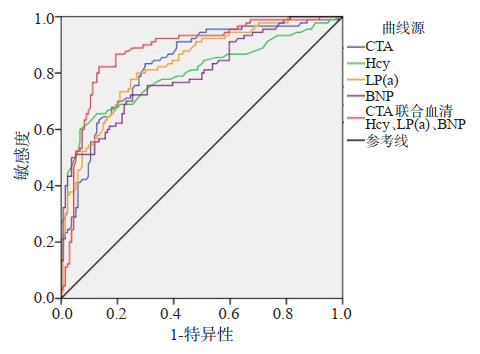
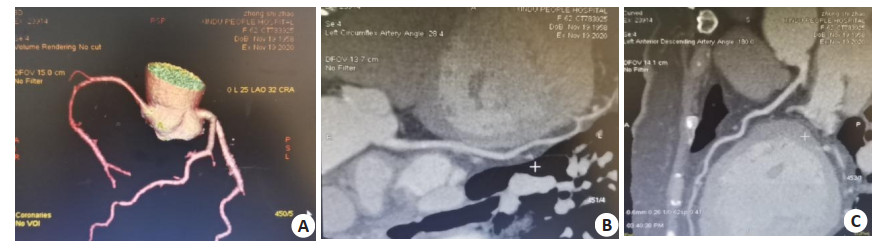
Objective To explore the value of CT angiography (CTA) combined with serum homocysteine (Hcy), lipoprotein (a)[LP(a)] and B-type natriuretic peptide (BNP) in evaluating coronary atherosclerosis stenosis. Methods A total of 150 patients with coronary heart disease admitted to the hospital between December 2020 and July 2021 were selected as the observation group. Meanwhile, 86 healthy individuals were selected as the control group. All patients were performed with CTA and detection of serum Hcy, BNP and LP(a) levels. The correlations of above- mentioned serum indicators with disease type, number of affected coronary arteries, coronary artery stenosis degree, character of plaque and CTA parameters were analyzed. Results Serum Hcy, BNP and LP(a) levels, plaque burden, vascular artery stenosis degree, and reconstruction index (RI) in the observation group were higher than those in the control group, while blood vessel volume was smaller than that in the control group (P < 0.05). Serum Hcy, LP(a) and BNP levels, and plaque burden decreased in sequence from patients with acute myocardial infarction, patients with unstable angina pectoris, patients with stable angina pectoris, to patients with latent coronary heart disease (P < 0.05). As affected coronary artery increased and coronary artery stenosis aggravated, the levels of serum indicators, plaque burden and RI in patients with coronary heart disease increased (P < 0.05). The levels of serum indicators, plaque burden, vascular stenosis degree and RI in patients with noncalcified plaques were higher than those in patients with calcified plaques (P < 0.05). The levels of serum indicators in patients with coronary heart disease were positively correlated with acute myocardial infarction, the number of affected coronary arteries, coronary artery stenosis degree, non-calcified plaque, plaque burden, vascular stenosis degree, and RI (P < 0.05). ROC curve showed that the sensitivity, specificity and area under the curve of CTA combined with the three serum indicators to diagnose severe coronary atherosclerosis stenosis (stenosis > 75%) were 0.81, 0.82 and 0.878, respectively. Conclusion Serum Hcy, LP(a) and BNP levels are increased in patients with coronary heart disease. The combined use of CTA parameters can better diagnose the degree of coronary atherosclerosis stenosis.
2022, 45(3): 400-403.
doi: 10.12122/j.issn.1674-4500.2022.03.18
Abstract:
Objective To investigate the diagnostic value of ultrasound-guided fine needle aspiration (US-FNAC) for papillary thyroid carcinoma. Methods The data of 235 patients with thyroid nodules examined in our hospital from January 2015 to March 2022 were selected. According to the maximum diameter of nodules, they were divided into four groups: group A (< 5 mm), group B (5.1-10 mm), group C (10.1-20 mm) and group D (≥20 mm). The pathological results were taken as the gold standard, and the positive rate of US- FNAC, undiagnosed rate, accuracy rate, positive predictive value, negative predictive value, and partition of metastatic nodules in four groups were recorded. Results US-FNAC examination showed that with the increase of nodule diameter, the positive rates of the four groups were continuously increased (29.41%, 39.50%, 41.51%, 61.82%), and the total positive rate was 42.45%. The difference in positive cases between groups was statistically significant (χ2=12.180, P=0.007). The total undiagnosed rate of US-FNAC cytology in the four groups was 1.80%, and the fluctuation decreased with the increase of nodules (1.96%, 2.52%, 1.89%, 0.00%). There was no significant difference between the four groups (χ2=1.370, P=0.713). The results of US-FNAC cytological examination showed that the total accuracy (coincidence rate) of the four groups was 95.32%, which fluctuated with the increase of nodule diameter, but the difference was not statistically significant (χ2=0.40, P=0.941). The total positive predictive value of the four groups of nodules reached 94.92%, and the total negative predictive value was slightly higher than the positive predictive value, which was 95.63%. The total sensitivity and total specificity were 94.12% and 96.23%, respectively. Among the 195 metastatic nodules, the coincidence rates of ultrasound regional localization and pathological examination were 95.45%, 98.11%, 86.21% and 97.89%, respectively. Conclusion The positive rate of US- FNAC cytology in the diagnosis of papillary thyroid carcinoma is affected by nodule size, but has little effect on accuracy, positive predictive value and negative predictive value.
2022, 45(3): 404-407.
doi: 10.12122/j.issn.1674-4500.2022.03.19
Abstract:
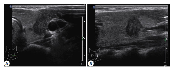
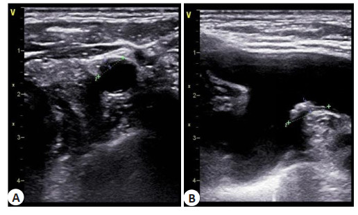
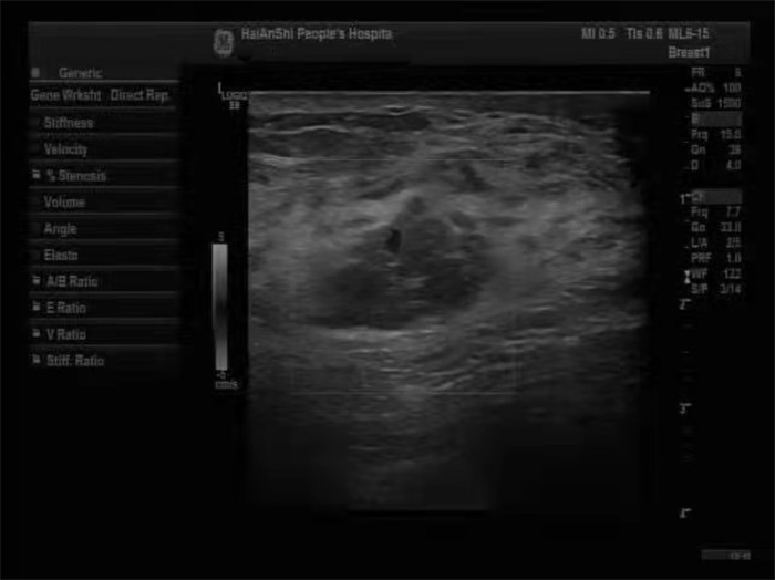
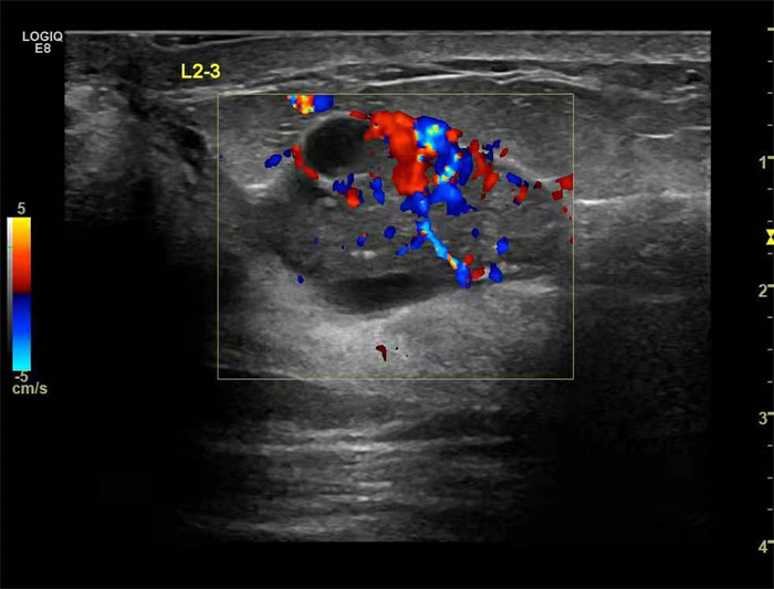
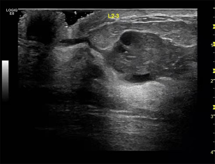
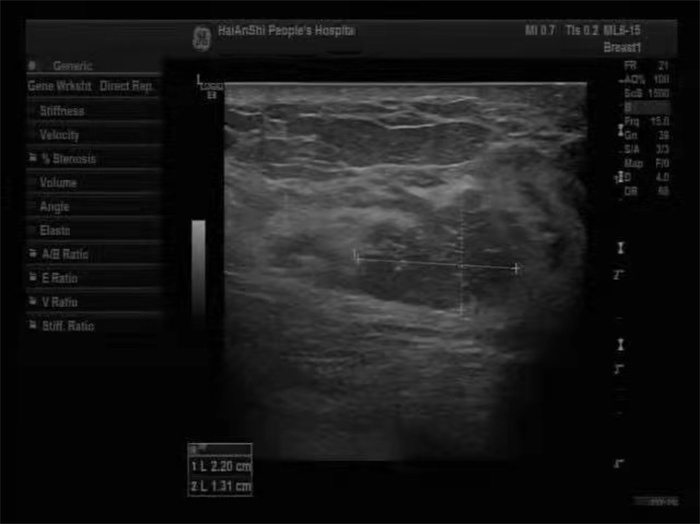
Objective To explore the differential diagnosis between ductal breast cancer (DBC) and intraductual papilloma (IDP) by ultrasonography. Methods This retrospective study enrolled 110 patients with breast diseases treated in our hospital from January 2017 to December 2020. These patients included 59 patients with DBC and 51 patients with IDP. The differences of the routine ultrasound features and ultrasound blood flow between the two groups were compared.The logistic regression diagnostic model was analyzed. Results There were significant differences in the catheter morphology (χ2=25.692, P < 0.001), catheter running (χ2=20.321, P < 0.001), catheter wall echo (χ2=6.052, P=0.014), calcification foci (χ2=34.552, P < 0.001) and blood flow distribution (χ2=22.441, P < 0.001) between patients with IDP and DBC, while there was no significant difference in blood flow grade between the two groups (P>0.05). Through multivariate analysis, the patient's catheter, catheter running, catheter wall echo, calcification foci and blood flow distribution were important factors in the diagnosis of DBC. Conclusion Ultrasound has good differential diagnostic significance for patients with DBC and IDP. The thickening of catheter, irregular routing of catheter, unclear echo of catheter wall, unclear calcification foci and blood flow distribution are the important basis for the diagnosis of DBC.
2022, 45(3): 408-412.
doi: 10.12122/j.issn.1674-4500.2022.03.20
Abstract:
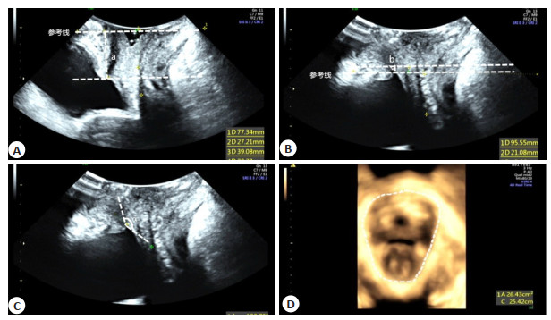
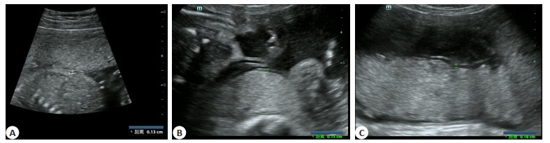
Objective To evaluate the transperineal color Doppler ultrasound parameters of women with postpartum stress urinary incontinence (SUI), and the effect of pelvic floor biofeedback electrical stimulation. Methods A total of 120 women with postpartum SUI admitted to the hospital from January 2019 to October 2021 were selected. They were divided into treatment group and control group by random number table method, with 60 cases in each group. The control group was treated with routine health education and postpartum health care. The treatment group was treated with biofeedback electrical stimulation. 1-hour pad test was conducted, pelvic floor ultrasound parameters were measured, and quality of life was evaluated before treatment and after 3 months of treatment. The treatment response rate, residual urine volume before and after treatment, frequency and volume of urine leakage were compared between the two groups. Results The total response rate of treatment group was higher than control group (95.00% vs 81.67%, P < 0.05). After treatment, the bladder neck descent, bladder lowest descent, retrovesical angle, levator hiatus area, residual urine volume, frequency and volume of urine leakage, the International Consultation Incontinence Questionnaire Short-Form (ICIQ-SF) scores and the Incontinence Impact Questionnaire (IIQ-7) scores were reduced, while levator thickness was increased in the two groups (P < 0.05). After treatment, bladder neck descent, bladder lowest descent, retrovesical angle, levator hiatus area, residual urine volume, frequency and volume of urine leakage, the ICIQ-SF scores and IIQ-7 scores of treatment group were smaller/lower than those of the control group, and levator thickness was larger than that of the control group (P < 0.05). Conclusion Applying pelvic floor biofeedback electrical stimulation to women with postpartum SUI can improve the curative effect and pelvic floor function, relieve symptoms of SUI. Transperineal color Doppler ultrasonography can provide objective evaluation indicators for pelvic floor function damage and rehabilitation in SUI patients.
2022, 45(3): 413-418.
doi: 10.12122/j.issn.1674-4500.2022.03.21
Abstract:
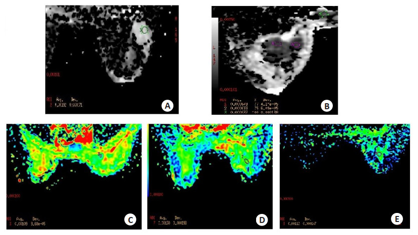
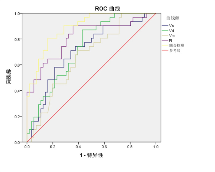
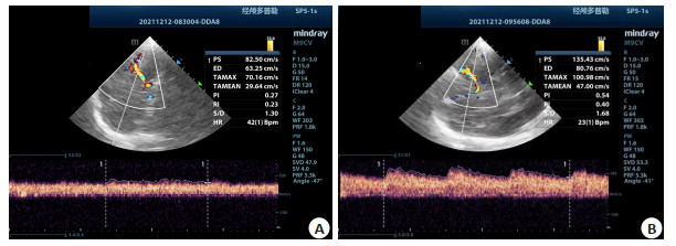
Objective To investigate the application value of transcranial Doppler ultrasonography in cerebral hemodynamic monitoring and prognosis evaluation of patients with extracorporeal membrane oxygenation. Methods The clinical data of 87 patients who underwent extracorporeal membrane oxygenation in our hospital from May 20 th, 2015 to January 3 rd, 2020 were retrospectively analyzed. The indexes of cerebral hemodynamics[peak systolic blood flow velocity (Vs), peak diastolic flow velocity (Vd), mean blood flow velocity (Vm), pulsatility index (PI)] and cardiac function indicators [left ventricular ejection fraction (LVEF), left ventricular outflow tract blood flow velocity time integral (VTI), mean arterial pressure (MAP)] were compared before and after treatment. After 1 year of follow-up, the prognosis status of 87 patients was counted. According to their different prognostic status, they were divided into good prognosis group (n=56) and poor prognosis group (n=31). The gender, age, disease classification, renal insufficiency, abnormal liver function, multiple organ failure, extracorporeal membrane oxygenation flow, inotrope score, Vs, Vd, Vm, PI, LVEF, MAP levels were compared between two groups. Results There was no significant difference in Vs, Vd, Vm and PI values before and after treatment (P>0.05). After extracorporeal membrane oxygenation treatment, LVEF, MAP and VTI increased, and the difference was statistically significant (P < 0.05). Univariate analysis showed that there were statistically significant differences in the rates of renal insufficiency, multiple organ failure, and Vs, Vd, Vm, PI, LVEF, VTI and MAP values between the two groups (P < 0.05). There was no significant difference in gender, age, disease classification, extracorporeal membrane oxygenation flow and inotrope score between the two groups (P>0.05). Vs, Vd, Vm, PI and MAP were independent risk factors for poor prognosis in patients with extracorporeal membrane oxygenation (P < 0.05). Conclusion Cerebral hemodynamic indexes (Vs, Vd, Vm, PI) are closely related to the prognosis of patients with extracorporeal membrane oxygenation. It is necessary to strengthen the detection of various indicators in clinical practice to assess the prognosis of patients with extracorporeal membrane oxygenation as soon as possible.
2022, 45(3): 419-423.
doi: 10.12122/j.issn.1674-4500.2022.03.22
Abstract:
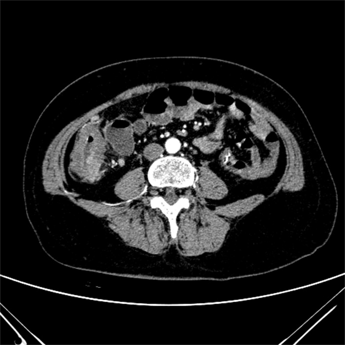
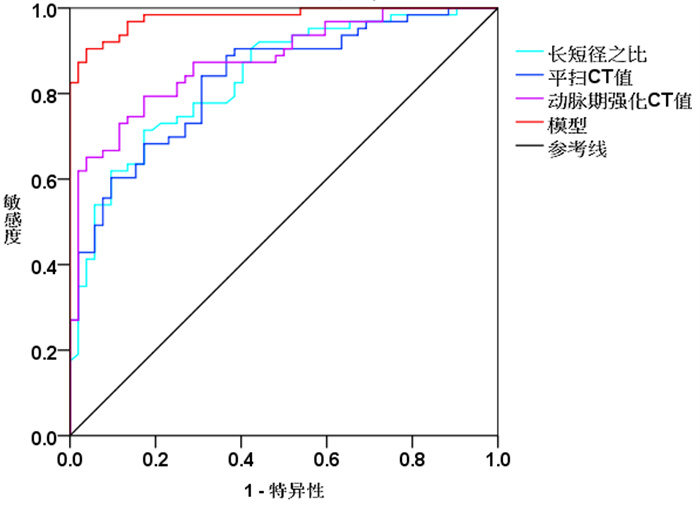
Objective To analyze the multi-slice spiral CT (MSCT) findings of pericolonic tumor deposits (TD) in patients with colorectal cancer, and its value in preoperative diagnosis. Methods The relevant data of 63 colorectal cancer patients with TD (TD group) and 52 colorectal cancer patients with lymph node metastasis (lymph node metastasis group) who were admitted to the hospital from December 2018 and December 2021 was retrospectively analyzed. All patients received MSCT examination before operation. MSCT findings of patients with TD and lymph node metastasis, and the value of MSCT in the diagnosis of TD were analyzed. Results The lesions in patients with TD were large, with irregular shapes, unclear boundaries with surrounding tissues, and uneven density on plain scan. Enhanced scan showed significant homogeneous enhancement which was similar to that of primary tumor. Arterial enhanced scan showed edge enhancement with liquefactive necrosis in a small amount of lesions. The lesions in patients with lymph node metastasis were small, with regular shape, mostly round or oval, relatively clear boundaries with surrounding tissues, and relatively uniform density. There were statistically significant differences in lesion density, boundary, shape, long-to-short diameter ratio, plain scan CT values, and arterial phase enhanced CT values between the TD group and the lymph node metastasis group (P < 0.05). Multivariate logistic regression analysis showed that long-to-short diameter ratio, plain scan CT value, and arterial phase enhanced CT value were related to TD (P < 0.05). The logistic regression model was constructed: Logit(P)=65.212-6.001×long-to-short diameter ratio-0.315×plain scan CT value-0.333×arterial phase enhanced CT value. The ROC curve showed that the area under the curve values of long-to-short diameter ratio, plain scan CT value, and arterial phase enhanced CT value to diagnose TD were 0.836, 0.832 and 0.878, respectively. The area under the curve of the regression model was 0.979. Conclusion MSCT examination provides an effective imaging basis for the diagnosis of TD in colorectal cancer. The MSCT-based regression model is helpful for the diagnosis of TD.
2022, 45(3): 424-428.
doi: 10.12122/j.issn.1674-4500.2022.03.23
Abstract:
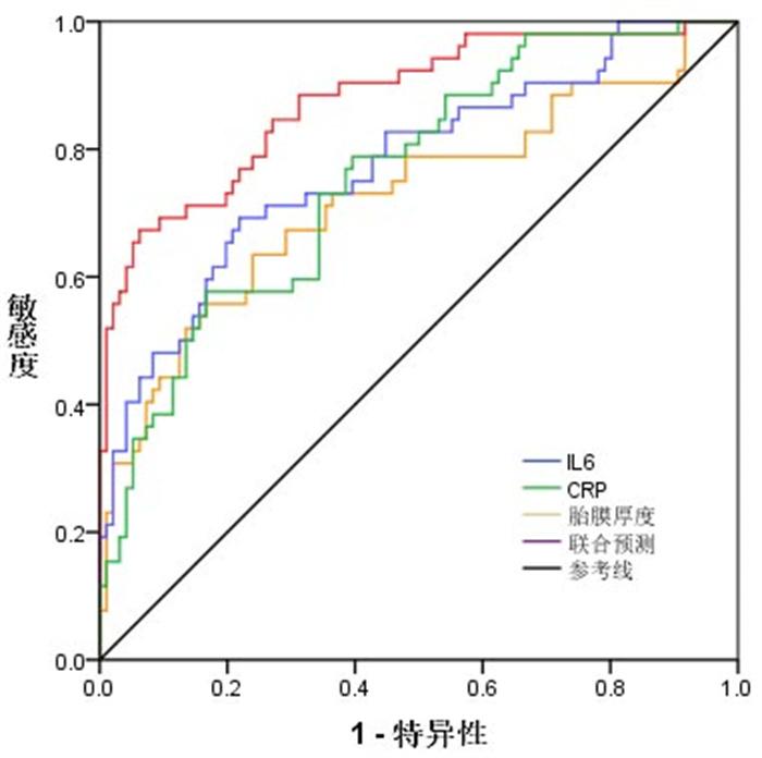
Objective To explore the clinical value of ultrasonic fetal membrane thickness combined with serum interleukin-6 (IL-6) and C-reactive protein (CRP) in predicting premature delivery in the second and third trimesters of pregnancy. Methods A total of 599 pregnant women undergoing prenatal examination and delivery in the hospital were enrolled as the research objects from January 1st, 2019 to December 31st, 2020. According to different delivery time, they were divided into premature delivery group (n=86) and full-term group (n=513). The serum IL-6, CRP and ultrasonic fetal membrane thickness in both groups were analyzed in the second and third trimesters of pregnancy. The predictive value of the above three indexes for premature delivery was analyzed by ROC curves. The positive rates of different indexes were compared between the two groups. Results Within 23-25 and 32-34 gestational weeks, levels of serum IL-6 and CRP in premature delivery group were significantly higher than those in full-term group (P < 0.05), while thickness of fetal membranes was thinner than that in fullterm group (P < 0.05). The ROC curves analysis showed that the area under the ROC curve of ultrasonic fetal membrane thickness combined with serum IL-6 and CRP for predicting premature delivery was greater than that of single index (P < 0.05). The accuracy, sensitivity and specificity of combined prediction were higher. The positive rates of premature delivery by single diagnosis and combined diagnosis in premature delivery group were higher than those in full-term group (P < 0.05). Conclusion Compared with full-term pregnant women, levels of serum IL-6 and CRP are significantly higher, and thickness of fetal membranes is thicker in pregnant women with premature deliver in the second and third trimesters. The combined detection of serum IL-6, CRP and fetal membrane thickness has higher predictive value for premature delivery.
2022, 45(3): 429-432.
doi: 10.12122/j.issn.1674-4500.2022.03.24
Abstract:
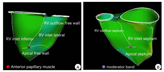
Objective To investigate the effect of complete right bundle branch block (CRBBB) on right ventricular volume and function assessed by three-dimensional speckle-tracking echocardiography. Methods A total of 106 patients with CRBBB in our hospital from January 2018 to January 2021 were enrolled, and another 50 patients without cardiac organic lesions confirmed by electrocardiogram, echocardiography and other laboratory examinations were selected as the control group. According to the absence or presence of right ventricular mechanical dyssynchrony presented in the images of three-dimensional speckletracking echocardiography, patients were classified into synchrony group and dyssynchrony group. The right ventricular volume and function of two groups were compared, then the value of three-dimensional speckle tracking echocardiography in CRBBB was analyzed. Results The time-to-peak area change ratio of RA inlet lateral, inlet inferior, inlet septum and outflow free wall was significantly higher in observation group than in control, apical free wall and apical septum showed no significant difference between groups (P>0.05). Among CRBBB patients, the QRS interval, left ventricular ejection fraction, right ventricular end- diastolic volume index, tricuspid regurgitation pressure gradient, and right ventricular end- systolic volume index of low standard deviation group were significantly lower than those of high standard deviation group (P < 0.05), while the left ventricular end-diastolic volume, left ventricular end- systolic volume, right ventricular fractional area change, right ventricular ejection fraction, and tricuspid annular plane systolic excursion were significantly higher than those of high standard deviation group (P < 0.05). Conclusion Application of three-dimensional speckle-tracking echocardiography can effectively reflect the contraction delay of each cardiac segment, right ventricular volume and function in patients with CRBBB, which is of great clinical value in guiding the clinical cardiac synchronization treatment and disease evaluation of CRBBB.
2022, 45(3): 433-436.
doi: 10.12122/j.issn.1674-4500.2022.03.25
Abstract:
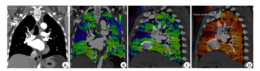
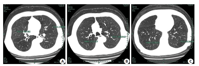
Objective To explore the characteristics of inspiratory and expiratory dual- phase CT quantitative parameters in patients with chronic obstructive pulmonary disease (COPD) and the early diagnosis value of CT quantitative parameters on COPD. Methods A total of 112 patients with COPD admitted to the hospital were retrospectively selected between March 2018 and December 2020 for inspiratory and expiratory dual-phase CT scan and pulmonary function examination. The pulmonary function indicators such as forced vital capacity (FVC), forced expiratory volume in one second (FEV1%) and ratio of forced expiratory volume in one second to maximum vital capacity (FEV1/FVC) were recorded. FACT-Digital lungTM data analysis software was used to obtain the CT quantitative parameters of lung volume (LV), mean lung density (MLD) and emphysema index (EI) of inspiratory and expiratory phases. The correlation between CT quantitative parameters and pulmonary function indicators was analyzed. Results The FVC, FEV1% and FEV1/FVC of patients with COPD were 2.95 ± 0.86 L, 2.06 ± 0.78 L and (61.76±11.93)%. Among patients with COPD, the LV of expiratory phase was significantly lower than that of inspiratory phase while the MLD and EI were significantly higher than those of inspiratory phase (P < 0.05). The expiratory phase LV was not significantly correlated with FVC (P > 0.05), but LV was significantly negatively correlated with FEV1% and FEV1/FVC (P < 0.05). MLD was significantly positively correlated with FVC, FEV1% and FEV1/FVC (P < 0.05). EI was significantly negatively correlated with FVC, FEV1% and FEV1/FVC (P < 0.05). Inspiratory phase LV was positively correlated with FVC and was negatively correlated with FEV1% and FEV1/FVC (P < 0.05). MLD was negatively correlated with FVC, and positively correlated with FEV1% and FEV1/FVC (P < 0.05), EI was significantly negatively correlated with FVC, FEV1% and FEV1/FVC (P < 0.05). Conclusion There is a good correlation between the quantitative parameters of inspiratory and expiratory dual-phase CT and pulmonary function indicators, which has a good guiding value for the early diagnosis of COPD.
2022, 45(3): 437-441.
doi: 10.12122/j.issn.1674-4500.2022.03.26
Abstract:
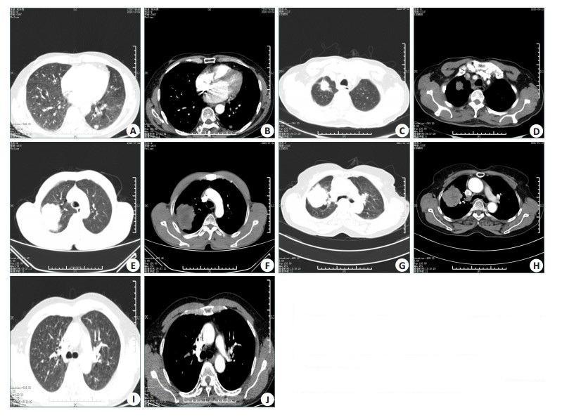
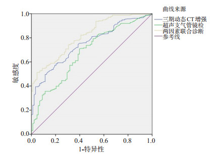
Objective To explore the diagnostic efficacy of three-phase dynamic CT enhanced scanning combined with ultrasound bronchoscopy in patients with peripheral lung cancer. Methods A retrospective study was conducted in 82 patients (observation group) with peripheral lung cancer diagnosed in our hospital from January 2018 to January 2020. In addition, 82 healthy volunteers who underwent physical examination in our hospital during the same period were selected as the control group. All patients received three-phase dynamic CT enhanced scanning and ultrasound bronchoscopy, and the CT value and net enhanced CT value of patients in the two groups were compared. The diagnostic efficacy of single detection and combined detection for peripheral lung cancer was analyzd. Results CT value, enhancement peak value and net enhancement value of CT in observation group were significantly higher than those in control group (P < 0.05). CT value, enhancement peak value and net enhancement value of CT in stage Ⅲ-Ⅳ were significantly higher than those in stage Ⅰ-Ⅱ (P < 0.05). There were statistically significant differences in CT value, enhancement peak value and net enhancement value of CT in patients with different pathological stages (P < 0.05). The specificity of combined diagnosis was significantly higher than that of single detection (P < 0.05). The area under the curve of combined diagnosis was significantly higher than that of single detection (P < 0.05). Conclusion Three-phase dynamic CT enhanced scanning combined with ultrasound bronchoscopy has good diagnostic efficacy in patients with peripheral lung cancer and is suitable for application
2022, 45(3): 447-453.
doi: 10.12122/j.issn.1674-4500.2022.03.28
Abstract:
Intervertebral disc degeneration is one of the main causes of low back pain. Imaging examination is an important means of diagnose intervertebral disc degeneration. Although traditional MRI technique can show the signal intensity and and morphological changes of the intervertebral disc, it is difficult to objectively quantify the degree of disc degeneration. T2 mapping technology can detect various biochemical components such as intervertebral disc water content, proteoglycan content and collagen fiber sequence, providing a new method for early clinical evaluation of intervertebral disc degeneration. This article discusses the research progress of quantitative magnetic resonance T2 mapping in disc degeneration is discussed from the comparison of T2 mapping with other quantitative magnetic resonance technologies, and its relationship with disc degeneration grade, Japanese Orthopaedic Association score, visual analog scale score, age and gender.
Intervertebral disc degeneration is one of the main causes of low back pain. Imaging examination is an important means of diagnose intervertebral disc degeneration. Although traditional MRI technique can show the signal intensity and and morphological changes of the intervertebral disc, it is difficult to objectively quantify the degree of disc degeneration. T2 mapping technology can detect various biochemical components such as intervertebral disc water content, proteoglycan content and collagen fiber sequence, providing a new method for early clinical evaluation of intervertebral disc degeneration. This article discusses the research progress of quantitative magnetic resonance T2 mapping in disc degeneration is discussed from the comparison of T2 mapping with other quantitative magnetic resonance technologies, and its relationship with disc degeneration grade, Japanese Orthopaedic Association score, visual analog scale score, age and gender.
2022, 45(3): 454-458.
doi: 10.12122/j.issn.1674-4500.2022.03.29
Abstract:
Thrombosis is the culprit of ischemic stroke, acute myocardial infarction and other ischemic diseases, causing a large number of deaths and disability every year, which seriously endangers human health and safety. Therefore, efficient and accurate detection of thrombosis has high research value and significance in clinic. Traditional imaging techniques such as CT, MRI and ultrasound mainly rely on non- specific imaging methods for disease examination, which cannot show the relationship between molecular changes and disease, so it is easy to miss the best treatment period. With the development of science and technology and the integration of interdisciplinary disciplines, the era of molecular imaging is coming, that is, using imaging methods to conduct qualitative and quantitative research on biological processes at the cellular and molecular levels. Molecular targeted probes show high contrast effect because of their high specificity to the target. In addition, the potential to realize the multimodal imaging is expected to diagnose diseases at the molecular level and truly achieve early diagnosis and treatment. This review will summarize the application of molecular targeted probes in the diagnosis of thrombosis.
Thrombosis is the culprit of ischemic stroke, acute myocardial infarction and other ischemic diseases, causing a large number of deaths and disability every year, which seriously endangers human health and safety. Therefore, efficient and accurate detection of thrombosis has high research value and significance in clinic. Traditional imaging techniques such as CT, MRI and ultrasound mainly rely on non- specific imaging methods for disease examination, which cannot show the relationship between molecular changes and disease, so it is easy to miss the best treatment period. With the development of science and technology and the integration of interdisciplinary disciplines, the era of molecular imaging is coming, that is, using imaging methods to conduct qualitative and quantitative research on biological processes at the cellular and molecular levels. Molecular targeted probes show high contrast effect because of their high specificity to the target. In addition, the potential to realize the multimodal imaging is expected to diagnose diseases at the molecular level and truly achieve early diagnosis and treatment. This review will summarize the application of molecular targeted probes in the diagnosis of thrombosis.
2022, 45(3): 459-464.
doi: 10.12122/j.issn.1674-4500.2022.03.30
Abstract:
With the rapid development of deep learning, big data and other technologies, artificial intelligence is the most promising technology in the field of medicine. In view of the key role of medical imaging in the diagnosis and timely treatment of diseases, the combination of medical imaging and artificial intelligence is becoming an essential interdisciplinary research direction. In clinical practice, doctors often need to refer to multimodal image data for comprehensive analysis and judgment in order to diagnose diseases more accurately and comprehensively. This paper first introduces the basic concept and working principle of multi-mode deep learning, and the representative research results of deep learning technology applied to multi-mode medical image-assisted diagnosis are summarized. Finally, the technical challenges of multi-mode deep learning technology in the medical image field are analyzed, and the application prospect of multi-mode deep learning technology is forecasted.
With the rapid development of deep learning, big data and other technologies, artificial intelligence is the most promising technology in the field of medicine. In view of the key role of medical imaging in the diagnosis and timely treatment of diseases, the combination of medical imaging and artificial intelligence is becoming an essential interdisciplinary research direction. In clinical practice, doctors often need to refer to multimodal image data for comprehensive analysis and judgment in order to diagnose diseases more accurately and comprehensively. This paper first introduces the basic concept and working principle of multi-mode deep learning, and the representative research results of deep learning technology applied to multi-mode medical image-assisted diagnosis are summarized. Finally, the technical challenges of multi-mode deep learning technology in the medical image field are analyzed, and the application prospect of multi-mode deep learning technology is forecasted.