


MRI signs of placenta accreta: the presence of at least two abnormal MRI signs has a high diagnostic value
-
摘要:
目的 评估产前MRI对胎盘植入诊断的准确性。 方法 回顾性分析2018年5月~2021年5月我院临床怀疑胎盘植入MRI图像,共47例孕妇图像符合标准纳入研究,分析2名医师对胎盘植入异常MRI征象检验的一致性,结合手术病理结果分析MRI胎盘植入诊断效能,比较评估胎盘植入的两种诊断方法:至少出现一种MRI征象(方法1)与至少出现两种MRI征象(方法2)。 结果 胎盘植入异常MRI图像中,2名医师对以下征象有较高一致性,T2WI序列低信号暗带(Kappa=0.874)、子宫肌层局部中断胎盘组织突入(Kappa=0.753)、胎盘内见异常增粗迂曲血管(Kappa=0.870)。MRI诊断胎盘植入总体准确度较高:Youden指数0.69,敏感度90.91%,特异性78.57%。诊断粘连性胎盘准确度低于植入性及穿透性胎盘。方法1诊断胎盘植入ROC曲线下面积为0.714,方法2诊断胎盘植入ROC曲线下面积为0.913,方法2的准确度高于方法1。 结论 MRI是评估胎盘植入的有效手段,至少出现两种异常MRI征象时诊断准确度最高。 Abstract:Objective To evaluate the accuracy of prenatal MRI for the diagnosis of placenta implantation. Methods MRI images of clinically suspected placenta implantation in our hospital from May 2018 to May 2021 were retrospectively analyzed. The images of 47 pregnant women were included in the study according to the criteria. The consistency of MRI signs of abnormal placenta implantation was analyzed. Combined with surgical pathological results, the diagnostic efficacy of MRI placenta implantation was analyzed. Two diagnostic methods were used to assess placenta implantation, with at least one MRI sign (method 1) and at least two MRI signs (method 2). Results Two physicians had high agreement with the following signs, hypointense band on T2WI sequence (Kappa=0.874), local interruption of placental tissue protrusion in the uterus (Kappa= 0.753), abnormal thickened tortuous vessels in placenta (Kappa=0.870). The overall accuracy of MRI in diagnosing placenta accreta was high: Youden index was 0.69, sensitivity was 90.91% and specificity was 78.57%. MRI diagnosis accuracy of adherent placenta is lower than that of implanted and penetrating placenta. The AUC area of method 1 was 0.714, and that of method 2 was 0.913. The accuracy of method 2 was higher than that of method 1. Conclusion MRI is an effective method to evaluate placenta implantation, and the diagnostic accuracy is highest when at least two abnormal MR signs occur. -
Key words:
- placental implantation /
- magnetic resonance imaging /
- placenta previa
-

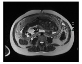
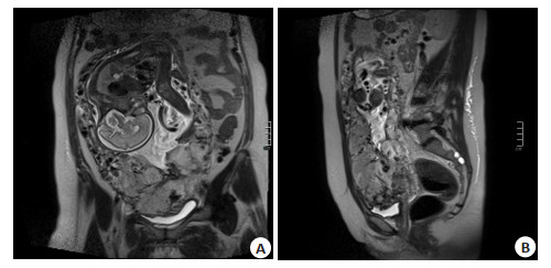
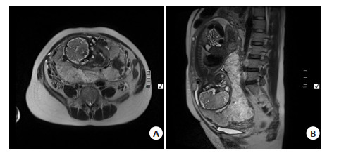
图 3 穿透性胎盘
A: 冠状位T2WI序列膀胱壁模糊, 幕状突起; B: 矢状位T2WI序列胎盘内异常增粗、迂曲血管.
Figure 3. Placenta percreta.

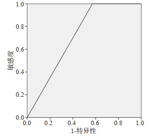
图 4 方法1诊断胎盘植入的ROC曲线(AUC=0.714)
Figure 4. ROC curve of method 1 for the diagnosis of placental implantation (AUC=0.714).

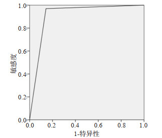
图 5 ROC curve of method 1 for the diagnosis of placental implantation (AUC=0.714).
Figure 5. ROC curve of method 2 for the diagnosis of placental implantation (AUC= 0.913).
表 1 2名医师胎盘植入MRI征象一致性分析
Table 1. Consistency analysis of MRI signs of placenta implantation in two physicians
MRI征象 Kappa值 胎盘内T2WI序列低信号暗带 0.874 子宫肌层变薄、模糊 0.573 子宫肌层中断、胎盘突入肌层 0.753 子宫、胎盘局部外凸 0.695 胎盘的异质性 0.637 膀胱壁模糊、膀胱幕状突起 0.670 胎盘内异常增粗迂曲血管 0.870 Kappa < 0.4为一致性较差,0.4≤Kappa≤0.75为中度一致性,Kappa>0.75为一致性较好.  下载: 导出CSV
表 2 MRI诊断胎盘植入结果与手术病理结果比较
Table 2. MRI diagnosis of placenta implantation results compared with surgery and pathology (n)
MRI诊断 手术及病理 合计 无植入胎盘 粘连性胎盘 植入性胎盘 穿透性胎盘 无植入 11 3 0 0 14 粘连性胎盘 2 3 3 0 8 植入性胎盘 1 3 10 1 15 穿透性胎盘 0 0 1 9 10 合计 14 9 14 10 47
下载: 导出CSV
表 3 MRI诊断胎盘植入的敏感度、特异性及约登指数
Table 3. Sensitivity, specificity, and Youden index in MRI diagnosis of placental implantation
MRI诊断 敏感度(%) 特异性(%) 约登指数 胎盘植入(总体) 90.91 78.57 0.69 粘连性胎盘 33.33 86.84 0.20 植入性胎盘 71.43 84.85 0.56 穿透性胎盘 90.00 97.30 0.87 约登指数范围为-1~1,值越大,诊断实验真实性越好,≤0时无临床应用价值.
下载: 导出CSV
表 4 MRI诊断胎盘植入与手术病理对照(方法1)
Table 4. MRI diagnosis of placenta implantation was compared with operation and pathology (method 1) (n)
MRI诊断 手术及病理 合计 阳性 阴性 阳性 33 8 41 阴性 0 6 6 合计 33 14 47
下载: 导出CSV
表 5 MRI诊断胎盘植入与手术病理对照(方法2)
Table 5. MRI diagnosis of placenta implantation was compared with operation and pathology (method 2) (n)
MRI诊断 手术及病理 合计 阳性 阴性 阳性 32 2 34 阴性 1 12 13 合计 33 14 47
下载: 导出CSV
-
[1] 吴柄钢, 姚强, 崔陶. 胎盘植入诊断国内外指南解读[J]. 现代妇产科进展, 2020, 29(1): 71-3. https://www.cnki.com.cn/Article/CJFDTOTAL-XDFC202001019.htm [2] 滕慧, 水旭娟, 焦岩, 等. 超声对胎盘植入的产前诊断意义及漏误诊原因分析[J]. 中国计划生育学杂志, 2020, 28(10): 1709-11. https://www.cnki.com.cn/Article/CJFDTOTAL-JHSY202010051.htm [3] Hobson SR, Kingdom JC, Murji A, et al. No. 383-screening, diagnosis, and management of placenta accreta spectrum disorders [J]. J Obstet Gynaecol Can, 2019, 41(7): 1035-49. [4] Jha P, Pōder L, Bourgioti C, et al. Society of abdominal radiology (SAR) and european society of urogenital radiology (ESUR) joint consensus statement for MR imaging of placenta accreta spectrum disorders[J]. Eur Radiol, 2020, 30(5): 2604-15. doi: 10.1007/s00330-019-06617-7 [5] 朱方玉, 漆洪波. 2018 FIGO胎盘植入性疾病指南解读[J]. 中国实用妇科与产科杂志, 2018, 34(12): 1353-9. https://www.cnki.com.cn/Article/CJFDTOTAL-ZGSF201812015.htm [6] Valentini AL, Gui B, Ninivaggi V, et al. The morbidly adherent placenta: when and what association of signs can improve MRI diagnosis? Our experience[J]. Diagn Interv Radiol, 2017, 23(3): 180- 6. doi: 10.5152/dir.2017.16275 [7] Jauniaux E, Hussein AM, Fox KA, et al. New evidence-based diagnostic and management strategies for placenta accreta spectrum disorders[J]. Best Pract Res Clin Obstet Gynaecol, 2019, 61: 75-88. doi: 10.1016/j.bpobgyn.2019.04.006 [8] 侯磊, 李光辉, 邹丽颖, 等. 全国剖宫产率及剖宫产指征构成比调查的多中心研究[J]. 中华妇产科杂志, 2014, 49(10): 728-35. [9] Woodward PJ, Kennedy A, Einerson BD. Is there a role for MRI in the management of placenta accreta spectrum?[J]. Curr Obstet Gynecol Rep, 2019, 8(3): 64-70. doi: 10.1007/s13669-019-00266-9 [10] Chen X, Shan RQ, Zhao LX, et al. Invasive placenta previa: Placental bulge with distorted uterine outline and uterine serosal hypervascularity at 1.5T MRI-useful features for differentiating placenta percreta from placenta accreta[J]. Eur Radiol, 2018, 28(2): 708-17. doi: 10.1007/s00330-017-4980-z [11] 金晶, 邹立巍, 赵红, 等. 产前MRI检查在胎盘植入中的诊断价值[J]. 临床放射学杂志, 2018, 37(4): 641-5. https://www.cnki.com.cn/Article/CJFDTOTAL-LCFS201804023.htm [12] 赵毅, 万军. MRI检查胎盘植入高危患者的结果分析及影像学表现[J]. 中国CT和MRI杂志, 2018, 16(3): 93-6. https://www.cnki.com.cn/Article/CJFDTOTAL-CTMR201803030.htm [13] 梁娜, 邓珍萍, 张羲娥, 等. 产前MRI对胎盘植入深度的诊断价值[J]. 四川医学, 2019, 40(8): 776-9. https://www.cnki.com.cn/Article/CJFDTOTAL-SCYX201908005.htm [14] Einerson BD, Rodriguez CE, Silver RM, et al. Accuracy and interobserver reliability of magnetic resonance imaging for placenta accreta spectrum disorders[J]. Am J Perinatol, 2021, 38(9): 960-7. doi: 10.1055/s-0040-1701196 [15] 邵明昱, 冯炜炜. 胎盘植入的风险评估与诊治策略[J]. 国际妇产科学杂志, 2018, 45(6): 676-80. https://www.cnki.com.cn/Article/CJFDTOTAL-GWVC201806016.htm [16] 韩鹏慧, 江魁明, 郭庆禄, 等. MRI在不同胎盘植入深度中的诊断价值[J]. 实用放射学杂志, 2018, 34(8): 1228-30, 1234. https://www.cnki.net/KCMS/detail/detail.aspx?dbcode=IPFD&filename=ZGZP201712001531&dbname=IPFDLAST2020 [17] 钱丽霞, 孟静文, 郭娟, 等. 产前MRI对于不同类型胎盘植入的诊断价值[J]. 中国CT和MRI杂志, 2017, 15(12): 96-9. https://www.cnki.com.cn/Article/CJFDTOTAL-CTMR201712030.htm [18] 洪静静, 黄伟康, 张和林, 等. MRI在产前胎盘植入诊断的应用与进展[J]. 分子影像学杂志, 2020, 43(2): 203-6. doi: 10.12122/j.issn.1674-4500.2020.02.05 [19] Khalaf LMR, Zeid HA, Othman ER. Reliability of Magnetic Resonance Imaging in diagnosis and assessment the depth of invasion of placental accreta in high risk gravid women[J]. Clin Imaging, 2019, 58: 5-11. doi: 10.1016/j.clinimag.2019.05.003 [20] 李晓凡, 余红军. MRI征象评分对胎盘种植异常的诊断价值[J]. 放射学实践, 2019, 34(12): 1358-63. https://www.cnki.com.cn/Article/CJFDTOTAL-FSXS201912020.htm [21] Bourgioti C, Zafeiropoulou K, Fotopoulos S, et al. MRI prognosticators for adverse maternal and neonatal clinical outcome in patients at high risk for placenta accreta spectrum (PAS) disorders [J]. J Magn Reson Imaging, 2019, 50(2): 602-18. doi: 10.1002/jmri.26592 [22] Millischer AE, Deloison B, Silvera S, et al. Dynamic contrast enhanced MRI of the placenta: a tool for prenatal diagnosis of placenta accreta?[J]. Placenta, 2017, 53: 40-7. doi: 10.1016/j.placenta.2017.03.006 [23] 丁治民, 施素华, 翟建, 等. 胎盘植入MRI诊断中增强扫描的价值[J]. 临床放射学杂志, 2018, 37(6): 989-93. https://www.cnki.com.cn/Article/CJFDTOTAL-LCFS201806026.htm [24] Mühler MR, Clément O, Salomon LJ, et al. Maternofetal pharmacokinetics of a gadolinium chelate contrast agent in mice[J]. Radiology, 2011, 258(2): 455-60. doi: 10.1148/radiol.10100652 -

 点击查看大图
计量
- 文章访问数: 306
- HTML全文浏览量: 105
- PDF下载量: 13
- 被引次数: 0