

 Find Duplicates
 Check Document
 Submission(new)
 Experts Office
 Editorial Office
2022 Vol. 45, No. 4
column
Display Method:
2022, 45(4): 465-469.
doi: 10.12122/j.issn.1674-4500.2022.04.01
Abstract:
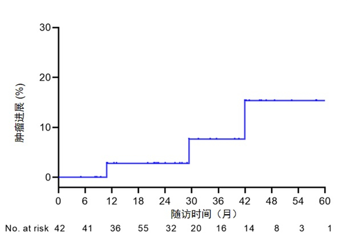
Objective To explore the interim efficacy of ultrasound-guided percutaneous microwave ablation for ≤5 cm breast cancer. Methods Forty-two women with invasive ductal carcinoma of the breast ≤5 cm treated by microwave ablation from January, 2014 to January, 2020 were retrospectively analyzed. The primary end point was tumor progression in the intention-to-treat population. Secondary end points included survival, cosmetic results, and complications. Results The patients received 48 ablation for 68 tumors in evaluated, The complete ablation rate was 100%. The tumor diameter was 2.5±1.2 (0.4-5.0) cm. Median follow-up was 29.2 months (range 5.0-70.5 months). All patients achieved technique effectiveness by contrast-enhanced ultrasound. One local tumor progression, one ipsilateral breast recurrence and one Brain metastases occurred at 42, 11 and 29 months after microwave ablation, respectively. There were no serious complications after ablation, and the patient's aesthetic satisfaction reached 100%. Conclusion Ultrasound-guided percutaneous microwave ablation for ≤ 5 cm breast cancer is safe. It offers a minimally invasive and tolerable local treatment option, especially for older women and patients with severe comorbiditions.

2022, 45(4): 470-474.
doi: 10.12122/j.issn.1674-4500.2022.04.02
Abstract:
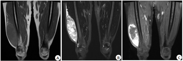
Objective To investigate the clinical, pathological and imaging features of extraskeletal myxoid chondrosarcoma in order to improve the cognition of the disease. Methods The clinical data, pathology and imaging findings of 5 cases with pathologically approved extraskeletal myxoid chondrosarcoma were retrospectively analyzed. Results There were 5 cases, including 3 males and 2 females, with the age range from 39 to 73 years old. 7 lesions were found in these cases, including 3 single lesions and 2 multiple lesions. X-ray images (n=3) showed that soft tissue mass was seen in all the lesions, of which 1 case showed patchy high-density. CT (n=2) showed the lesions were low-density masses with clear boundaries. Spotted and nodular calcification shadows were observed in 1 case. The enhanced scan showed different degree of enhancement in both cases. MRI examination (n=4) showed hypo- and iso-intense on T1WI and heterogeneous hyper-intense on T2WI, that contains strip-like septums. On enhanced scanning, 1 case showed obvious marginal enhancement, 3 cases showed lobular and nodular enhancement, and the internal septums was obviously enhanced. General pathology showed that masses were mostly lobular while myxoid stroma and oncocyte among strip-like fibrous tissue were seen in microscope pathology. Conclusion The incidence of extraskeletal myxoid chondrosarcoma is extremely low, and the imaging findings are corresponding to the histomathological structure, which has certain diagnostic significance.


2022, 45(4): 475-479.
doi: 10.12122/j.issn.1674-4500.2022.04.03
Abstract:
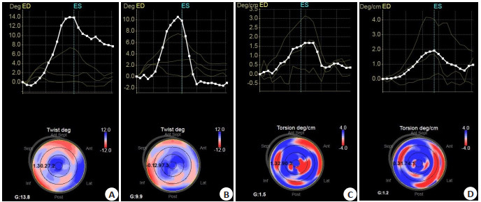
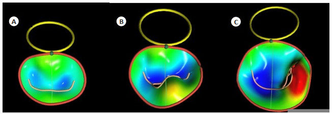
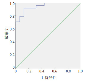
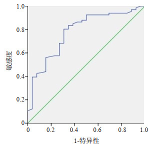
Objective To evaluate the changes of left ventricular torsion in patients with breast cancer after anthracycline chemotherapy by three-dimensional speckle tracking imaging, and detect the occurrence of early cardiotoxicity, and explore the correlation between left ventricular torsion and high sensitive cardiac troponin T (Hs-cTnT). Methods A total of 82 patients with breast cancer after anthracycline chemotherapy were selected. The parameters of left ventricular torsion and strain function were measured at four time points: before chemotherapy (Ⅰ), after receiving two cycles (Ⅱ), four cycles (Ⅲ) and six cycles (Ⅳ). The sensitivity of each parameter to evaluate early cardiotoxicity was compared. then correlation analysis was conducted between Hs-cTnT concentration and left ventricular torsion. Results Comparing the four groups, left ventricular torsion parameters (Ptw, Tor) decreased (P < 0.05). The areas under ROC curve of Ptw and Tor were 0.954 and 0.912, they are both with high specificity and sensitivity. After six cycles chemotherapy, the Hs-cTnT was lower than the normal, however, it increased with the dose accumulation of drug during the chemotherapy (P < 0.05). Pearson correlation analysis showed that there was a significant negative correlation between the left ventricular torsion and Hs-cTnT concentration in different chemotherapy stages (Ptw: r=-0.467, P < 0.001; Tor: r=-0.419, P < 0.001). Conclusion Left ventricular torsion can be used as sensitive parameters to evaluate the early cardiotoxicity induced by anthracycline chemotherapy after breast cancer.

2022, 45(4): 480-485.
doi: 10.12122/j.issn.1674-4500.2022.04.04
Abstract:
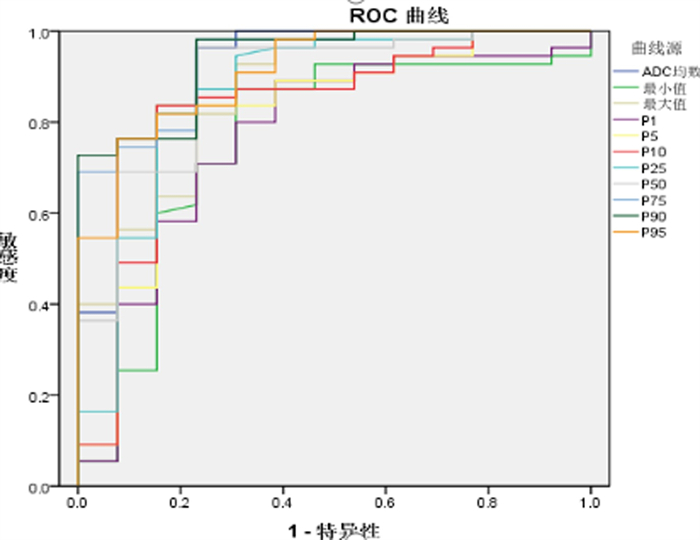
Objective To explore the correlation between the whole focus apparent diffusion coefficient (ADC) histogram analysis and CA125 and the degree of differentiation, microsatellite and Ki-67 of endometrioid adenocarcinoma, respectively. Methods Sixty-eight patients with endometrial carcinoma confirmed by preoperative pathology. All patients underwent superconducting MRI scans. The ROIs were drawn by FireVoxel software and histogram analysis was carried out. The correlation between the ADC histogram parameters and CA125 and differentiation, microsatellite and Ki-67 of endometrioid adenocarcinoma were analyzed. Results The ADC histogram parameter and Ki-67 were statistically significant in different tissue differentiation degrees(P < 0.05). The ADC histogram parameters, Ki-67 and CA125 differences were not statistically significant between the stable and unstable microsatellite groups(P>0.05). ROC curve analysis showed that P90 achieved the highest AUC of 0.937. Histogram parameters were negatively correlated with the degree of tissue differentiation with low or middle degree. Mean, maximum, P10, P25, P50, P75, P90, P95 and Ki-67 were negatively correlated with low degrees. Conclusion ADC histogram parameters and serum CA125 help to evaluate the histopathological characteristics of endometrial adenocarcinoma.

2022, 45(4): 486-492.
doi: 10.12122/j.issn.1674-4500.2022.04.05
Abstract:
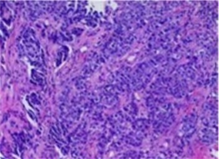
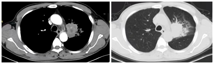
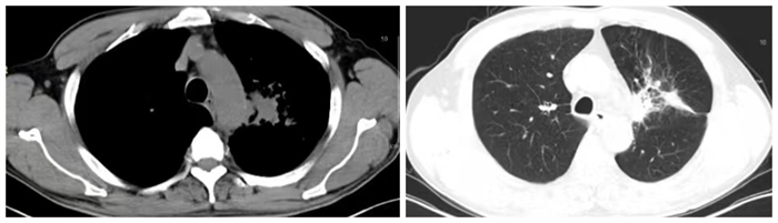
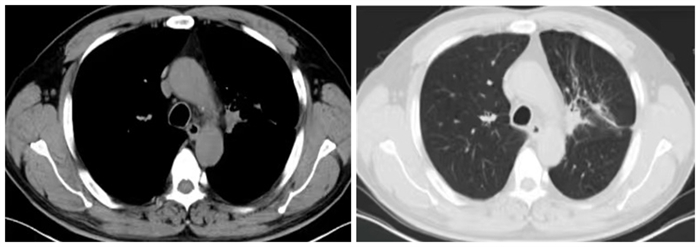
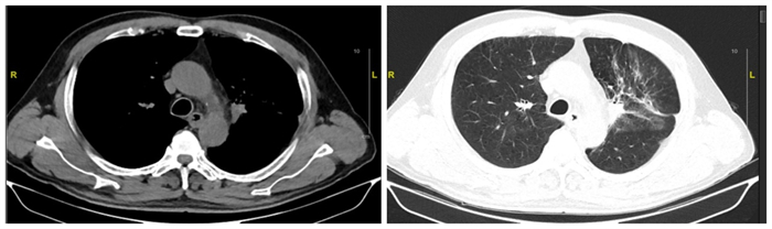
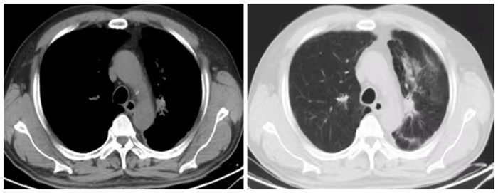
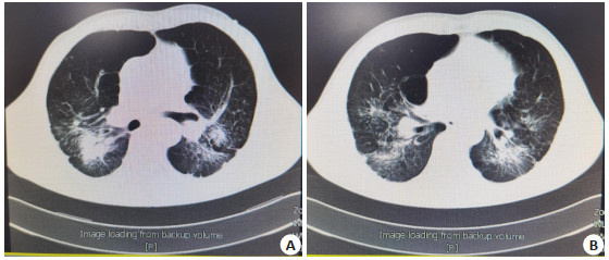
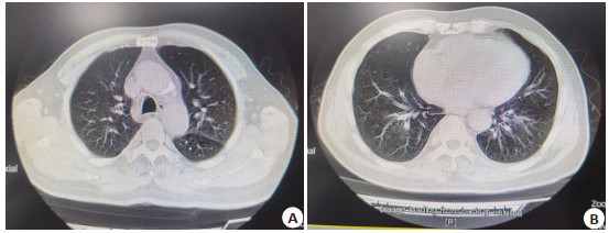
Objective To investigate the effect of pembrolizumab combined with induction chemotherapy and radiotherapy on the clinical treatment effect and lung CT findings of patients with intermediate and advanced lung squamous cell carcinoma. Methods A total of 80 patients with intermediate and advanced lung squamous cell carcinoma who were treated in our hospital from November 2019 to December 2020 were selected. They were randomly divided into the control group (n=40) and the observation group (n=40). The control group was given induction chemotherapy + radiotherapy, the observation group was given pembrolizumab on the basis of the control group. The clinical efficacy, lung CT findings, T lymphocyte subsets (CD3+, CD4+, CD8+, CD4+/CD8+ ratio), serum tumor markers (CYFRA21-1, CEA, SCC), adverse reactions and quality of life (SF-36) between the two groups were compared. Results The objective response rate and disease control rate of the observation group were significantly higher than those of the control group (P < 0.05). Lung CT can be used to evaluate the clinical efficacy of the patients. Compared with the control group after treatment, the objective response rate and disease control rate of the observation group were significantly higher (P < 0.05). CD4+ and CD4+/CD8+ ratios were significantly higher (P < 0.05), CD8+ was significantly lower (P < 0.05), CYFRA21-1, CEA, and SCC levels were significantly lower (P < 0.05), the scores of SF-36 in various fields were significantly higher (P < 0.05), and the difference of overall incidence of adverse reactions was not statistically significant (P>0.05). Conclusion Pembrolizumab combined with induction chemotherapy and radiotherapy has good efficacy in the treatment of patients with advanced lung squamous cell carcinoma. It improves T lymphocyte subsets and tumor marker levels and has better security.
Multimodal magnetic resonance was used to determine the value of muscular invasion in bladder cancer
2022, 45(4): 493-496.
doi: 10.12122/j.issn.1674-4500.2022.04.06
Abstract:
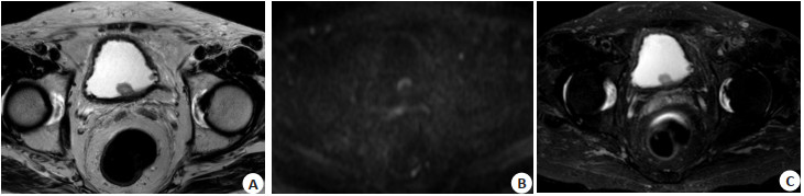
Objective To evaluate the value of T2 SPAIR sequence combined with T2WI + DWI in determining muscle layer invasion of bladder cancer based on multimodal MRI (mpMRI). Methods Sixty patients with bladder tumor who underwent magnetic resonance examination and obtained pathological results in our hospital from March 2020 to March 2022 were collected. T2WI combined with DWI sequence, T2WI combined with T2 SPAIR, DWI combined with T2 SPAIR and T2WI combined with DWI + T2 SPAIR sequence were detected. The results of different combination sequences were calculated to evaluate the efficacy and consistency of muscularis invasion stage of bladder cancer. Results The specificity, sensitivity and accuracy of T2WI combined with DWI+T2 SPAIR sequences for muscle layer infiltration were significantly higher than T2WI combined with DWI, T2WI combined with T2 SPAIR and DWI combined with T2 SPAIR(P < 0.05). SPAIR sequence of T2WI combined with DWI+T2 had the highest consistency with pathological diagnosis. Conclusion T2 SPAIR combined with DWI+ T2 SPAIR sequence scanning can significantly improve the diagnostic accuracy of T stage of bladder cancer, and T2WI combined with DWI+T2 SPAIR sequence has the highest consistency with pathological diagnosis, which is more conducive to the evaluation of muscle layer infiltration of lesions.
2022, 45(4): 497-505.
doi: 10.12122/j.issn.1674-4500.2022.04.07
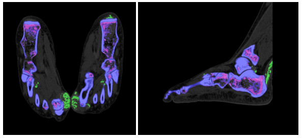
Abstract:
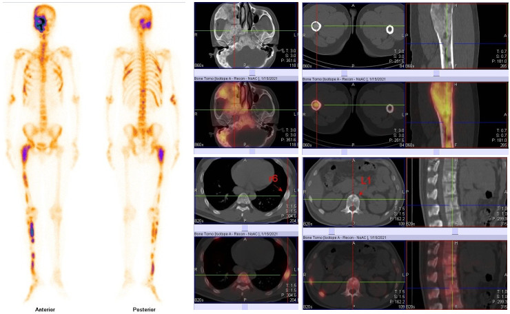
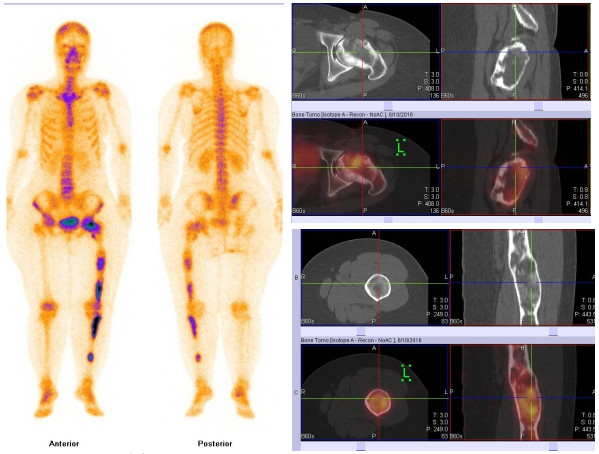
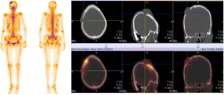
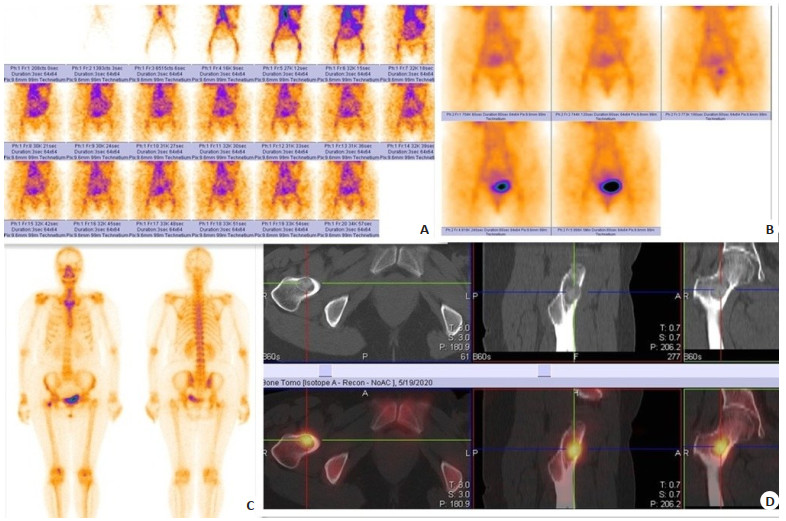
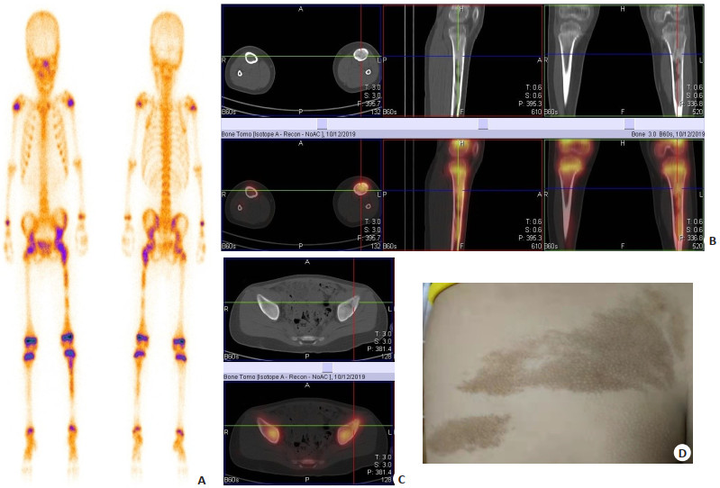
Objective To explore the imaging characteristics and clinical features of whole-body SPECT/CT for bone scintigraphy in patients with osteofibrous dysplasia. Methods The characteristics of SPECT/CT imaging were analyzed retrospectively in 34 patients with osteofibrous dysplasia. According to the number of lesions, the cases were divided into multi-lesion group(n=25)and single-lesion group(n=9). The indicators of two groups were compared. Results The symptom of pain was present in 20 cases (58.8%). The difference between the two groups was not significant (χ2=1.045, P=0.307). The age of the multi-lesion group was younger than that of the single-lesion group(t=17.315, P=0.018). The serum WBC levels, neutrophil levels, C-reactive protein, alkaline phosphatase and all the tumor markers (CEA, AFP, CA199, CA724, CYFRA21-1, NSE, ProGRP) had no statistically significant differences between two groups (P>0.05). The lesions were more common in the long bones of extremities, which showed ground glass and cystic changes. The probability of ground glass changes was higher in the multi-lesion group (χ2=8.579, P=0.003), while no significant difference in loofah, map, and cystic changes between the two groups (P>0.05). The SPECT imaging showed all the lesions in the thirty-four patients by high uptake of radiotracer, especially when focusing on one limb in polyostotic osteofibrous dysplasia. Conclusion Bone scintigraphy with SPECT/CT can be used as an important supplementary examination method for the differentiation and diagnosis of fibrous dysplasia by observing multiple bone lesions in the whole body, analyzing the anatomical structure and metabolic status of each lesion through one-time whole-body imaging.


2022, 45(4): 506-510.
doi: 10.12122/j.issn.1674-4500.2022.04.08
Abstract:
Objective To explore quantitative parameters and pulmonary function test results of multislice spiral CT in chronic obstructive pulmonary disease (COPD) patients. Methods A totao of 112 COPD patients who were admitted to the hospital were enrolled between August 2018 to September 2021. According to different clinical phenotype of COPD, the patients with bronchitis COPD were divided into bronchitis group (n=73) and the patients with emphysema COPD were divided into emphysema group (n=39). All patients underwent two-time chest multislice spiral CT examinations and pulmonary function tests, with an interval of 4 months. The pulmonary volume indexes [Vex/Vin], pulmonary density indexes [Mean lung density at the end of deep inspiration (MLDin), mean lung density at the end of deep exhalation (MLDex), MLDex-MLDin] and pulmonary function indexes [FEV1, FVC, FEV1/FVC] were compared between the two groups. The correlation between pulmonary volume indexes, pulmonary density indexes and disease severity was analyzed by spearman method. Results At the last time, FEV1 and FEV1/FVC of the two groups of patients were lower than the first time, and the FVC was higher than the first time (P < 0.05). Vin, Vex, Vex/Vin and MLDin, MLDex, MLDex-MLDin of the two groups of patients were lower than the first time (P < 0.05). The measured difference FEV1/FVC (%) was positively correlated with the lung density index MLDex-MLDin, and the difference FEV1 and FEV1/FVC of the two pulmonary function measurements in patients with emphysema were positively correlated with Vex/Vin and MLDex-MLDin (P < 0.05). Conclusion Multislice spiral CT combined with pulmonary function test can show the changes of emphysema and bronchus in COPD patients with different phenotypes, which has certain clinical value.
2022, 45(4): 511-517.
doi: 10.12122/j.issn.1674-4500.2022.04.09
Abstract:
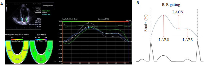
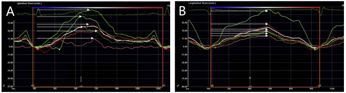
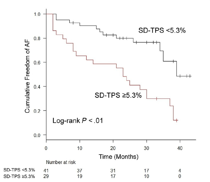
Objective To evaluate the relationship between left atrial mechanical dispersion and new-onset atrial fibrillation by using ultrasonic strain imaging, and its incremental value in predicting atrial fibrillation beyond left atrial enlargement and left atrial dysfunction was discussed. Methods A total of 376 general population at risk of atrial fibrillation were examined by electrocardiogram from 2018-2021. After 3 years of follow-up, 35 subjects with new-onset atrial fibrillation were selected as the study group. In the same cohort, subjects without atrial fibrillation who had the same number of cases as the study group and matched with age and sex were selected as the control group. Ultrasound strain imaging was applied to measure 12-segment left atrial strain values in both groups. The differences of left atrial volume index (LAVI), left atrial pump strain (LAPS), left atrial reservoir strain (LARS), and left atrial mechanical dispersion (SD-TPS) between the two groups were obtained and analyzed. SD-TPS was defined as the standard deviation of peak time of reservoir strain in each segment of left atrial standardized by R-R interval. The baseline data, LAVI, LAPS and LARS were adjusted. The independent correlation between SD-TPS and new-onset atrial fibrillation, and its incremental predictive value were analyzed. Results There was no significant difference in left atrial volume index (32.5±9.2 mL/m2 vs 29.5±8.3 mL/m2, P=0.16). Patients with new-onset atrial fibrillation had significantly worse left atrial pump strain [(16.6±4.3)% vs (20.6±4.3)%, P < 0.01)and reservoir strain[(31.4±7.7)% vs (38.0±7.3)%, P < 0.01)than those without atrial fibrillation. SD-TPS was significantly higher in patients with atrial fibrillation than in those without it[(6.3 ± 2.3)% vs (3.9 ± 1.6)%, P < 0.01). SD-TPS was independently associated with new-onset atrial fibrillation after adjustment for CHARGE-AF score, LAVI and LARS (hazard ratio=1.26, 95%CI: 1.10-1.45, P < 0.01). In the nested Cox models, the model based on the left atrial volume and strain for predicting new onset atrial fibrillation was significantly improved by adding SD-TPS (P < 0.01). Conclusion The left atrial mechanical dispersion obtained from ultrasound strain imaging can be used as an effective predictor of risk stratification of new-onset atrial fibrillation. It can provide incremental value beyond left atrial volume and left atrial dysfunction in predicting new-onset atrial fibrillation.
2022, 45(4): 518-525.
doi: 10.12122/j.issn.1674-4500.2022.04.10
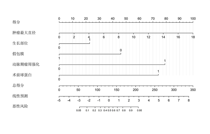
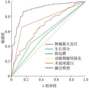
Abstract:
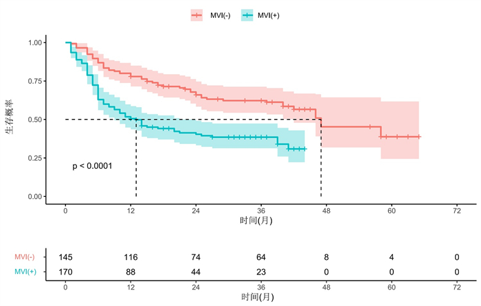
Objective To investigate the value of a preoperative nomograph and prediction model based on imaging and serological characteristics for predicting microvascular invasion (MVI) in hepatocellular carcinoma (HCC). Methods Clinical data of 548 patients with HCC who underwent liver resection or liver transplantation from January 2015 to December 2020 in our Hospital were retrospectively included. A total of 315 patients with HCC (MVI+ or MVI-) with an average age of 53.2±11.5 years old and a maximum direct tumour of 3.7-7.0 cm were included. Clinical and imaging data were analyzed. Univariate and multivariate logistic analyses were used to screen out independent risk factors that could predict MVI, and a nomograph model was constructed to predict MVI in HCC, which was evaluated using a subject operating curve, calibration curve and decision curve. Results The median survival time of patients with MVI(+) was 13 months (95% CI: 8.1-17.9) while that of patients with MVI(-)was 47 months (95%CI: 32.7-61.3). The 1-year, 3-year and 5-year disease-free survival rates of patients with MVI(+) were 50.6%、38.5% and 30.9%, while that of patients with MVI(-) were 77.9%、62.3% and 38.8%, respectively. Multivariate logistic regression analysis showed that larger tumour size, extrahepatic growth, absence or incomplete pseudocapsule, presence of arterial peritumoral enhancement and high preoperative globulin value were independent risk factors for MVI(+). The final model efficacy were as follows: AUC=0.895, 95%CI: 0.859-0.930, accuracy: 85.1%, sensitivity: 85.9%, specificity: 84.1%. The calibration curve showed that the predicted probability was in good agreement with the MVI(+)/MVI(-) probability of the pathological results. Thus, the decision curve model displayed good clinical application value. Conclusion The constructed Nomograph and prediction model can better predict the probability of MVI(+) before surgery. It can aid in adjusting the treatment plan of HCC according to the risk of MVI to optimize the survival outcome.


2022, 45(4): 526-532.
doi: 10.12122/j.issn.1674-4500.2022.04.11
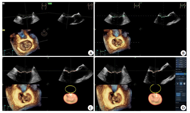
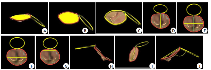
Abstract:
Objective To evaluate the correlation between valve structure changes and the degree of mitral regurgitation, and predict the risk factors for aggravating the degree of regurgitation in patients with mitral valve prolapse via real-time three-dimensional transesophageal echocardiography (RT-3D-TEE). Methods Forty patients who were diagnosed as mitral valve prolapse with mitral valve regurgitation by transthoracic echocardiography, and 10 volunteers in our hospital were involved. All the 50 enrolled patients underwent RT-3D-TEE and the three-dimensional images of the mitral valve were collected. Then the 4D Auto MVQ offline workstation was used for image post-processing and quantitative analysis. Results Annulus area 3D, annulus area 2D, annulus perimeter, anterior-to-posterior diameter, anterolateral to posteromedial diameter, non-planar angle, total leaflet area (A Total), posterior leaflet area increased and annular height to commissural width ratio decreased with the increase of the degree of regurgitation. The differences between two groups were statistically significant (P < 0.05). Annulus area 3D、annulus area 2D、annulus perimeter、anterior-to-posterior diameter、A Total were extremely strongly correlated with the degree of mitral regurgitation (r=0.847, 0.843, 0.845, 0.854, 0.854 respectively, P < 0.05). A Total was a risk factor for severe mitral regurgitation (P < 0.01, B=1.576, OR=4.834). The sensitivity of A Total (cut point value=8.9 cm2) in predicting severe mitral regurgitation was 91.7%, the specificity was 87.5%, and the area under the curve was 0.948 (P < 0.01). Conclusion The size of the mitral valve annulus, the degree of flattening and the size of the valve leaflet were positively correlated with the degree of regurgitation. Among them, A Total is a risk factor for the aggravation of mitral regurgitation in patients with mitral valve prolapse.


2022, 45(4): 533-541.
doi: 10.12122/j.issn.1674-4500.2022.04.12
Abstract:
Objective To investigate the application value of low tube voltage, low concentration iodine contrast agent combined with adaptive statistical iterative reconstruction-V (ASIR-V) in CT imaging of left atrium (LA) and pulmonary vein (PV) in patients with atrial fibrillation before radiofrequency catheter ablation. Methods All atrial fibrillation patients diagnosed with radiofrequency catheter ablation in our Hospital from January 2019 to June 2021 were retrospectively analyzed. The patients were divided into experimental group (group A) and control group (group B) according to tube voltage, with 143 cases in each group. Group A used low tube voltage 100 kVp, contrast agent iohexol 300 mgI/mL, ASIR-V 10%-100% interval 10% reconstruction. Group B used conventional tube voltage 120 kVp, contrast agent iopamidol 370 mgI/mL, ASIR-V 50% reconstruction.The objective evaluation of LA and PV image quality was performed by comparing the signal-to-noise ratio and contrast-to-noise ratio, and the subjective score was evaluated by a double-blind method on a 5-point scale. The general clinical data, radiation dose, iodine intake, display rate of LA and PV anatomical variation, related measurement indexes and image quality were evaluated. Results There was no significant difference in CT value, anatomical variation display rate and related measurement indexes of LA and PV between groups A and B (P > 0.05). Compared with group B, the effective radiation dose and iodine intake of group A were decreased (P < 0.05). With the increase of ASIR-V reconstruction ratio, the SD value of group A gradually decreased, while the signal-to-noise ratio and contrast-to-noise ratio gradually increased (P < 0.05). In the reconstructed images of group A, the subjective scores of 70% and 80% ASIR-V were the highest (P < 0.05). Conclusion Low tube voltage (100 kVp), low concentration iodine contrast agent (300 mgI/mL) combined with 70% or 80% ASIR-V scanning scheme can be used to evaluate LA and PV anatomy and related indicators before radiofrequency catheter ablation in atrial fibrillation patients. Significant reductions in ionizing radiation and iodine intake can be achieved without sacrificing image quality.


2022, 45(4): 542-545.
doi: 10.12122/j.issn.1674-4500.2022.04.13
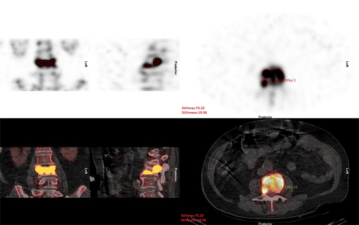
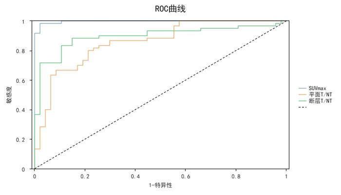
Abstract:
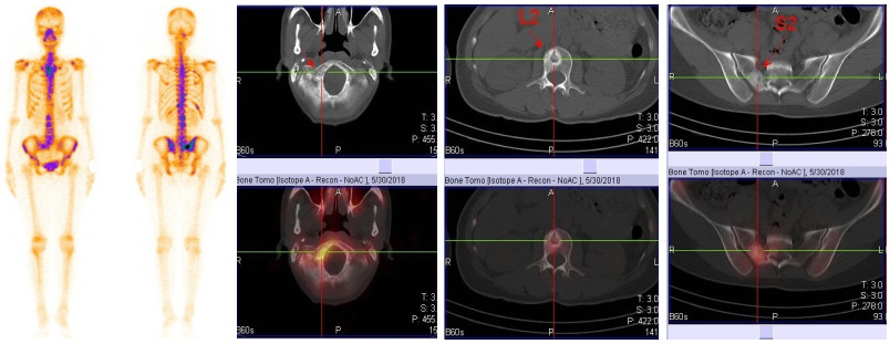
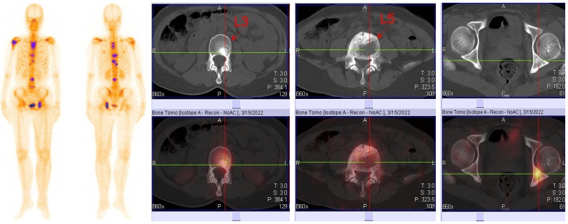
Objective To investigate the clinical value of standard uptake value obtained by quantitative single-photon-emission computed tomography (SPECT)/CT in elderly osteoporotic spinal compression fractures. Methods Sixty-five patients with osteoporotic compression fractures who underwent 99mTc-MDP whole body bone scan, SPECT/CT scan and magnetic resonance imaging(MRI) from August 2019 to June 2021 were enrolled. Based on the clinical history, 107 fractured vertebrae were divided into fresh group (n=60) and old group (n=47), the maximum standard uptake value (SUVmax) and the target/non-target ratio (T/NT) between the two groups was compared. Analyze the correlation between the SUVmax of the fractures and the injury time. ROC curves were plotted to obtain the cut-off values of SUVmax and T/NT and to compare their diagnostic value. Results The vertebral SUVmax was 43.20 ± 17.17, 10.71 ± 3.17 and 9.78 ± 2.95 in the fresh, old and normal groups, respectively, with significant differences between the fresh and old groups (P < 0.01) and no significant differences between the old and normal groups (P > 0.05). The planar T/NT was 2.22 ± 0.70 and 1.41 ± 0.45 in the fresh and old groups, the difference was statistically significant (P < 0.01). The tomo T/NT was 5.06±3.52 and 2.08±0.73 in the fresh and old groups, the difference was statistically significant (P < 0.01). The cut-off values of SUVmax, planar T/NT and tomo T/NT were obtained by plotting ROC curves for differential diagnosis, and the diagnostic sensitivity, specificity and diagnostic agreement of SUVmax were higher than planar T/ NT and tomo T/NT. Conclusion The standard uptake value of quantitative SPECT/CT has important reference value in the diagnosis of elderly osteoporotic spinal compression fractures, it can provide an objective basis for clinical treatment.

2022, 45(4): 546-550.
doi: 10.12122/j.issn.1674-4500.2022.04.14
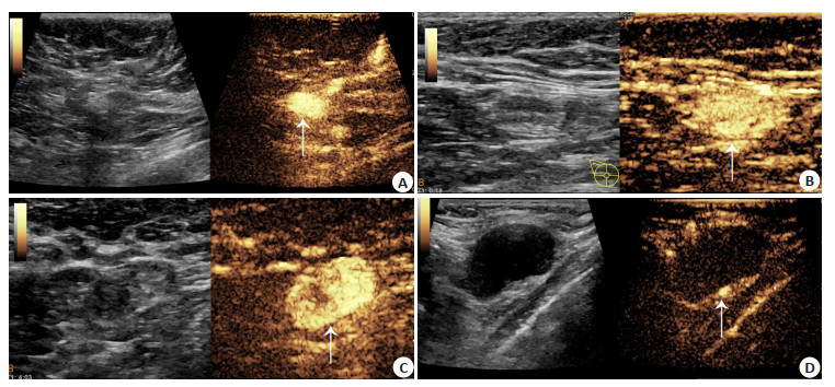
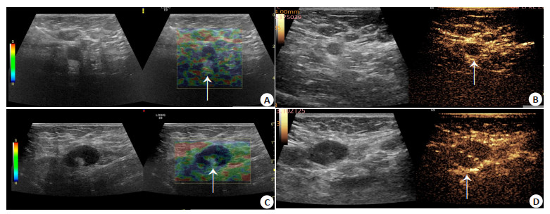
Abstract:
Objective To investigate the diagnostic value of conventional two-dimensional ultrasound, ultrasound elastography and percutaneous contrast ultrasound (CEUS) in ipsilateral axillary sentinel lymph node in breast cancer patients. Methods We retrospectively analyzed 158 cases of breast cancer patients. The patients undewent preoperative lines of conventional two-dimensional ultrasound, ultrasound elasticity imaging, CEUS and coarse needle puncture, with pathological biopsy results as the gold standard, and Kappa test for consistency with pathological results. Logistic regression model was established with pathological diagnosis of sentinel lymph node metastasis as dependent variable and routine two-dimensional ultrasound, ultrasound elastography and CEUS as independent variables. ROC curve was used to evaluate the diagnostic value of conventional two-dimensional ultrasound, ultrasound elastography and CEUS in sentinel lymph nodes of breast cancer. Results A total of 206 lymph nodes were detected by pathological diagnosis in 158 patients with breast cancer, including 76 metastatic nodes and 130 non-metastatic nodes. The sensitivity, specificity and accuracy of conventional two-dimensional ultrasonography for sentinel lymph node metastasis of breast cancer were 69.7%, 94.6% and 85.4%, respectively. The sensitivity, specificity and accuracy of ultrasound elastography were 84.0%, 96.9% and 92.2%, respectively. The sensitivity, specificity and accuracy of CEUS were 88.3%, 98.4% and 94.7% respectively. The sensitivity, specificity and accuracy were 93.4%, 99.2% and 97.1% respectively. The consistency of elastography and CEUS is higher than that of conventional two-dimensional ultrasound, the consistency of combined diagnosis of the three is the best (Kappa=0.683, 0.828, 0.884, 0.937, P < 0.05). ROC curve showed that the AUC of conventional two-dimensional ultrasound, ultrasound elastic imaging, CEUS and their combination in the diagnosis of sentinel lymph node metastasis of breast cancer were 0.668, 0.712, 0.738 and 0.756. Conclusion The diagnostic value of ultrasound elastic imaging and CEUS for sentinel lymph nodes of breast cancer is higher than conventional two-dimensional ultrasound. The combined diagnosis of the three has higher sensitivity, specificity and accuracy, which is of great value for the preoperative diagnosis of ipsilateral metastatic sentinel lymph nodes in breast cancer patients.

2022, 45(4): 551-554.
doi: 10.12122/j.issn.1674-4500.2022.04.15
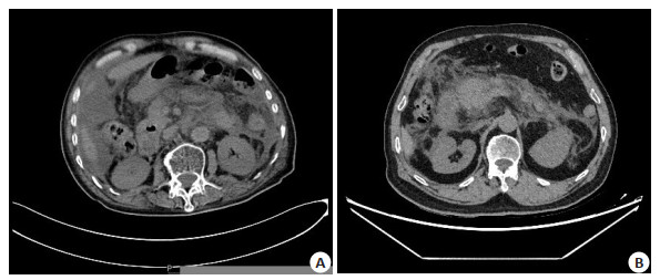
Abstract:
Objective To investigate the prognostic value of pancreatic CT density and maximum cross-sectional area combined with neutrophil lymphocyte ratio (NLR) detection in severe pancreatitis. Methods Forty-six patients with severe pancreatitis who admitted to our hospital from November 2019 to November 2021 were selected, and 40 healthy subjects were selected as the control group during the same period. CT examination was performed on all subjects, combined with Image J to calculate the CT density and maximum cross-sectional area of the pancreas, and NLR was detected. Results The CT density of the pancreas in the observation group was significantly lower than that in the control group (P < 0.05). The maximum cross-sectional area was significantly larger than that in the control group (P < 0.05). The NLR in the observation group was significantly higher than that in the control group (P < 0.05), and the score was higher. The area under the curve of CT density and maximum cross-sectional area for evaluating the prognosis of severe pancreatitis was 0.786, the sensitivity was 80.31%, and the specificity was 80.84%. The area under the curve of NLR for evaluating the prognosis of severe pancreatitis was 0.775, and the sensitivity was 62.51%, specificity was 56.27%. The area under the curve of CT density and maximum cross-sectional area combined with NLR to evaluate the prognosis of severe pancreatitis was 0.884, the sensitivity was 87.51%, and the specificity was 82.15%. Conclusion The use of pancreatic CT density and maximum cross-sectional area combined with NLR detection has a certain clinical value in evaluating the prognosis of severe pancreatitis.

2022, 45(4): 555-559.
doi: 10.12122/j.issn.1674-4500.2022.04.16
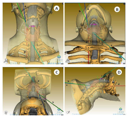
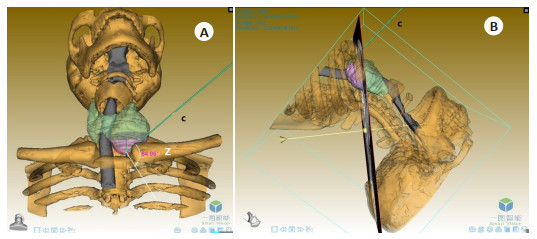
Abstract:
Objective To evaluate the clinical application of three-dimensional reconstruction of head and neck with neck extension in thyroid surgery. Methods The unenhanced CT images which were scan with neck extension of 20 patients proved to be thyroid disease by pathology were collected. The thyroid gland, thyroid nodules, thyroid cartilage, mandible, clavicle, and skin were reconstructed with the software of 3DVWorks. Measuring different angles and diameters for disease evaluation and treatment base on the reconstruction model. Results In the 3D reconstruction model of head and neck, intersectoral angles and distances between different tracheal planes and anatomic structures of endoscopic thyroidectomy were measured. Based on these 3D data, 3 of 11 patients with thyroid nodule < 3 cm were not suitable for endoscopic thyroidectomy via a transoral approach. Meantime, 3 patient was not suitable for the thoracic and breast approach or axillo-breast approach and 1 patient was only suitable for open surgery. The other 4 of 11 patients were suitable for all surgical approaches. 6 patients with thyroid nodule > 3 cm were not diagnosed with a substernal goiter by the model, which could be accessible via a transcervical approach but not a transsternal operative approach. The model showed residual thyroid after surgery in 3 cases could be located accurately and directed the operation. Conclusion Three-dimensional reconstruction of head and neck with neck extension has unique advantages in choice of operative approach, diagnosis, and treatment of substernal goiter and intraoperative localization of residual thyroid tissue, which help to make a better surgical decision and benefit physician-patient communication.
2022, 45(4): 560-565.
doi: 10.12122/j.issn.1674-4500.2022.04.17
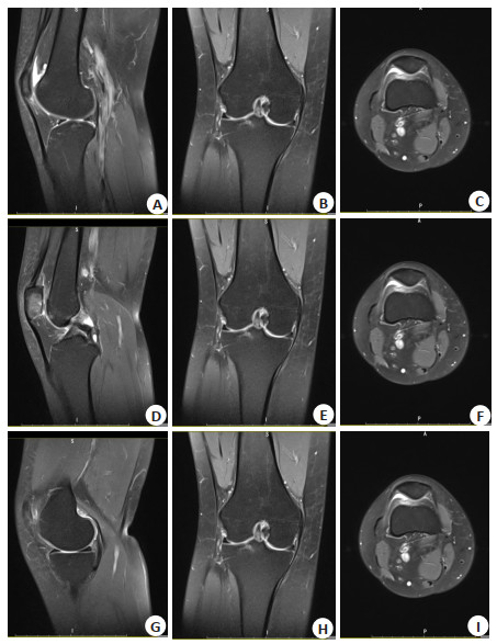
Abstract:
Objective To investigate the correlation between serum inflammatory factors and MRI features of knee osteoarthritis (KOA). Methods A total of 120 patients with knee osteoarthritis admitted to our hospital from January 2020 to May 2021 were selected as the research group, and another 90 healthy persons during the same period were selected as the control group. Patients in research group were divided into group Ⅰ (synovitis) and group Ⅱ (no synovitis) according to whether there was synovitis. MRI findings and serum levels of inflammatory factors [tumor necrosis factor-α (TNF-α), interleukin-1 (IL-1), interleukin-6 (IL-6) and interleukin-1β (IL-1β)] in all patients were compared. The correlation between MRI imaging features and serum inflammatory factors in KOA patients was analyzed. Results All patients in group Ⅰ had chondropathy, and 66.20% had grade 2 chondropathy, while 5 patients in group Ⅱ had no chondropathy, 59.18% had grade 1 chondropathy. The thickness of cartilage in group Ⅰ was lower than that in group Ⅱ (P < 0.05). The expression levels of IL-1β, IL-6 and TNF-α in serum were lower than those in control group < group Ⅱ < group Ⅰ (P < 0.05). The expression levels of IL-10 were lower than those in group Ⅱ < group Ⅰ < control group (P < 0.05). Il-1β, IL-6 and TNF-α were positively correlated with MRI cartilage grading in group Ⅰ (r= 0.387, 0.289, 0.426, P < 0.05), while IL-10 was not correlated with MRI cartilage grading (P > 0.05). Only IL-1β was positively correlated with MRI cartilage grading in group Ⅱ (r=0.509, P < 0.05), while IL-6, IL-10 and TNF-α were not correlated (P > 0.05). MRI synovial thickness was positively correlated with IL-1β, IL-6 and TNF-α in serum inflammatory cytokines in patients with KOA combined with synovitis (r=0.497, 0.425, 0.506, P < 0.05). The thickness of MRI cartilage was negatively correlated with il-1β and TNF-α in serum inflammatory cytokines (r=-0.503, -0.313, P < 0.05). Conclusion MRI abnormalities in patients with KOA are significantly correlated with serum inflammatory factors. The combination of knee imaging features and serum inflammatory factors can provide guidance for the clinical diagnosis and treatment of KOA.

2022, 45(4): 566-571.
doi: 10.12122/j.issn.1674-4500.2022.04.18
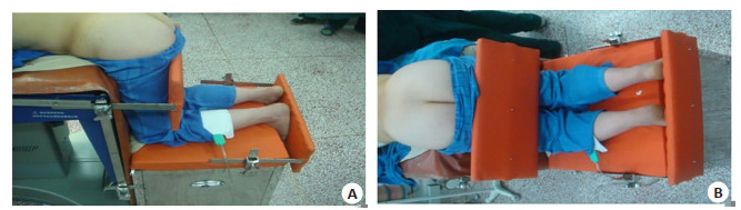
Abstract:
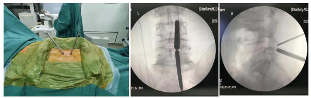
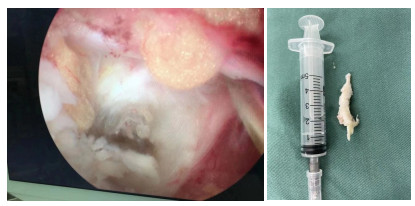
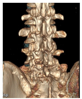
Objective To compare the clinical effect of two kinds of open operation in the treatment of lumbar disc herniation under different position, and discuss the clinical advantage of simple kneeling operation table in the lamina decompression and decompression of nucleus pulpotomy under kneeling position. Methods A total of 132 patients with single lumbar disc herniation were treated with unilateral laminectomy decompression of Lumbar core. Patients were randomly divided into two groups. We used a numerical table extraction method to compare the surgical outcomes of patients treated with a simple kneeling table in the kneeling position (observation group, n=67) and patients treated with a conventional table in the prone position (control group, n=65). The operative time, intraoperative blood loss, postoperative bed time, postoperative hospital stay and complications of two groups were compared. The Oswestry dysfunction index (ODI), visual analogue score (VAS) and clinical efficacy (MacNab excellent and good rate) of the two groups before operation, 1 week after operation and 1, 3, 6 months after operation were compared. Results The operative time, intraoperative blood loss, bed time, hospital stay and the number of complications in the observation group were significantly less than those in the control group (P < 0.05). Compared with before surgery, ODI index and VAS score of 2 groups 1 week and 1, 3, 6 months after surgery were significantly lower than before surgery (P < 0.05). Compared with control group, ODI index and VAS score of observation group were lower than control group at 1 week and 1, 3, 6 months after surgery (P < 0.05). After 6 months of follow-up, the clinical efficacy of observation group was significantly better than control group (MacNab excellent and good rate: 91.04% vs 84.62%, P < 0.05). Conclusion Both methods are feasible for unilateral lamina decompression and removal of nucleus pulposus. Postoperative MRI showed that the responsible disc and nerve compression operation was satisfactory. But kneeling position of unilateral fenestration of the lamina and decompression of the nucleus pulposus can significantly relieve the postoperative pain of patients, improve the surgical effect, and postoperative patients recover quickly.
2022, 45(4): 572-575.
doi: 10.12122/j.issn.1674-4500.2022.04.19
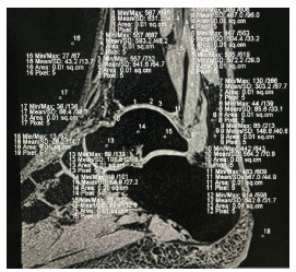
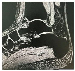
Abstract:
Objective To analyze the advantages and disadvantages of 3D MR sequences in displaying articular cartilage by comparing the signal to noise ratio (SNR) and contrast to noise ratio (CNR) of cartilage in three kinds of ankle 3D water excitation (3D-WE) sequences. Methods Thirty volunteers (aged 18-25 years old) were selected to perform three kinds of 3D MRI scans of ankle cartilage, using Siemens skyra 3.0T MR and head/neck 18 channel coil. Dual echo steady state (DESS) sequence, multi echo data image combination (MEDIC) sequence, three-dimensional volume interpolation fast phase scrambling, volume interpolated body examination (VIBE). The SNRca-x and signal intensity of ankle cartilage, fat, bone, muscle, fluid were measured at the central level of the tibiotalar joint of the ankle joint. The cartilage of SNR and CNRca-Fat, CNRca-Bone, CNRca-Muscle and CNRa-Fluid were calculated by computer. At the same time, the ability of different sequences to display ankle cartilage is evaluated. Results The value of SNR of MEDIC and VIBE were significantly higher than that of DESS (P < 0.05). The values of CNRca-Fat between VIBE, DESS and MEDIC were significantly different(12.75±2.78 vs 6.72±2.09 vs 15.07±3.89, P < 0.05). The value of CNRca-Bone between VIBE, DESS and MEDIC were significantly different(12.88±2.46 vs 7.32±2.07 vs -1.75± 1.95, P < 0.05).The value of CNRca-Muscle between VIBE, DESS and MEDIC were significantly different(2.28 ± 0.71 vs 1.17 ± 1.27 vs -1.15±2.17, P < 0.05). The CNRca-Fluid value between VIBE, DESS and MEDIC were significantly different(1.37±1.31 vs -5.62±6.01 vs -3.28 ± 3.06, P < 0.05). Conclusion Compared with DESS and MEDIC sequences, VIBE can more clearly display the fine anatomical structure and edge of ankle cartilage. The boundary with the surrounding joint cavity effusion is clearer. It can clearly and accurately display various three-dimensional data of ankle cartilage. It is an accurate tool for measuring and calculating the thickness and volume of cartilage.


2022, 45(4): 576-579.
doi: 10.12122/j.issn.1674-4500.2022.04.20
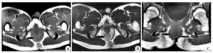
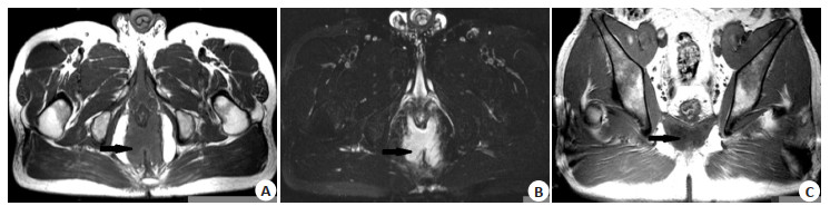
Abstract:
Objective To analyze the causes of long-term clinical neglect of perianorectal muscle tissue abscess, and summarize the recovery of anal function in patients after operation. Methods The MRI features of 39 patients with abscess of external anal sphincter and/or puborectalis muscle and levator ani muscle were analyzed retrospectively. The anal defecation control and defecation ability were followed up. Results There were 18 cases of single muscle tissue abscess and 21 cases of complicated with more than two groups of muscle tissue abscess. There were 28 cases of deep abscess of external anal sphincter, 26 cases of superficial abscess, 9 cases of puborectal abscess, 4 cases of levator ani abscess and 1 case of subcutaneous abscess of external anal sphincter. The infected muscle tissue swelled and thickened, basically maintaining the anatomical shape. The deep part of the external sphincter showed a typical "U" shape, and the levator ani muscle showed a symmetrical "butterfly wing" appearance. 34 cases had clear perianal fat space, 5 cases had unclear fat space and 26 cases had internal openings. 35 patients without involvement of levator ani muscle recovered well after operation. Three patients with abscess of levator ani muscle had different poor ability of fecal control and defecation after operation, and other patients with levator ani muscle abscess lost contact. Conclusion The external anal sphincter, puborectal muscle and levator ani muscle sometimes can not stop the spread of perianal infection, and even form an abscess inside the muscle tissue. The patients with external anal sphincter and puborectal muscle abscess have normal ability of fecal control and defecation after operation. Patients with levator ani abscess may have decreased ability of fecal control and defecation after operation.
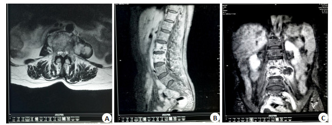
2022, 45(4): 580-583.
doi: 10.12122/j.issn.1674-4500.2022.04.21
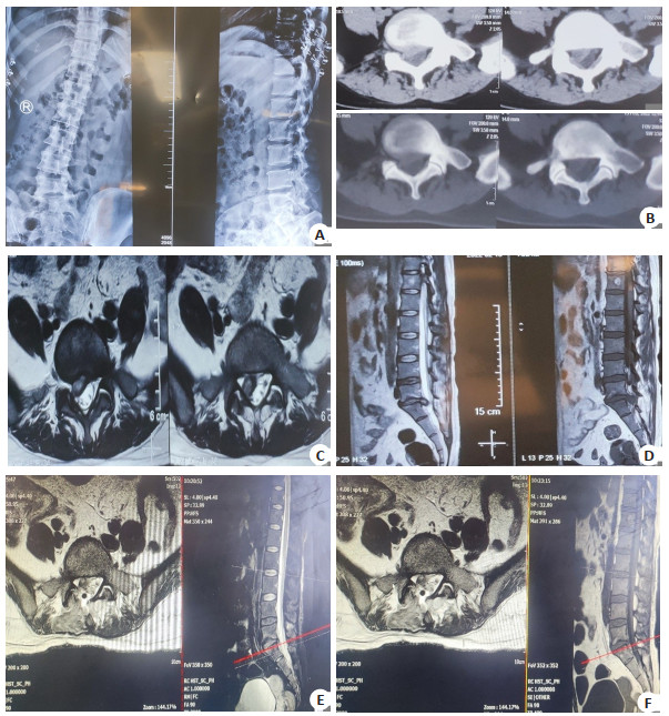
Abstract:
Objective To compare the value of enhanced CT and MRI in diagnosis of lumbar tuberculosis. Methods Sixty patients with suspected lumbar tuberculosis admitted to the hospital were enrolled between June 2019 and June 2021. All underwent examinations by contrast-enhanced CT and MRI images. Taking pathological or treatment follow-up results as the golden standard, their imaging manifestations were observed. The diagnostic efficiency of the two methods for lumbar tuberculosis was compared. Results There were 42 cases confirmed with lumbar tuberculosis out of 60 cases. The contrast-enhanced CT showed that there were 36 cases with lumbar tuberculosis. Compared with the golden standard, its sensitivity, specificity, accuracy and Kappa value were 85.71%, 77.77%, 83.33% and 0.615, respectively. MRI showed that there were 40 cases with lumbar tuberculosis. Compared with the golden standard, its sensitivity, specificity, accuracy and Kappa value were 95.24%, 88.89%, 93.33% and 0.841, respectively. The detection rates of intervertebral disc involvement, paravertebral abscess and spinal canal involvement by MRI were higher than those by contrast-enhanced CT (P<0.05). The detection rate of sequestrum formation by CT was higher than that by MRI (P<0.05). Conclusion Both contrast-enhanced CT and MRI are of certain clinical diagnostic efficiency for lumbar tuberculosis. MRI is superior to contrast-enhanced CT. MRI has obvious advantages in the diagnosis of intervertebral disc involvement, paravertebral abscess and spinal canal involvement, while enhanced CT is superior to MRI in the detection of sequestrum formation.

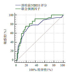
2022, 45(4): 584-589.
doi: 10.12122/j.issn.1674-4500.2022.04.22
Abstract:
Objective To investigate the effect of intracranial artery calcification on the clinical prognosis of patients with acute cerebral infarction who received thrombolytic therapy, in order to predict the prognosis positively. Methods A total of 124 patients with acute anterior circulation cerebral infarction were retrospectively collected. The 90-day modified Rankin scale was used to divide the patients into a good prognosis group (n=87) and a poor prognosis group (n=37). We collected the perfect brain CT at the time of the patient's visit, and used the image processing software ImageJ to measure the calcification volume of the siphon segment of the intracranial carotid artery on the side of the responsible artery. Logistic regression analysis was further applied to the factors affecting the prognosis of patients to determine whether intracranial artery calcification was an independent risk factor for poor prognosis of patients. Finally, the combined predictors of the calcification volume and the NIHSS score before thrombolysis were calculated. ROC curve was drawn by the predictor and pre-thrombolysis NIHSS scores, AUCs were calculated, and the significance of the difference in AUC between two indicators was compared. Results Calcified volume was an independent risk factor for patients with acute anterior circulation cerebral infarction treated with intravenous thrombolysis (OR=1.10, 95%CI=1.05-1.21). The pre-thrombolytic NIHSS score showed that AUC was 0.77, 95%CI was 0.69-0.85, cut-off value was 5, sensitivity was 75.7% and specificity was 72.4%. The pre-thrombolytic NIHSS score combined with calcified volume showed that AUC was 0.83, 95% CI was 0.75-0.89, sensitivity was 75.7% and specificity was 80.5%. The combined predictor was more predictive of patient prognosis than the pre-thrombolysis NIHSS score alone (Z=2.21, P=0.027). Conclusion Calcification volume is independently associated with poor prognosis in patients with acute anterior circulation cerebral infarction receiving thrombolysis, which can constitute an indicator for judging the prognosis of patients after thrombolysis. The pre-thrombotic NIHSS score combined with the calcification volume has a better predictive value than the pre-thrombotic NIHSS score alone.

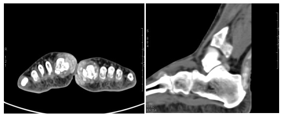
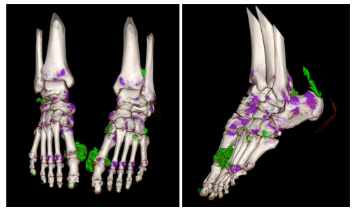
2022, 45(4): 590-594.
doi: 10.12122/j.issn.1674-4500.2022.04.23
Abstract:
Objective To explore the value of dual-source CT dual-energy imaging (DECT) combined with serum C-reactive protein (CRP) in the diagnosis of suspected gout. Methods The clinical data of 92 patients with suspected gout treated in outpatient department of the hospital were retrospectively analyzed from January 2019 to July 2021. The patients were diagnosed by DECT. Among them, 59 patients were diagnosed by DECT and serum CRP at the same time, and the results were compared with the final standard diagnosis. The level of serum CRP was compared between patients diagnosed with gout and patients without gout among 59 cases with serum CRP examination. Results The level of serum CRP of patients with gout was significantly higher than that of patients without gout (t=6.090, P<0.001). The sensitivity, specificity, positive predictive value, negative predictive value and Kappa value of DECT in diagnosing gout were 100.00%, 75.00%, 83.87%, 100.00%, 0.772. The serum CRP were 79.31%, 100.00%, 100.00%, 73.91%, 0.739. The DECT combined with serum CRP were 91.22%, 100.00%, 100.00%, 87.50%, 0.887. Conclusion DECT combined with serum CRP has good consistency in the diagnosis of gout, which is higher than DECT or serum CRP alone.
2022, 45(4): 595-598.
doi: 10.12122/j.issn.1674-4500.2022.04.24
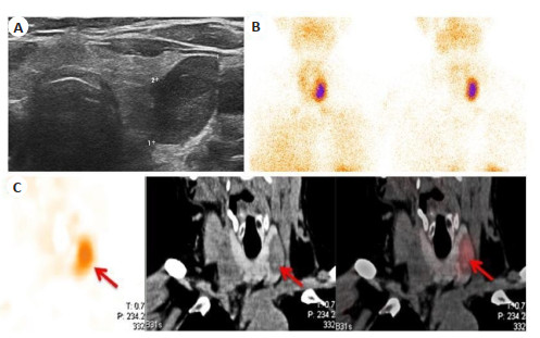
Abstract:
Objective To investigate the application of 99mTc-MIBI SPECT/CT and neck ultrasound in the preoperative diagnosis of primary hyperparathyroidism. Methods The data of 92 patients with suspected primary hyperparathyroidism were collected. The diagnostic efficiency of 99mTc-MIBI SPECT/CT and neck ultrasound was evaluated. The relationship between 99mTc-MIBI uptake and preoperative parathyroid hormone (PTH), the maximum diameter of lesions was analyzed. Results The sensitivity values of neck ultrasound, 99mTc-MIBI plane imaging, SPECT/CT imaging and combined detection were 58.70%, 71.74%, 85.87% and 93.48%, respectively. The sensitivity of SPECT/CT imaging and combined detection was higher than that of ultrasound and 99mTc-MIBI plane imaging (P<0.05). The preoperative PTH and postoperative maximum diameter of lesions in positive 99mTc-MIBI plane imaging group were higher than those in negative group (P<0.05). The uptake ratios T/Ne and T/Nd in early phase lesions, delayed phase lesions and normal tissues were positively correlated with preoperative PTH and postoperative maximum diameter of lesions (P<0.05). ROC curves showed that the AUC, 95%CI, sensitivity and specificity of preoperative PTH for predicting positive 99mTc-MIBI plane imaging were 0.780, 0.675-0.886, 80.30% and 69.23%, respectively. Conclusion The preoperative PTH and lesion size are positively correlated with 99mTc-MIBI uptake. The combination of 99mTc-MIBI SPECT/CT and ultrasound has high application value in the preoperative diagnosis of PHPT.
2022, 45(4): 599-603.
doi: 10.12122/j.issn.1674-4500.2022.04.25
Abstract:
Objective To explore the relationship between chronic stress and neuroimaging and prognosis in patients with depression. Methods A total of 120 patients with depression were selected from February 2019 to February 2021, chronic stress was assessed by life events scale (LES). They were classified into chronic stress group (n=78) and non-chronic stress group (n= 42) according to the score of LES. Thirty healthy volunteers were selected in control group. The subjects underwent cranial MRI scans, FreeSurfer automatic processing was used to calculate and compare the cortical thickness of the whole brain region. Depressive symptoms were assessed with the 17-item hamilton depression scale (HAMD-17). The total cortical thickness and HAMD-17 scores were compared among the three groups. Pearson correlation analysis was used to analyze the correlation between LES scores and the total cortical thickness, HAMD-17 scores. Patients with depression were followed up for 6-12 months after discharge. Time of depressive episode was counted, the depressive episode time and clinical cure rate within 6 months were compared between chronic stress group and non-chronic stress group. Results The thickness of left frontal triangle, bilateral superior frontal gyrus, left frontal margin, bilateral precuneus, bilateral middle temporal gyrus, left superior parietal margin gyrus, left cingulate margin cortex, right insular lid of frontal lobe and right inferior frontal gyrus in control group, non-chronic stress group, chronic stress group showed a decreasing trend, the difference between the groups was significant (P<0.05), no increase in cortical thickness was observed in brain regions. HAMD-17 scores in control group, non-chronic stress group, chronic stress group showed a decreasing trend (P<0.05). Pearson correlation analysis showed that LES scores was negatively correlated with the total cortical thickness (P<0.05), and positively correlated with HAMD-17 scores (P<0.05). The 6-month clinical cure of chronic stress group was lower than that of non-chronic stress group (P<0.05). Depressive episodes lasted longer than those in the non-chronic stress group (P<0.05). Conclusion Chronic stress exacerbates cortical atrophy in patients with depression. It is closely related to the degree of disease and prognosis of patients.

2022, 45(4): 604-608.
doi: 10.12122/j.issn.1674-4500.2022.04.26
Abstract:
Objective To explore and analyze the effete of unilateral biportal endoscopic (UBE) surgery in lumbar disc herniation (LDH) and its effect on JOA score. Methods A total of 69 patients with lumbar disc herniation admitted from October 2020 to December 2021 were randomly divided into observation group (n=34) and control group (n=35) according to random number table method. The patients in the control group were treated with Posterior incision of nucleus pulposus, removal of pedicle, internal fixation and interbody fusion, and the patients in the observation group were treated with UBE. The general data of the two groups (gender, age, lesion location, course of disease, operation time, blood loss, etc. were observed, and the visual analog scale (VAS) score, JOA score and Oswestry disability index (ODI) score before and after operation were evaluated. Results There was no significant difference in VAS score between two groups before operation (P> 0.05), but the VAS score of the two groups after operation was significantly lower than that before operation (P<0.05), and the VAS score of the observation group was significantly lower than that of control group at 1, 24, 72 h after operation (P<0.05); There was no significant difference between the two groups before operation (9.34±0.33 vs 9.23±0.43, P>0.05), and the JOA score of the two groups after operation was significantly higher than that before operation (P<0.05), which in the observation group was significantly higher than that in control group 6 months after operation (15.54±0.52 vs 14.23±0.43, P<0.05); There was no significant difference of ODI score between the two groups before operation (15.43±3.54 vs 15.74±3.34, P>0.05), but the ODI score of the two groups 1 month and 6 months after operation was significantly higher than that before operation (P<0.05), and the ODI score of the observation group 6 months after operation was significantly higher than that of control group (51.43±3.73 vs 48.75±3.64, P<0.05); There was no significant difference in the levels of serum HMGB1 and IL-6 between the two groups before operation (4.43±0.54 vs 4.41±0.65, 44.74±4.25 vs 45.54±4.54, P>0.05), but the levels of serum HMGB1 and IL-6 in the two groups 1 month and 6 months after operation were significantly higher than those before operation (P<0.05), and the levels of patients in the observation group were significantly lower than those in control group 6 months after operation (5.4±0.54 vs 6.54±0.32, 46.85±4.45 vs 52.63±4.41, P<0.05). Conclusion Compared with traditional disc arthroscopy, UBE surgery can effectively reduce the degree of pain and improve lumbar stability in patients with Posterior incision of nucleus pulposus, removal of pedicle, internal fixation and interbody fusion, which is worthy of clinical promotion.
2022, 45(4): 609-614.
doi: 10.12122/j.issn.1674-4500.2022.04.27
Abstract:
Artificial intelligence technology has been increasingly widely used in the diagnosis and treatment of benign and malignant identification and prediction of axillary lymph node metastasis and has become a research hotspot. Imaginomics uses deep learning and computer-assisted diagnosis to provide more information for the diagnosis of breast nodules and to predict the metastasis of lymph nodes. Breast nodules are one of the common diseases in women for physical examination. How to identify the nature of nodules is related to the treatment and prognosis of patients. The most often metastatic site of malignant nodules in the axillary lymph nodes, and predicting the metastasis of lymph nodes is of great significance to the choice of the treatment regimen. This paper provides a review of artificial intelligence techniques in breast nodule elastography and in predicting lymph node metastasis.
Artificial intelligence technology has been increasingly widely used in the diagnosis and treatment of benign and malignant identification and prediction of axillary lymph node metastasis and has become a research hotspot. Imaginomics uses deep learning and computer-assisted diagnosis to provide more information for the diagnosis of breast nodules and to predict the metastasis of lymph nodes. Breast nodules are one of the common diseases in women for physical examination. How to identify the nature of nodules is related to the treatment and prognosis of patients. The most often metastatic site of malignant nodules in the axillary lymph nodes, and predicting the metastasis of lymph nodes is of great significance to the choice of the treatment regimen. This paper provides a review of artificial intelligence techniques in breast nodule elastography and in predicting lymph node metastasis.
2022, 45(4): 615-620.
doi: 10.12122/j.issn.1674-4500.2022.04.28
Abstract:
With the accelerating aging of our society, the incidence of senile dementia is increasing. It is also an important cause of death in the elderly. Mild cognitive impairment is a cognitive impairment state between normal brain aging and senile dementia. Amnestic mild cognitive impairment, as its main classification, has become a high risk factor for senile dementia. Compared with the elderly with normal cognition, the risk of dementia in patients with mild cognitive impairment is 4-10 times higher. Therefore, effective diagnosis and treatment of patients in this state stage can greatly delay the progression of patients' disease to dementia. Currently, amnestic mild cognitive impairment diagnosis relies mainly on the clinical neural electrophysiological examination and imaging examination, with the continuous development of magnetic resonance imaging technology in recent years, greatly improve the early detection of amnestic mild cognitive impairment, in this paper, In this paper, several MRI techniques, such as diffusion tensor imaging, voxel-based morphometry, arterial spin labeling, magnetic resonance spectroscopy, amide proton transfer imaging, and resting- state functional magnetic resonance imaging, have been investigated in amnestic mild cognition. The current status of application in disorders is reviewed in order to provide assistance to clinicians.
With the accelerating aging of our society, the incidence of senile dementia is increasing. It is also an important cause of death in the elderly. Mild cognitive impairment is a cognitive impairment state between normal brain aging and senile dementia. Amnestic mild cognitive impairment, as its main classification, has become a high risk factor for senile dementia. Compared with the elderly with normal cognition, the risk of dementia in patients with mild cognitive impairment is 4-10 times higher. Therefore, effective diagnosis and treatment of patients in this state stage can greatly delay the progression of patients' disease to dementia. Currently, amnestic mild cognitive impairment diagnosis relies mainly on the clinical neural electrophysiological examination and imaging examination, with the continuous development of magnetic resonance imaging technology in recent years, greatly improve the early detection of amnestic mild cognitive impairment, in this paper, In this paper, several MRI techniques, such as diffusion tensor imaging, voxel-based morphometry, arterial spin labeling, magnetic resonance spectroscopy, amide proton transfer imaging, and resting- state functional magnetic resonance imaging, have been investigated in amnestic mild cognition. The current status of application in disorders is reviewed in order to provide assistance to clinicians.
2022, 45(4): 621-626.
doi: 10.12122/j.issn.1674-4500.2022.04.29
Abstract:
Breast cancer is one of the most common malignant tumors in women in the world, with the highest incidence and mortality of cancer. Early diagnosis and treatment can improve the prognosis of the disease and reduce the disease-related mortality. Ultrasound and MRI imaging are the non-invasive examination methods recommended by the guidelines for the diagnosis and treatment of breast cancer for the evaluation of primary tumors. With the development of science and technology, more and more new imaging technologies are applied in clinical practice, and the detection rate and diagnostic accuracy of breast cancer have been greatly improved. The emergence of artificial intelligence has expanded the application of imaging omics and promoted the development of precision medicine for breast cancer. This article reviews the application of ultrasound and MRI imaging and imaging omics in clinical diagnosis and treatment of breast cancer.
Breast cancer is one of the most common malignant tumors in women in the world, with the highest incidence and mortality of cancer. Early diagnosis and treatment can improve the prognosis of the disease and reduce the disease-related mortality. Ultrasound and MRI imaging are the non-invasive examination methods recommended by the guidelines for the diagnosis and treatment of breast cancer for the evaluation of primary tumors. With the development of science and technology, more and more new imaging technologies are applied in clinical practice, and the detection rate and diagnostic accuracy of breast cancer have been greatly improved. The emergence of artificial intelligence has expanded the application of imaging omics and promoted the development of precision medicine for breast cancer. This article reviews the application of ultrasound and MRI imaging and imaging omics in clinical diagnosis and treatment of breast cancer.
2022, 45(4): 627-630.
doi: 10.12122/j.issn.1674-4500.2022.04.30
Abstract:
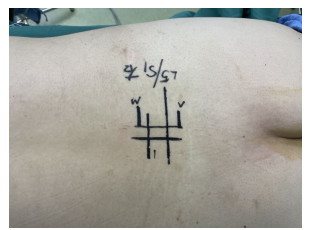
Adolescent scoliosis is a common spinal disease that affects adolescent physical development. With the development of medical imaging technology, imaging measurements are vital to the diagnosis and classification, treatment selection, rehabilitation efficacy and follow-up monitoring of scoliosis. This paper reviews the application and progress of several different imaging methods in the diagnosis of scoliosis, analyzes the advantages and disadvantages of various methods, in order to make these imaging technologies more safe, convenient, effective, accurate and repeatable in the measurement of adolescent scoliosis in the future, It provides some reference for clinical application research and selection.
Adolescent scoliosis is a common spinal disease that affects adolescent physical development. With the development of medical imaging technology, imaging measurements are vital to the diagnosis and classification, treatment selection, rehabilitation efficacy and follow-up monitoring of scoliosis. This paper reviews the application and progress of several different imaging methods in the diagnosis of scoliosis, analyzes the advantages and disadvantages of various methods, in order to make these imaging technologies more safe, convenient, effective, accurate and repeatable in the measurement of adolescent scoliosis in the future, It provides some reference for clinical application research and selection.