

 Find Duplicates
 Check Document
 Submission(new)
 Experts Office
 Editorial Office
2022 Vol. 45, No. 2
column
Display Method:
2022, 45(2): 157-166.
doi: 10.12122/j.issn.1674-4500.2022.02.01
Abstract:
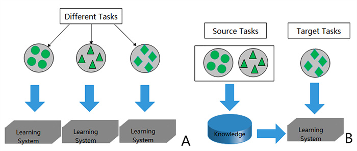
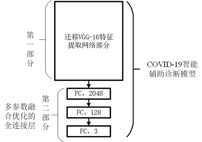
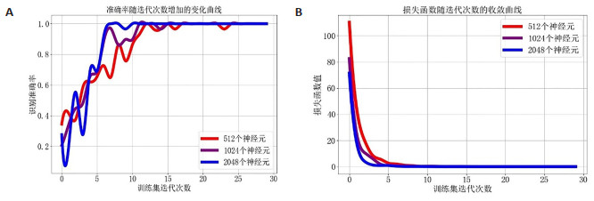
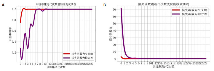
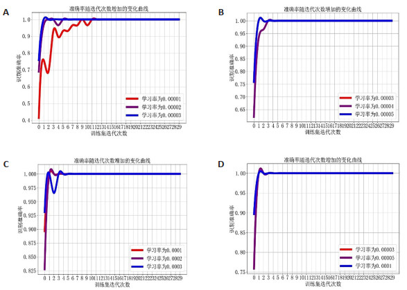
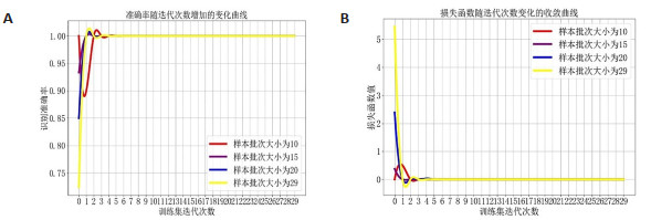
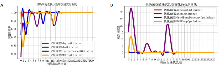
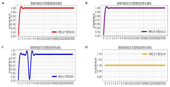
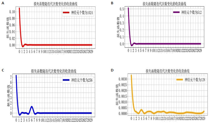
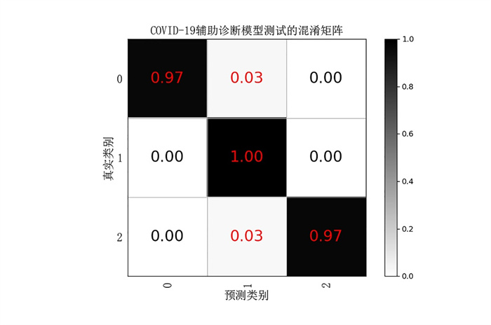
Objective To address the problem of low accuracy and efficiency of traditional CT image diagnosis, this paper discusses the model of deep learning technology in assisting the diagnosis of covid-19 in imaging. Methods First of all, a COVID-19 imaging dataset was constructed for three categories: early stage, progressive stage and critical stage. Then, an initial model for diagnosing COVID-19 based on VGG-16 transfer learning was constructed. Finally, a COVID-19 aided diagnosis model is designed by gradually optimizing the multi parameter fusion of the full connection layer network structure, activation function, loss function, optimization algorithm, learning rate and sample batch size. Results The accuracy of the COVID-19 auxiliary diagnosis model on the COVID-19 imaging test set was 98.10% with sensitivities of 0.97, 1.00 and 0.97 for the early, progressive and severe samples, and F1-scores of 0.98, 0.97 and 0.99, respectively. Conclusion Through migration learning and multi-parameter fusion optimization strategies, the designed COVID-19 aided diagnosis model had high accuracy on the test set. The assisted diagnosis model can help medical workers to improve their efficiency when preventing and controlling epidemics.
2022, 45(2): 167-174.
doi: 10.12122/j.issn.1674-4500.2022.02.02
Abstract:
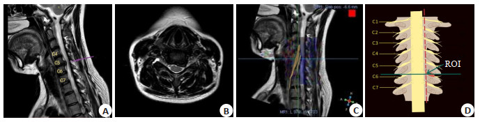
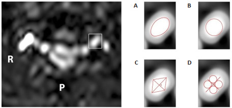
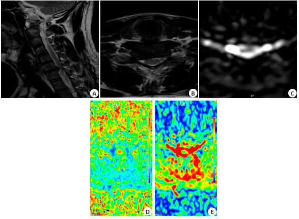
Objective To establish a reliable method for the region-of-interest (ROI) mapping of cervical nerve roots and to verify its clinical relevance. Methods Twenty healthy volunteers and 20 patients with single-segment neurological cervical spondylopathy were selected. Four ROI mapping methods were applied by two imaging physicians to measure the fractional anisotropy (FA) and apparent diffusion coefficient (ADC) values of bilateral cervical 5-8 nerve roots in healthy volunteers as well as the FA and ADC values of the corresponding nerve roots on the affected lesioned segment and the healthy side of the patient to determine the best consistent method for the ROI mapping of the cervical nerve roots, while recording each subject's age, gender, BMI, the patient's VAS score and ISNCSCI score, and then analyzed the clinical characteristic correlations using the best method to measure the obtained diffusion tensor imaging values. Results The maximum circle method had the best consistency. The difference between FA and ADC values of the cervical nerve roots in healthy patients had no statistical significance compared with those of the healthy side of the patients (P>0.05), but had statistical significance compared with those of the affected side. The difference between FA and ADC values on the healthy side of patients and on the affected side was statistically significant (P < 0.05); measured FA and ADC values were correlated with age (P < 0.05), and the older the age, the smaller the FA value and the larger the ADC value, but there was no correlation with BMI. In terms of patients, VAS scores were positively correlated with ADC values on the affected side (P < 0.05) and negatively correlated with FA/ADC ratio on the affected side (P < 0.05), and ISNCSCI scores were positively correlated with FA values (P < 0.05). Conclusion The maximum circle ROI mapping method is a reliable method for diffusion tensor imaging measuring cervical nerve roots. The more obvious the radicular pain, the greater the ADC value and the smaller affected side FA/ ADC ratio. The better the cervical nerve function, the greater the FA value.
2022, 45(2): 175-182.
doi: 10.12122/j.issn.1674-4500.2022.02.03
Abstract:
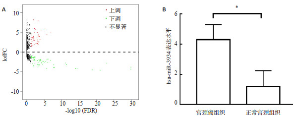
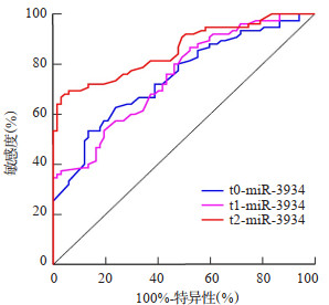
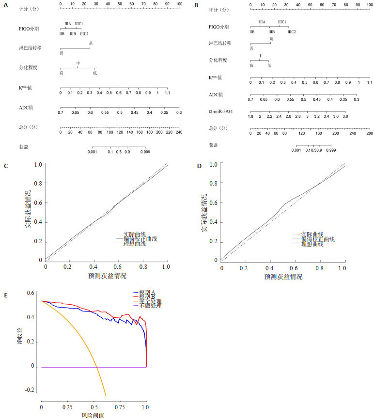
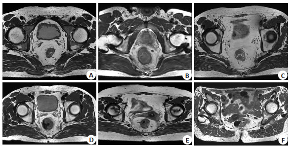
Objective To investigate the value of microRNA-3934 (miR-3934) combined with MRI to assess the benefit of concurrent chemoradiotherapy in stage IIB~III cervical cancer. Methods A total of 142 patients with stage IIB~III cervical cancer diagnosed at the Affiliated Hospital of North China University of Technology from August 2019 to September 2021 were selected as the study subjects, given concurrent chemoradiotherapy and divided into the benefit group (n = 67) and non-beneficiary group (n = 75) according to the treatment benefit. The miR-3934 level was detected by real-time fluorescent quantitative PCR. Receiver operating characteristic (ROC) curve was used to evaluate the value of miR-3934 in determining the benefit of concurrent chemoradiotherapy for stage IIB~III cervical cancer. Logistic regression was used to analyze the risk factors for the benefits of concurrent chemoradiotherapy in stage IIB~III cervical cancer, and nomogram regression model of risk factors was constructed. The consistency index, calibration curve and decision curve analysis were used to evaluate the value of the model. Results The miR-3934 levels in the beneficiary group were lower than in the non-beneficiary group before (t0), on day 2th (t1) and on day 8th (t2) of concurrent chemoradiotherapy (2.71±0.30 vs 3.05±0.40, 2.64±0.28 vs 2.97±0.35, 2.44± 0.24 vs 2.91±0.36, P < 0.05). The miR-3934 levels in both groups tended to decrease with time progression, and miR-3934 levels at time point t2 were lower than t0 in both the benefit and non-benefit groups (P < 0.05). The values of reflux rate constant, volume transfer constant (Ktrans) and extravascular extracellular gap volume ratio in the beneficiary group were lower than those in the non-beneficiary group [(1.17±0.11)/min vs (1.22±0.11)/min, (0.50±0.15)/min vs (0.67±0.12)/min, (0.37±0.09)% vs (0.47±0.12)%, P < 0.05], and the apparent diffusion coefficient (ADC) value was higher than that in the non-beneficiary group [(0.55±0.06)×10-3 mm2/s vs (0.48±0.07)×10-3 mm2/s, P < 0.05]. The miR-3934 at time point t2 determined the benefit of synchronous radiotherapy for stage IIB~III cervical cancer under the ROC curve area was higher than t0 and t1 (P < 0.05). International Federation of Gynecology and Obstetrics (FIGO) stage III, lymph node metastasis, low differentiation, miR-3934>2.66 at t2 and Ktrans>0.59 were independent risk factors for concurrent chemoradiotherapy of stage IIB~III cervical cancer(P < 0.05). ADC>0.52 was an independent protective factor for concurrent chemoradiotherapy of stage IIB~III cervical cancer (P < 0.05). The C-index of model A (consisting of FIGO staging, lymph node metastasis, differentiation, Ktrans and ADC) was 0.969, which was lower than that of model B (consisting of FIGO staging, lymph node metastasis, differentiation, miR-3934 at t2, Ktrans and ADC, 0.986). The mean square error and mean absoluteerror of model A were 0.00046 and 0.02, respectively, which were lower than those of model B (0.00098 and 0.03). When the threshold probability was 0.88~0.92, the clinical application value of model A was greater than that of model B. When the threshold probability was 0~0.88 or 0.92~1.00, the clinical application value of model B was greater than that of model A. Conclusion Model B consisting of FIGO stage, lymph node metastasis, differentiation, miR-3934 at t2, Ktrans and ADC has high value in assessing the benefits of concurrent chemoradiotherapy for stage IIB~III cervical cancer, which can assist physicians in decision making.

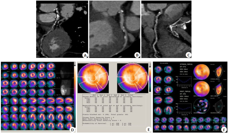
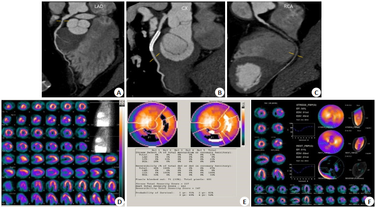
2022, 45(2): 183-188.
doi: 10.12122/j.issn.1674-4500.2022.02.04
Abstract:
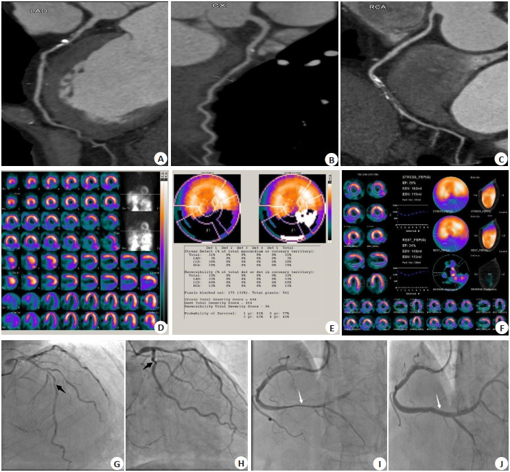
Objective To investigate the clinical application value of one-day dobutamine loading/resting single-photon emission computed tomography (SPECT) myocardial perfusion imaging in guiding the treatment of coronary heart disease. Methods A retrospective collection of 68 patients with coronary heart disease who presented to our hospital with chest tightness and chest pain in our hospital from May 2019 to July 2021, routinely underwent coronary computed tomography angiography (CCTA), 34 of them underwent one-day dobutamine loading/resting SPECT myocardial perfusion imaging at the same time, and the therapy was selected based on SPECT results as the study group. The other 34 cases selected the therapy based on CCTA results as the control group. After determining the treatment regimen, the two groups were followed up to compare the rates of symptom relief and cardiovascular adverse events, and to analyse the differences between the treatment regimens recommended by SPECT and CCTA and the reasons for them. Results There were no significant differences in clinical condition and the stenosis degree, lesion location and number of coronary artery between the two groups (P <0.05). In the study group, the patient who was treated with revascularization method accounted for 3% (1/34), the remaining patients treated with drugs alone accounted for 97% (33/34), and the symptom remission rate was 100%. In the control group, those patients which were treated by revascularization method accounted for 50%(17/34), the remaining patients which were treated with drugs alone accounted for 50% (17/34), and the symptom remission rate was 100%. The revascularization rate of the study group was lower than that of the control group (χ2 = 19.342, P <0.05). At 7-33 months follow-up (median 28 months), the incidence of adverse cardiovascular events was 3% in the control group (1/34) and 0 in the study group. 14 patients in the study group had treatment recommendations for SPECT that differed from those for CCTA. In the end, these 14 patients chose drug therapy alone based on SPECT results, which was approximately 41% less than the rate of coronary angiography where decisions were made based on CCTA results (14/34) and a saving of approximately 50% in the cost of a single hospital visit. Conclusion One-day dobutamine loading/resting SPECT myocardial perfusion imaging facilitates the screening of patients with coronary heart disease who do not need interventional therapy. It is safe and effective using a drug-only regimen and reduces the need for unnecessary invasive tests and interventional procedures compared to the currently commonly used CCTA screening, lowering the total cost of care and shortening the length of hospital stay.
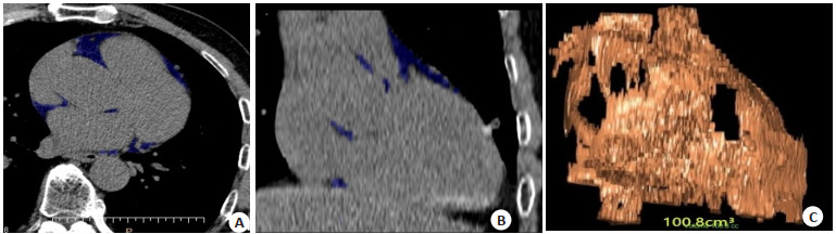
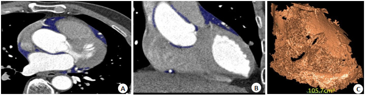
2022, 45(2): 189-193.
doi: 10.12122/j.issn.1674-4500.2022.02.05
Abstract:
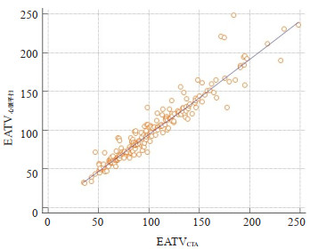
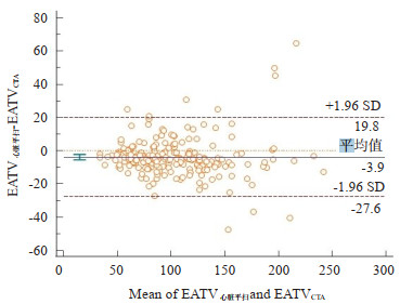
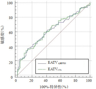
Objective To study the consistency between low-dose cardiac plain scanning and coronary artery CT angiography (CTA) in quantification of epicardial adipose tissue volume (EATV). Methods A total of 208 patients with clinically suggestive signs of coronary heart disease were examined by low-dose cardiac plain scan and coronary artery CTA. Epicardial fat volume was measured on the images of both examinations respectively. The correlation between epicardial fat volume measured by the two examinations EATVcardiac plain scan and EATVCTA was evaluated by Pearson correlation coefficient. Bland-altman analysis was used to evaluate their consistency. Patients were divided into the plaque free group and the plaque groups, and the differences of EATVcardiac plain scan and EATVCTA between the two groups were compared. ROC curve was used to evaluate the value and difference of EATVcardiac plain scan and EATVCTA in predicting coronary atherosclerosis. Results There was a significant correlation between low-dose EATV cardiac sweep of 103.12±44.01 cm3 and EATVCTA of 106.72±44.14 cm3 (r = 0.955, P< 0.001). The mean consistency between EATVcardiac plain scan and EATVCTA was -3.5, with 4.8% (10/208) points falling outside the 95%CI (95%CI: 24.0930- 29.2116, P< 0.001). The area under the curve for predicting by EATVcardiac plain scan and EATVCTA were 0.601 and 0.605 respectively, with no statistical difference between the two groups (P= 0.935). Conclusion Low-dose cardiac plain scan and coronary artery CTA are equivalent in the measurement of EAT, and both are of equal value in the prediction of coronary atherosclerosis.
2022, 45(2): 194-198.
doi: 10.12122/j.issn.1674-4500.2022.02.06
Abstract:
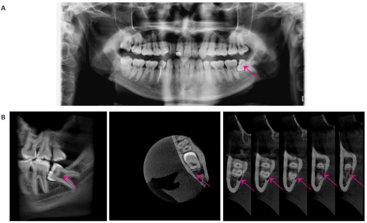
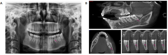
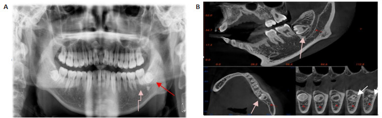
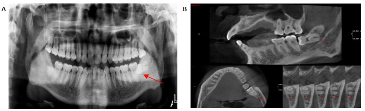
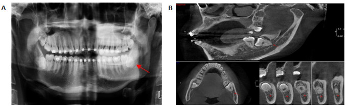
Objective To analyze the clinical value of cone- beam CT (CBCT) combined with panoramic radiography in the preoperative examination of impacted mandibular teeth extraction. Methods A total of 102 patients with mandibular impacted teeth (126 impacted teeth) treated in our hospital from January 2019 to January 2020 were selected. All patients underwent preoperative oral imaging, and the related variables of impacted teeth, including root angulation, root number, distal and mesial root morphology, and relationship with mandibular neural tube were recorded. and the above variables were evaluated as gold standard intraoperatively and postoperatively. Results The panoramic images could not clearly show the threedimensional relationship between the root and the inferior alveolar neural tube, which was clearly shown in the CBCT images. There were no significant differences in angle formation, root number, distal root bending, proximal root bending or distal root bending, proximal root bending or proximal root bending (P>0.05) in panoramic image, CBCT and CBCT combined panoramic image. There were statistically significant differences in the relationship between root and mandibular canal (P < 0.05). The extraction time was 15.93±3.24 min, the bleeding volume was 26.45±3.15 mL, only one (0.79%) had root breakage. During the operation, only one patient developed root breakage and root fracture, and one patient developed lingual bone plate fracture. there were no patients with bleeding or infection postoperatively, and two patients showed numbness and one showed swelling and limitation. Conclusion CBCT combined with panoramic radiography has great advantages in determining the relationship between the mandibular third molars and the mandibular canal, which has positive significance in improving intraoperative situation and intraoperative and postoperative complications of patients.
2022, 45(2): 199-203.
doi: 10.12122/j.issn.1674-4500.2022.02.07
Abstract:
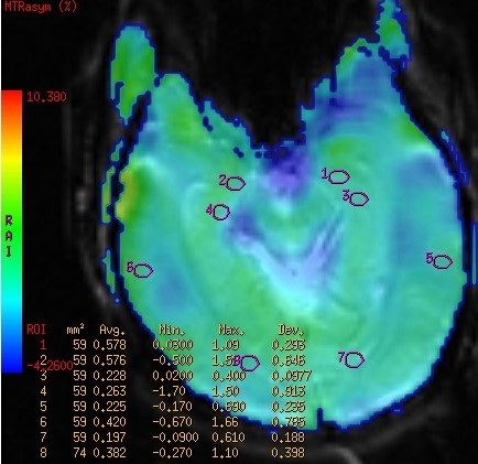
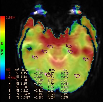
Objective To investigate the feasibility of using magnetic resonance amide proton transfer imaging (APT) to study the pathophysiological changes in various brain regions of interest in patients with amnestic mild cognitive impairment (aMCI) and its correlation with some clinical cognitive score scales. Methods Twenty-one patients with aMCI diagnosed in the Department of Neurology from January 2021 to January 2022 were selected as the case group, and another twenty-one healthy subjects in the physical examination center of our hospital during the same period were selected as the control group. The clinical data of the two groups were analyzed, the clinical psychological scale was assessed, and routine magnetic resonance imaging and amide proton transfer imaging were performed in each brain region of interest. Results Compared with the control group, APT values in the right hippocampus and bilateral amygdala were significantly increased (P < 0.05), and left hippocampus had statistical significance (P < 0.01). The clinical scores of MoCA and MMSE in case group were significantly lower than those in control group (P < 0.05). MMSE score scale was negatively correlated with APT values of right hippocampus and left amygdala (P < 0.05). In the comparison of the APT values in each subitem score of the Montreal Cognitive Assessment score and each brain region, the APT values in the right hippocampus were negatively correlated with visual spatial and executive function scores in the aMCI group (P < 0.05), and the APT value of left amygdala and bilateral hippocampus were negatively correlated with memory scores (P < 0.05). Conclusion Forgetting in patients with aMCI part of the brain is a series of pathophysiological changes. APT technique, as an emerging functional magnetic resonance imaging technique, can help clinicians to make an early diagnosis of patients with aMCI to a certain extent.
2022, 45(2): 204-209.
doi: 10.12122/j.issn.1674-4500.2022.02.08
Abstract:
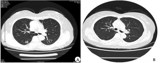
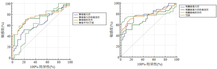
Objective To analyze the diagnostic value on imaging features and CT quantitative parameters of pulmonary microinfiltrating adenocarcinoma (MIA) and infiltrating adenocarcinoma (IAC). Methods Eighty patients with lung adenocarcinoma admitted to our hospital from January 2019 to January 2020 were selected as the study subjects. According to pathology, they were divided into MIA group (n=36) and IAC group (n=44). The CT signs of the two groups were analyzed. Receiver operating characteristic curve (ROC) was used to evaluate the sensitivity and specificity of lung window maximum diameter, vertical diameter of lung window maximum diameter, lung window lesion volume, lung window average CT value, mediastinal window maximum diameter, vertical diameter of mediastinal window maximum diameter, mediastinal window lesion volume, TDR indicators in the identification of IAC and MIA. Results There were no significant differences in lesion location, lesion morphology, vacuolar sign and lung tumor interface in the CT images of the IAC MIA group (P >0.05). There were significant differences in pleural depression, burr sign, lobulation sign, vascular cluster sign and bronchial inflation sign (P < 0.05). The maximum diameter of lung window, vertical diameter of maximum diameter of lung window, volume of lung window lesion, maximum diameter of mediastinal window, vertical diameter of maximum diameter of mediastinal window and lesion volume of mediastinal window in IAC group were significantly higher than those in MIA group (P < 0.05), and the mean CT value of the lung window and the disappearance rate of the tumor shadow were significantly lower than those in the MIA group (P < 0.05). ROC curve showed that the highest differential diagnostic value among the CT quantitative parameters was the volume of the mediastinal window lesion, followed by the maximum diameter of the mediastinal window, the disappearance rate of the tumor shadow, the vertical diameter of the maximum diameter of the lung window, the vertical diameter of the lung window, and the maximum diameter of the lung window. Conclusion When the imaging features of the lesion have pleural indentation, burr sign, lobulation sign, vascular cluster sign and bronchial inflatation sign, the possibility of IAC and mediastinal window lesion volume and mediastinal window maximum diameter are of great value in differentiating IAC from MIA.
2022, 45(2): 210-213.
doi: 10.12122/j.issn.1674-4500.2022.02.09
Abstract:
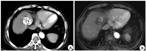
Objective To evaluate the clinical value of MRI combined with multi-slice spiral CT (MSCT) in evaluating the efficacy of interventional therapy for primary liver cancer. Methods A total of 130 patients with primary liver cancer undergoing interventional therapy from May 2019 to May 2021 were selected as the study subjects, of which 50 patients received single MSCT examination, 40 patients received single MRI examination, and 40 patients received combined MRI and MSCT examination. All patients were verified by digital subtraction angiography. The sensitivity, specificity and accuracy of MRI single examination, MSCT single examination and combined examination of residual lesions and recurrent lesions after interventional therapy were compared using the results of digital subtraction angiography as the gold standard, and the lesion detection rates of three examination methods were compared. Results The sensitivity and accuracy of MRI and MSCT combined examination of residual lesions were 89.3% and 96.2%, which were higher than those of MSCT single examination (77.9%, 91.4%) and MRI alone (83.5%, 95.9%) (P < 0.05). The sensitivity and accuracy of MRI combined with MSCT were 88.6% and 95.1%, which were higher than those of MSCT single examination (70.7%, 91.4%) and MRI single examination (74.2%, 93.9%) (P < 0.05). Conclusion MRI combined with MSCT can better detect residual and recurrent lesions in patients with primary liver cancer undergoing interventional therapy, and the ability to detect positive lesions is more in line with the actual clinical needs than single imaging examination.
2022, 45(2): 214-218.
doi: 10.12122/j.issn.1674-4500.2022.02.10
Abstract:
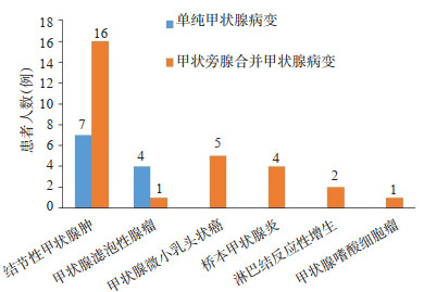
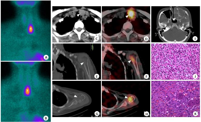
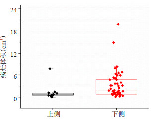
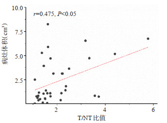
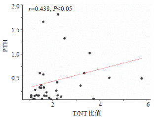
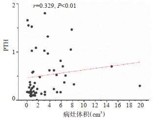
Objective To investigate the value of 99Tcm-MIBI SPECT/CT imaging combined with T/NT semi-quantitative analysis in pre-operative of primary parathyroid adenoma. Methods The 99Tcm-MIBI SPECT/CT imaging and clinical data of 81 patients diagnosed with primary parathyroid adenoma were retrospectively analyze, to investigate the detection rate of parathyroid adenoma in different sites. Results The detection rate of 99Tcm-MIBI SPECT/CT tomography imaging, 99Tcm-MIBI SPECT/CT planar imaging were 95.7%(67/70) and 90%(63/70), respectively. There was no significant difference in detection rate between 99Tcm-MIBI SPECT/CT tomography imaging and planar imaging ( χ2=1.723, P=0.189). Adenomas are classified as superior and inferior according to their location, the detection rate of 99Tcm-MIBI SPECT/CT tomography imaging, 99Tcm-MIBI SPECT/CT planar imaging for superior adenomas were 92.8%(13/14)and 85.7%(12/14), and for inferior adenomas were 100%(53/53)and 100%(53/53), respectively. There was no significant difference in detection rate of adenomas between the upper and lower sides on 99Tcm-MIBI SPECT/CT tomography imaging ( χ2=3.84, P=0.20). The detection rate of inferior adenomas in 99Tcm-MIBI SPECT/CT planar imaging was higher on the lower side than on the upper side ( χ2=7.80, P=0.04). Adenoma volume on the inferior side was greater than on the superior side (Z=-3.19, P=0.001), and there was a weak correlation between adenoma volume and early phase T/NT ratio and serum parathyroid hormone (r=0.475, 0.329, P < 0.05). Conclusion By performing preoperation 99Tcm-MIBI SPECT/CT imaging in patients with primary parathyroid adenoma combined with T/NT semi-quantitative analysis, the accuracy of adenoma localization at different sites is improved and could provide a valuable reference for surgery.
2022, 45(2): 219-222.
doi: 10.12122/j.issn.1674-4500.2022.02.11
Abstract:
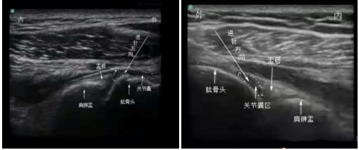
Objective To analyze the improvement effect of ultrasound-guided compound betamethasone and lidocaine hydrochloride injection on shoulder pain after stroke under ultrasound guidance. Methods Sixty patients with shoulder pain after stroke treated in our hospital from January 2020 to June 2021 were selected as the research objects, and the patients were divided into treatment group and control group according to random number table method, with 30 patients in each group. The control group was treated with freehand localization drug injection on the basis of routine rehabilitation therapy. The treatment group was treated with ultrasound-guided compound betamethasone and lidocaine hydrochloride injection under ultrasound guidance on the basis of routine rehabilitation. The pain degree, range of motion of shoulder joint, functional motion of upper limb and the occurrence of adverse reactions were observed and compared between the two groups before and after treatment. Results After 2 and 4 weeks of treatment, the patients in the treatment group had lower visual analogue scores than those in the control group (P < 0.05), greater shoulder forward flexion, abduction and external rotation mobility than those in the control group (P < 0.05), and higher upper limb component scores on the Fugl-Meyer Motor Function Scale than those in the control group (P < 0.05). There was no statistical significance in the total incidence of adverse reactions between the two groups (P>0.05). Conclusion Compared with hands- free localization drug injection, ultrasound- guided compound betamethasone and lidocaine hydrochloride injection is more effective in improving pain in patients with post-stroke shoulder pain, and it can effectively improve shoulder mobility.
2022, 45(2): 223-228.
doi: 10.12122/j.issn.1674-4500.2022.02.12
Abstract:
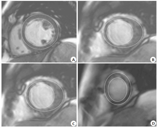
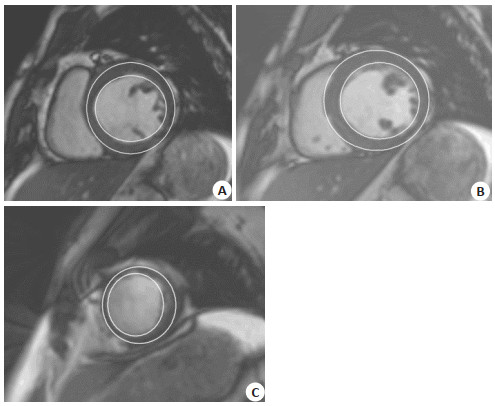
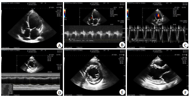
Objective To access the effect of salbutamol on cardiac structure and function by cardiac magnetic resonance imaging (CMR) in T2DM patients without heart failure. Methods In this prospective study, patients with T2DM without heart failure were treated with salbutamol as part of routine guideline-guided treatment. Clinical evaluation, CMR imaging and biomarkers were evaluated in a blinded manner and compared after 6 months of continuous treatment. The changes of left ventricular ejection fraction, changes of right ventricular end-diastolic volume, ventricular mass, global strain and cardiac biomarkers (NT-proBNP and hsCRP) were analyzed after 6 months of treatment. Results The average age of patients (n=16) in this study was 59.9 years old, 69% of whom were male. The average HbA1c was (8.3±0.9)%, and the mean pretreatment score was (57.4±3.4)%, with no significant change after 6 months [0.2%, 95% CI: (-2.5, 2.1), P=0.86]. CMR showed no change including left/right ventricular volumes, left ventricular mass and characteristic track-derived strain (P>0.05). Levels of N-terminal pro-B-type natriuretic peptide and high-sensitivity C-reactive protein did not change significantly (P>0.20). Conclusion According to the evaluation of CMR and biomarkers, salbutamol is not associated with adverse ventricular remodeling after 6 months of treatment in this stable type 2 diabetic non-heart failure patient.
2022, 45(2): 229-234.
doi: 10.12122/j.issn.1674-4500.2022.02.13
Abstract:
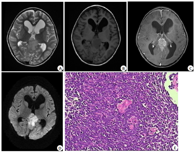
Objective To investigate the CT and MRI features of pineal parenchymal cell tumors. Methods CT, MRI and clinical data of 34 cases of pineal parenchymal cell tumor in our hospital were collected. The patients were divided into three groups according to the pathological results: pineocytoma (PC group), moderately differentiated pineal parenchymal cell tumor (PPTID group) and pineoblastoma (PB group). Results Pineal parenchymal tumors were mainly characterized by well-defined, regular shape of low signal intensity on T1WI, high signal intensity on T2WI, and significant enhancement, high signal intensity on diffusion-weighted imaging, and elevated Cho/NAA ratio on magnetic resonance spectroscopy. Among the three groups cases, there were significant differences in age (P=0.009) and cystic degeneration (P=0.047). We found that the age of patients with PB were younger than that in both PPTID group (P=0.007) and PC group (P=0.007), while cystic changes occurred less frequently in the PC group than in the PB group (P=0.019). Conclusion With certain clinical and imaging features, pathological types of pineal parenchymal tumors can be identify with the help of CT and MRI technology
2022, 45(2): 235-238.
doi: 10.12122/j.issn.1674-4500.2022.02.14
Abstract:
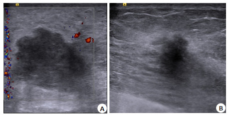
Objective To analyze the relationship between the efficacy of neoadjuvant chemotherapy and ultrasound breast image reporting and the BI-RADS signs in breast cancer patients. Methods Eighty-six patients who admitted to our hospital between June 2019 and December 2020 were selected and divided into ineffective group (n=23) and effective group (n=63) according to their efficacy. All patients were treated with standard neoadjuvant chemotherapy regimens. The patients were examined by ultrasonography, and the risk level of lesions was recorded and assessed using BI-RADS method. Results After treatment, the tumor volume and tumor area of patients in the effective group were significantly lower than those in the ineffective group (P < 0.05). The proportion of patients with clear edge, low elastic score and posterior echo enhancement in the effective group was significantly higher than that in the ineffective group (P < 0.05). The BI-RADS grading of patients in both groups was significantly better than that before treatment (P < 0.05), and the BI-RADS grading of patients in the effective group was significantly better than that of the ineffective group (P < 0.05). Conclusion ultrasound BI- RADS sign analysis can effectively improve the evaluation of the efficacy neoadjuvant chemotherapy in breast cancer patients.
2022, 45(2): 239-243.
doi: 10.12122/j.issn.1674-4500.2022.02.15
Abstract:
Objective To analyze the myocardial energy expenditure (MEE) and related factors of poor prognosis in heart failure patients with preserved ejection fraction. Methods Clinical data of 107 patients with heart failure with retained ejection fraction admitted to our hospital from January 2020 to January 2021 were retrospectively collected. All patients underwent echocardiography and were followed up for 6 months after discharge. Patients were divided into the good prognosis group (n=72) and the poor prognosis group (n=35) according to whether adverse cardiovascular events (MACE) occurred during followup. Single factors for poor prognosis in patients with heart failure with preserved ejection fraction were statistically analyzed using multifactorial logistic regression and echocardiographic images were analysed in typical cases. Results A total of 35 of 107 heart failure patients with preserved ejection fraction developed MACE, with an incidence of poor prognosis of 32.71%. Univariate analysis showed that the levels of erythrocyte count, hemoglobin and left ventricular ejection fraction (LVEF) in the poor prognosis group were lower than those in the good prognosis group (P<0.05), while the levels of serum brain natriuretic peptide and MEE were higher than those in the good prognosis group (P<0.05). Multivariate Logistic regression analysis showed that high level of serum brain natriuretic peptide, high MEE and low LVEF were independent risk factors for poor prognosis in heart failure patients with retained ejection fraction (OR=2.457, 3.083, 2.986, P<0.05). Conclusion The independent risk factors for poor prognosis in patients with retained ejection fraction include high level of serum brain natriuretic peptide, high MEE and low LVEF. Echocardiographic MEE and LVEF could be used to predict the development of MACE in patients with heart failure with preserved ejection fraction.
2022, 45(2): 244-247.
doi: 10.12122/j.issn.1674-4500.2022.02.16
Abstract:
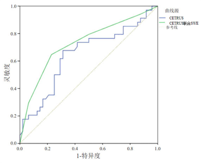
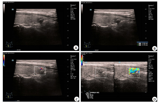
Objective To analyze the value of transrectal contrast-enhanced ultrasound (CETRUS) combined with shear wave elastography in the differential diagnosis of benign and malignant prostate lesions. Methods A total of 102 patients with prostate diseases admitted to our hospital from May 2020 to December 2021 were selected, and divided into prostate cancer group (n=37) divided into prostate cancer group (n=37). All patients underwent transrectal contrast-enhanced ultrasound combined with shear wave elastography. Eration value and Emax were compared between the two groups, and the diagnostic accuracy of different methods was compared. Results The Eration value and Emax in prostate cancer group were significantly higher than those in benign prostatic hyperplasia group(P < 0.05). The sensitivity, specificity and accuracy of CETRUS combined with shear wave elastography in diagnosing benign and malignant prostate lesions were significantly higher than those of CETRUS alone(81.08%、80.00%、80.39% vs 56.76%、64.62%、61.76%, P < 0.05). The area under the curve of CETRUS combined with shear wave elastography was 0.748 (95%CI: 0.534-0.760), which was larger than that of CETRUS alone 0.685(95%CI: 0.266-0.851). Conclusion Transrectal contrast-enhanced ultrasound combined with shear-wave elastography can reflect tissue hardness through Young's modulus value, and then diagnose benign and malignant prostate lesions, which has certain clinical value.

2022, 45(2): 248-251.
doi: 10.12122/j.issn.1674-4500.2022.02.17
Abstract:
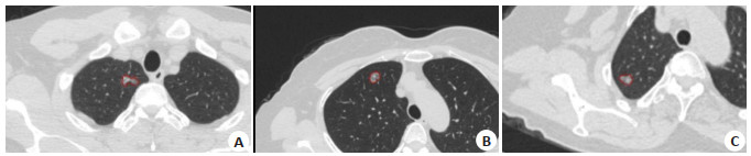
Objective To explore the value of its application in the diagnosis of patients with subsolid pulmonary nodules by means of artificial intelligence automatic recognition combined with DenseNet network model CT examination. Methods Ninty-eight patients with subsolid pulmonary nodules admitted to our hospital from June 2018 to December 2019 were selected and examined by CT, including 32 patients in the group of ≤10 mm in diameter, 33 patients in the group of 10 mm < diameter≤20 mm and 33 patients in the group of > 20 mm in diameter. All patients were diagnosed by high-resolution CT, and the CT examination data were recorded into an artificial intelligence system based on DenseNet network deep learning so as to control the quality of the training data set of artificial intelligence imaging diagnosis. All patients were diagnosed as benign or malignant by the artificial intelligence group and the manual film reading group. The predictive value of CT value, volume and malignant probability in CT plain scan, enhanced arterial phase and delayed phase of patients with different diameters of pulmonary nodules by AI were compared and analyzed to test the sensitivity, specificity and coincidence rate in diagnosis. Results There were statistically significant differences in predictive values of CT value, volume and malignant probability of patients with different diameters of pulmonary nodules (P < 0.05). The sensitivity of the AI group was 94.61%, the specificity (93.12%) and compliance rate (92.08%) were higher than those of the conventional manual reading group (P < 0.05). In patients with diameters 10 mm < diameter ≤20 mm and >20 mm group, there was no statistical difference in the diagnostic sensitivity between the artificial intelligence group and the manual reading group (P>0.05), but the diagnostic specificity and compliance rate were higher than those of the manual reading group (P < 0.05). Conclusion Artificial intelligence recognition combined with Densenet network model CT have higher sensitivity and specificity for pulmonary nodules recognition, and plain CT can assist in predicting malignant probability of pulmonary nodules, which can assist clinicians in diagnosis and improve work efficiency.
2022, 45(2): 252-255.
doi: 10.12122/j.issn.1674-4500.2022.02.18
Abstract:
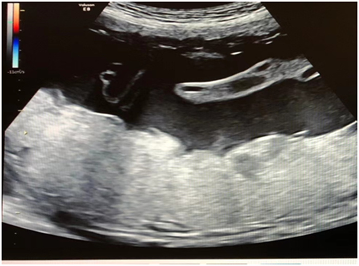
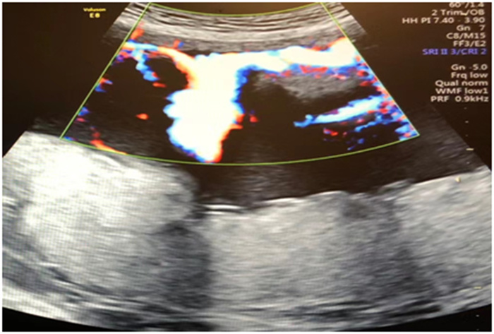
Objective To analyze the relationship between the prognosis of pregnant women with velar placenta and ultrasonographic parameters of fetal umbilical artery blood flow. Methods The clinical data and prenatal ultrasound examination results of 102 pregnant women with Vela placenta admitted to our hospital from October 2018 to October 2021 were retrospectively analyzed. According to pregnancy outcomes, they were divided into good pregnancy outcome group (n=60) and poor pregnancy outcome group (n=42). At the same time, fifty normal pregnant women who underwent pregnancy examination in our hospital at the same period were selected as control group. The ratio of end-systolic peak flow velocity to end- diastolic peak flow velocity (S/D) and resistance index of fetal umbilical artery were measured using pulsed Doppler ultrasonography, and the S/D ratio and resistance index of each group were compared. Pearson correlation was used to analyze the relationship between S/D ratio and resistance index and gestational week of delivery and birth weight of fetus. Spearman correlation analysis was used to analyze the relationship between S/D ratio and resistance index, fetal adverse outcome rate and postpartum haemorrhage rate. Results The S/D ratio and resistance index in poor pregnancy outcome group were higher than those in good pregnancy outcome group and control group (P < 0.05), with the increase of S/D ratio and resistance index, the shorter gestational age of pregnant women, the lower the birth weight of the fetus, and the higher the rate of poor fetal outcome and postpartum hemorrhage (P < 0.05). Further correlation analysis showed that S/D ratio and resistance index were inversely correlated with gestational week of delivery and fetal birth mass and positively proportional to the rate of poor fetal outcome and postpartum hemorrhage (P < 0.05). Conclusion Fetal umbilical artery flow ultrasound parameters S/D ratio and resistance index are significantly correlated with the prognosis of pregnant women with velar placenta. The higher the S/D ratio and resistance index, the earlier the gestational week of delivery, the lower the birth weight of the fetus, and the higher the rate of poor outcomes of the fetus.
2022, 45(2): 256-260.
doi: 10.12122/j.issn.1674-4500.2022.02.19
Abstract:
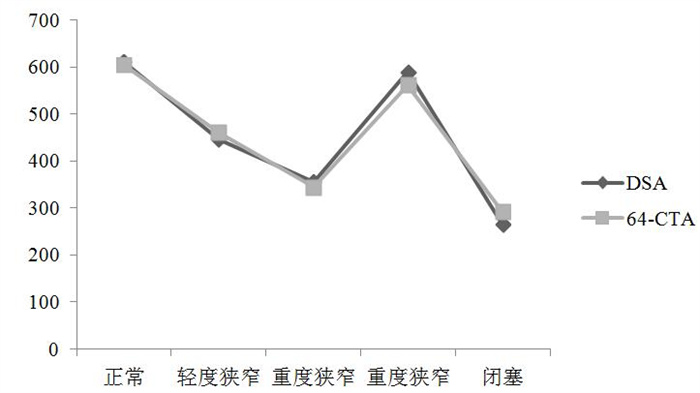
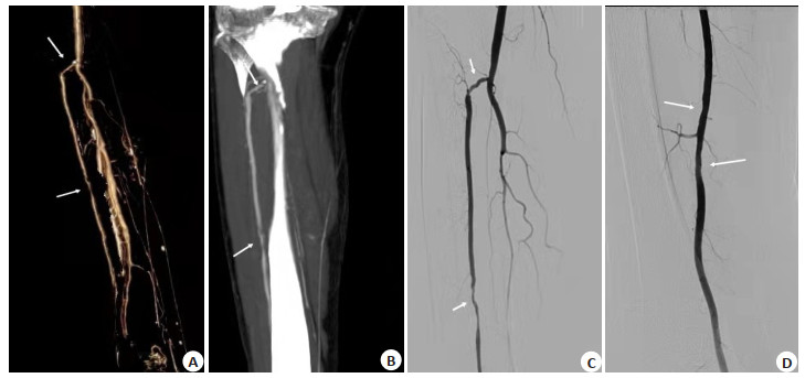
Objective To analyze the imaging value of 64-slice CT angiography (64-CTA) and digital subtraction angiography (DSA) in lower limb arteriosclerosis obliterans in diabetic patients. Methods The data of 64-CTA and DSA of 200 patients with diabetic lower limb arterial occlusive disease diagnosed in our hospital from May 2019 to May 2021 were collected. All patients underwent 64-CTA examination, and DSA examination was performed after 24 h. The interval between the two examinations was not more than 2 weeks. The sensitivity, specificity, accuracy, positive predictive value and negative predictive value of 64-CTA in the diagnosis of diabetic lower extremity arteriosclerosis obliterans were calculated using DSA as the gold standard. The diagnostic coincidence rates of the three treatment methods of 64-CTA were calculated. Results A total of 2265 segments of 200 patients were compared between 64-CTA and DSA. Taking DSA as the gold standard, 2115 segments of 64-CTA were consistent with DSA, with a diagnostic coincidence rate of 93.38%. 64-CTA overestimated grade 1 by 86 times and underestimated grade 1 by 53 times. The distribution curve of 64-CTA and DSA showing the grade of lower limb arterial stenosis was drawn. The results of kappa agreement test showed that the kappa value=0.915 (P < 0.01). Taking DSA as the gold standard, 64-CTA had a sensitivity of 97.27%, specificity of 98.17%, accuracy of 97.62%, positive predictive value of 98.83% and negative predictive value of 95.76% for diagnosing diabetic lower extremity arteriosclerosis obliterans, with a high diagnostic efficacy for each diagnostic cut-off point. The coincidence rate of 64-CTA-MIP was 88.74%, the coincidence rate of 64-CTA-MPR was 92.98%, and the coincidence rate of 64-CTA-VR was 88.74%, and the diagnostic coincidence rate of 64-CTA-MIP and 64-CTA-VR was significantly lower than that of 64-CTA (P < 0.05). Image analysis showed that the degree of stenosis displayed by 64-CTA was closer to the true situation than that of DSA. Conclusion 64-CTA can accurately show the degree of stenosis in diabetic lower extremity arteriosclerosis obliterans, with high consistency with DSA examination.
2022, 45(2): 261-264.
doi: 10.12122/j.issn.1674-4500.2022.02.20
Abstract:
Objective To explore the value of high-resolution MRI (HR-MRI) in evaluating preoperative stage and lymph node metastasis of rectal cancer. Methods We retrospectively reviewed 60 patients with rectal cancer who underwent surgery in our hospital from April 2020 and July 2021, and their preoperative HR-MRI data and intraoperative sample pathological test results were collected to compare HR-MRI test and surgical pathological test results. Results The surgical pathological results showed 20, 32 and 8 patients with stage T1-2, T3 and T4, respectively, and the Preoperative HR-MRI findings showed 17, 38 and 5 patients, respectively with no significant difference (HC=0.012, P=0.914).The sensitivity/specificity of HR-MRI was 85.00% and 100.00% for the diagnosis of stage T1-2, 93.75% and 71.43% for the detection of stage T3 tumours, and 62.50% and 96.15% for stage T4, respectively, with no statistically significant differences in sensitivity and specificity between the three stages (specificity: χ2=0.302, P=0.289; sensitivity: χ2=0.408, P=0.378). Surgery and pathological examination showed that there were 289 lymph nodes were simultaneously present in 60 rectal cancer patients by pathology and HR-MRI, 129 (44.64%) lymph nodes were positive and 160 (55.36%) were negative for metastasis in pathological examination. 113 (39.10%) lymph nodes were positive for metastasis and 176 (60.90%) were negative for metastasis. The specificity and sensitivity of HR-MRI in diagnosing whether lymph nodes had metastasis were 72.87% and 88.13%. The diagnostic coincidence rate was 81.31%. Conclusion HR-MRI can accurately diagnose and evaluate the preoperative stage and lymph node metastasis of rectal cancer, and the results are similar to surgical pathological results. It can provide a reliable basis for clinical planning of rectal cancer surgery and deserve clinical attention.
2022, 45(2): 265-269.
doi: 10.12122/j.issn.1674-4500.2022.02.21
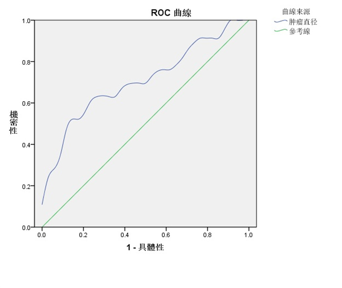
Abstract:
Objective To investigate the factors associated with preoperative ultrasound suspicion of lymph node metastasis (LLNM) in the lateral cervical region of papillary thyroid cancer. Methods From January 2010 to January 2021, our hospital line gland regular ultrasound examination of 80 patients with high suspicion of PTC were selected and treated with preoperative ultrasound and confirmed to have PTC by postoperative pathology. 80 patients were divided into a group with lymph node metastasis in the lateral cervical region (LLNM group, n=46) and a group without lymph node metastasis in the lateral cervical region (NLLNM group, n=34). NLLNM group, n=34), their clinical features and ultrasound sonographic features were collected, Independent factors related to preoperative ultrasound suspicion of LLNM were analyzed by multifactor Logistic analysis. The best critical value of LLNM was calculated from ROC curve by patient tumor diameter prediction. Results Univariate analysis showed statistically significant differences (P < 0.05) in gender, film violation, lymph node transfer, tumor diameter, and tumor diameter, nodule microcalification in the central area, and nodules of nodules were independent risk factors associated with LLNM. The ROC curve showed a tumour diameter cut-off point of 1.404 cm, with a sensitivity of 52.2% and specificity of 88.2%, and an area under the curve of 0.709. Conclusion Male, film violation, lymph node transfer, tumor diameter ≥1.404 cm, nodule microcetocrification on the lymph node metastasis of thyroid pillar carcinoma neck side zone has important predictive value, and attention to the above risk factors may increase the detection rate of the lymph nodes of the lateral cervical region.
2022, 45(2): 270-274.
doi: 10.12122/j.issn.1674-4500.2022.02.22
Abstract:
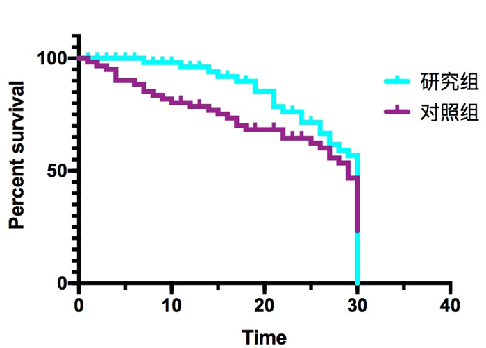
Objective To explore the clinical effects of percutaneous transluminal coronary intervention (PCI) under the guidance of quantitative blood flow fraction (QFR) in the treatment of very elderly patients with acute coronary syndrome with multivessel disease. Methods Eighty super-aged patients who were treated in our hospital from March 2019 to April 2020 and met the inclusion criteria and planned to undergo acute coronary syndrome were selected as the research objects. They were divided into observation group and control group, with 40 cases in each group. The control group used coronary angiography alone to diagnose PCI surgery. The observation group used QFR to guide PCI. The plasma brain natriuretic peptide, NT-proBNP, cardiac troponin I levels, changes in cardiac function indicators before and after surgery (left ventricular ejection fraction, left ventricular end diastolic diameter, left ventricular end systolic diameter, ejection fraction), NYHA grading changes and adverse events were detected. Results The observation group had 83 coronary angiography vessel diameter stenosis≥50%, of which 43 were positive for QFR≤0.8 (51.81%) and 40 were negative (48.19%). Among them, 41 positive and 7 negative were treated with PCI combined with coronary artery revascularization surgery, which was consistent with the results of QFR analysis. The rates were 95.35% and 82.50%, respectively. The control group had 79 vessels with coronary angiography vessel diameter stenosis≥50%, 70 of which were treated with PCI combined with coronary artery revascularization. The plasma brain natriuretic peptide of the observation group was 1 month after the operation. The levels of NT- proBNP, left ventricular end systolic diameter and left ventricular end diastolic diameter were significantly lower than those of the control group, and the ejection fraction value was higher than that of the control group (P < 0.05). The adverse event rate of 27.50% (n=11) in observation group was significantly lower than that of control group within 1 month after surgery 50.00% (n=20, P < 0.05). Conclusion Quantitative blood flow scores are safe and feasible to guide surgical percutaneous coronary intervention in the treatment of acute coronary syndrome in very elderly patients with multivessel disease. It can reduce the incidence of postoperative adverse events. However, its effectiveness still needs to be further expanded and validated by prospective clinical studies.
2022, 45(2): 275-278.
doi: 10.12122/j.issn.1674-4500.2022.02.23
Abstract:
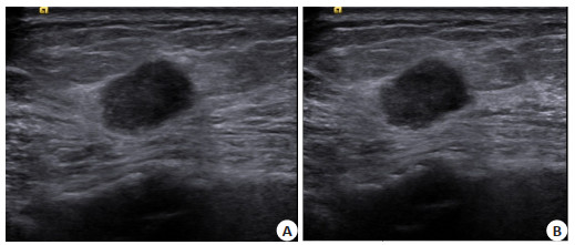
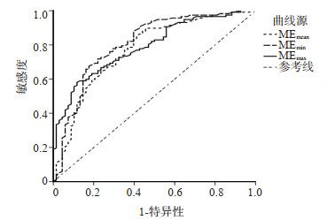
Objective To investigate the diagnostic value of gray scale ultrasound, Doppler ultrasound and shear wave elastography Young's modulus in papillary thyroid microcarcinoma. Methods A total of 136 patients with papillary thyroid microcarcinoma in our hospital from February 2018 to May 2020 were enrolled. Gray scale ultrasound, Doppler ultrasound, shear wave elastography Young's modulus and pathological examination were performed, respectively. Taking pathological examination results as golden standards, the value of remaining three methods in the diagnosis of papillary thyroid microcarcinoma was analyzed. Results The sensitivity, specificity and accuracy of gray-scale ultrasound were 43.21%, 68.30% and 57.30%, respectively, and those of Doppler ultrasound were 75.32%, 90.74% and 84.32%, respectively. Based on pathological examinations, patients were divided into benign group and malignant group for ME value comparison. Values of MEmean, MEmin and MEmax in benign group were significantly lower than those in malignant group (P < 0.05). The optimal ROC curve cutoff points of MEmean, MEmin and MEmax for predicting thyroid microcarcinoma were 40.25, 16.85 and 65.92 kPa, respectively, and the corresponding sensitivity and specificity were 85.5%, 68.8%, 67.5%, 82.4%, 58.1%, and 88.2%, respectively. The accuracy of combined examinations was significantly higher than that of single examination (P < 0.05). Conclusion Combined examinations of gray scale ultrasound, Doppler ultrasound and shear wave elastography Young's modulus in papillary thyroid microcarcinoma has a high diagnosis accuracy.
2022, 45(2): 279-283.
doi: 10.12122/j.issn.1674-4500.2022.02.24
Abstract:
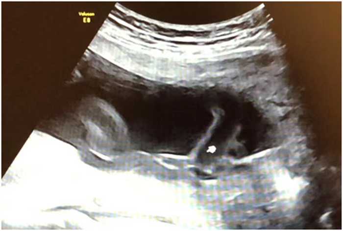
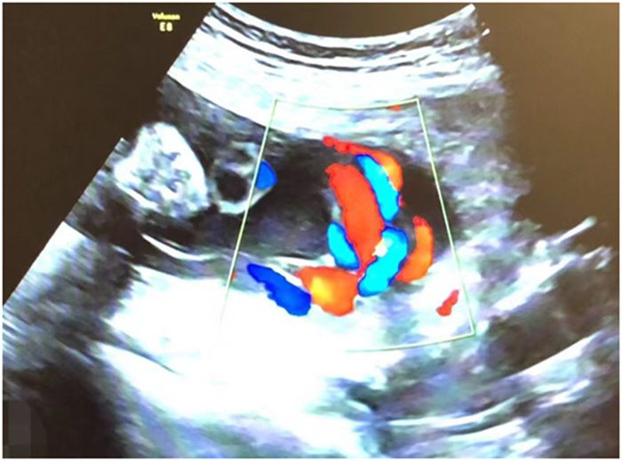
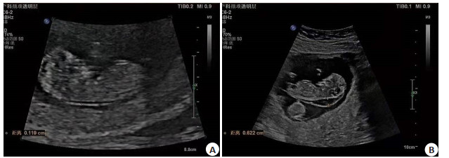
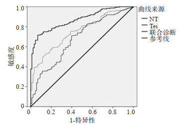
Objective To study the diagnostic of fetal cardiac malformations in early pregnancy by ultrasound detection of nuchal translucency (NT) thickening combined with Tei index and the factors influencing the diagnostic accuracy. Methods A total of 108 cases of pregnant women who underwent early pregnancy screening for congenital diseases in our hospital from January 2015 to February 2020 were selected as the research object. 101 cases of normal pregnant women who underwent pregnancy examination at the same time were selected as the control group. The differences in NT and Tei index between the two groups were compared to study the diagnostic efficacy of combined diagnosis of NT and Tei index for congenital heart disease and to analyse the factors influencing diagnostic accuracy. Results NT (t=16.780, P < 0.001) and Tei index (t=7.406, P < 0.001) in the observation group were significantly higher than those in the control group. The diagnostic specificity of combined diagnosis of NT and Tei index for congenital heart disease was significantly higher than that of individual tests, and the area under the curve of combined diagnosis of NT and Tei index was significantly higher than that of individual tests by ROC curve analysis. At the same time, through critical value analysis, for the diagnosis of congenital heart disease, the critical value of NT was 2.56 mm and the critical value of Tei index was 0.51. There were significant differences between the timing of screening, doctor's expertise and fetal position for accurate diagnosis and false-positive and false-negative patients (P < 0.05). The multifactorial analysis showed that the timing of screening, doctor's expertise and fetal position were all influential factors in the false positives and false negatives. Conclusion Ultrasonic detection of NT thickening combined with Tei index has positive implications for the diagnosis of fetal cardiac malformation in early pregnancy. In the diagnosis, the smaller timing of screening, the low expertise of the physician and lack of cooperation of the fetal position are all factors affecting the diagnostic accuracy.
2022, 45(2): 284-288.
doi: 10.12122/j.issn.1674-4500.2022.02.25
Abstract:
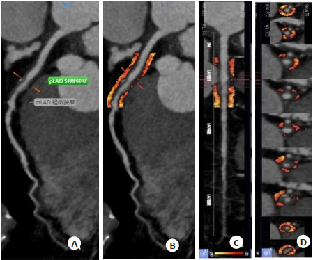
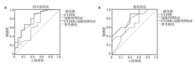
Objective To investigate the evaluation and prediction of coronary atherosclerotic heart disease in high altitude area by artificial intelligence machine learning-based coronary CT fractional flow reserve (CT-FFR) combined with coronary perivascular fat attenuation index (FAI). Methods Sixty-three patients with suspected coronary artery disease who underwent coronary CT angiography (CTA) in our hospital from October to December 2021 were selected, and the patients were divided into stenosis rate ≥50% group and stenosis rate < 50% group using coronary CTA as the gold standard. The degree of coronary stenosis, CT-FFR and perivascular FAI were compared. Patients were re-divided into two groups according to the degree of coronary artery calcification, mild-to-moderate calcification group and severe calcification group. Then the diagnostic efficacy of CT-FFR and perivascular FAI alone and in combination for abnormal coronary blood flow was evaluated. Results A total of 79 coronary vessels were examined in 63 patients, of which 24 vessels with stenosis rate ≥50% and 55 vessels with stenosis rate < 50%. The lesion length yielded no statistical difference between stenosis rate ≥50% group and stenosis rate < 50% group (P > 0.05), while the proportion of patients with single-vessel coronary stenosis showed statistical difference between two groups (P < 0.05). The CT-FFR in stenosis rate ≥50% group was lower than that in stenosis rate < 50% group, and the perivascular FAI was higher than that in stenosis rate < 50% group (P < 0.05). Among the 63 patients, 47 had mild-to-moderate calcification and 16 had severe calcification. Within mild-to-moderate calcification group, the coronary stenosis rate in stenosis rate ≥50% group was higher than that in stenosis rate < 50% group, and CT-FFR was lower than that in stenosis rate < 50% group. Within severe calcification group, the CT-FFR in stenosis rate ≥50% group was lower than that in stenosis rate < 50% group, and perivascular FAI was higher than that in stenosis rate < 50% group, and no significant difference was found in coronary stenosis rate between stenosis rate < 50% group and stenosis rate ≥50% group (P > 0.05). ROC curve showed that the diagnostic efficiency of CT-FFR+ perivascular FAI for vascular calcification was significantly improved compared with two separate examinations (P < 0.05). Conclusion CT-FFR has low diagnostic efficiency for severe calcification of patients with coronary heart disease, while its combination with perivascular FAI examination achieves high diagnostic value in coronary heart disease patients with blood flow abnormalities.
2022, 45(2): 289-293.
doi: 10.12122/j.issn.1674-4500.2022.02.26
Abstract:
In recent years, the development of imaging technology, especially magnetic resonance special sequence imaging, has made it possible to obtain information on functional changes, metabolism, localization, signal transmission and so on. It is of great significance for the study of the pathogenesis of hepatic encephalopathy, early diagnosis and monitoring of treatment effects, and is a hot spot for current research. We searched for hepatic encephalopathy and image manifestations as keywords in China National Knowledge Internet and PubMed, and made a summary for current status of brain MRI, special sequence imaging and nuclear medicine in the diagnosis, severity classification, pathogenesis and therapeutic effect monitoring.
In recent years, the development of imaging technology, especially magnetic resonance special sequence imaging, has made it possible to obtain information on functional changes, metabolism, localization, signal transmission and so on. It is of great significance for the study of the pathogenesis of hepatic encephalopathy, early diagnosis and monitoring of treatment effects, and is a hot spot for current research. We searched for hepatic encephalopathy and image manifestations as keywords in China National Knowledge Internet and PubMed, and made a summary for current status of brain MRI, special sequence imaging and nuclear medicine in the diagnosis, severity classification, pathogenesis and therapeutic effect monitoring.
2022, 45(2): 294-298.
doi: 10.12122/j.issn.1674-4500.2022.02.27
Abstract:
Traumatic penumbra is a reversible area present in the periphery of the injured core area after traumatic brain injury. It is an important factor affecting the prognosis and later quality of life of patients with brain injury. Through early diagnosis and timely treatment of traumatic penumbra, the brain tissue in it can be effectively prevented from developing in a harmful direction, so as to reduce the high disability rate caused by traumatic brain injury. Multimodal magnetic resonance imaging is widely used in the diagnosis of brain diseases, which has the advantages of high resolution, high accuracy and noninvasiveness. Multimodality magnetic resonance technology (diffusion weighted imaging, cerebral perfusion imaging, arterial spin labeling, susceptibility-weighted imaging and magnetic resonance spectroscopy) can be used to understand the presence and extent of traumatic penumbra and its substance and energy metabolism, providing an objective imaging basis for clinical treatment selection and evaluation of curative effect and prognosis. In recent years, with the development of multi-modal magnetic resonance technology, the magnetic resonance methods to define the traumatic penumbra have also been diversified. This review focuses on the pathophysiological mechanisms of traumatic penumbra and brain injury, multimodality magnetic resonance imaging sequences, and multimodality magnetic resonance imaging to assess traumatic penumbra.
Traumatic penumbra is a reversible area present in the periphery of the injured core area after traumatic brain injury. It is an important factor affecting the prognosis and later quality of life of patients with brain injury. Through early diagnosis and timely treatment of traumatic penumbra, the brain tissue in it can be effectively prevented from developing in a harmful direction, so as to reduce the high disability rate caused by traumatic brain injury. Multimodal magnetic resonance imaging is widely used in the diagnosis of brain diseases, which has the advantages of high resolution, high accuracy and noninvasiveness. Multimodality magnetic resonance technology (diffusion weighted imaging, cerebral perfusion imaging, arterial spin labeling, susceptibility-weighted imaging and magnetic resonance spectroscopy) can be used to understand the presence and extent of traumatic penumbra and its substance and energy metabolism, providing an objective imaging basis for clinical treatment selection and evaluation of curative effect and prognosis. In recent years, with the development of multi-modal magnetic resonance technology, the magnetic resonance methods to define the traumatic penumbra have also been diversified. This review focuses on the pathophysiological mechanisms of traumatic penumbra and brain injury, multimodality magnetic resonance imaging sequences, and multimodality magnetic resonance imaging to assess traumatic penumbra.
2022, 45(2): 299-302.
doi: 10.12122/j.issn.1674-4500.2022.02.28
Abstract:
Ultrasound elastography, an emerging ultrasound technology, is becoming increasingly sophisticated in the diagnosis of breast, thyroid, and prostate diseases. Currently, the main ultrasound elastography techniques used in the diagnosis of breast diseases include static/quasi- static elastography, acoustic radiation force pulse imaging and shear wave elastography. In this paper, we briefly introduce the principles of these elastography techniques and summarize and analyze the progress and clinical reliability of their application in differential diagnosis of benign and malignant breast nodules to provide some reference significance for clinical diagnosis.
Ultrasound elastography, an emerging ultrasound technology, is becoming increasingly sophisticated in the diagnosis of breast, thyroid, and prostate diseases. Currently, the main ultrasound elastography techniques used in the diagnosis of breast diseases include static/quasi- static elastography, acoustic radiation force pulse imaging and shear wave elastography. In this paper, we briefly introduce the principles of these elastography techniques and summarize and analyze the progress and clinical reliability of their application in differential diagnosis of benign and malignant breast nodules to provide some reference significance for clinical diagnosis.
2022, 45(2): 303-307.
doi: 10.12122/j.issn.1674-4500.2022.02.29
Abstract:
Prostate cancer is one of the common malignant tumors of the urinary system, and its incidence is increasing year by year in China. The early stage of prostate cancer has no obvious clinical manifestations, and the disease is insidious and prone to deteriorate and metastasize. When patients show symptoms and seek treatment, it is often in the middle and late stage or even distant metastasis, missing the best opportunity for treatment. The prognosis of prostate cancer is closely related to whether it is diagnosed in time. Ultrasound molecular imaging is a targeted acoustic contrast agent prepared by attaching targeted molecular markers to the surface of an acoustic contrast agent with microbubbles carrier, which can significantly improve the diagnosis and therapeutic effects of disease with fewer toxic side effects on normal tissues and organs. The special structure and properties of ultrasound contrast agents allow them to undergo a series of chemical modifications to deliver therapeutic genes and chemotherapeutic drugs to the tumor region, improving the efficiency of gene transfection and drug efficacy. This article describes several commonly used targeting ligands, therapeutic modalities for lesions, and recent advances in multimodality imaging and treatment of prostate cancer with targeted ultrasound contrast agents in the diagnosis and treatment of prostate cancer.
Prostate cancer is one of the common malignant tumors of the urinary system, and its incidence is increasing year by year in China. The early stage of prostate cancer has no obvious clinical manifestations, and the disease is insidious and prone to deteriorate and metastasize. When patients show symptoms and seek treatment, it is often in the middle and late stage or even distant metastasis, missing the best opportunity for treatment. The prognosis of prostate cancer is closely related to whether it is diagnosed in time. Ultrasound molecular imaging is a targeted acoustic contrast agent prepared by attaching targeted molecular markers to the surface of an acoustic contrast agent with microbubbles carrier, which can significantly improve the diagnosis and therapeutic effects of disease with fewer toxic side effects on normal tissues and organs. The special structure and properties of ultrasound contrast agents allow them to undergo a series of chemical modifications to deliver therapeutic genes and chemotherapeutic drugs to the tumor region, improving the efficiency of gene transfection and drug efficacy. This article describes several commonly used targeting ligands, therapeutic modalities for lesions, and recent advances in multimodality imaging and treatment of prostate cancer with targeted ultrasound contrast agents in the diagnosis and treatment of prostate cancer.
2022, 45(2): 308-312.
doi: 10.12122/j.issn.1674-4500.2022.02.30
Abstract:
Bladder, as one of the main organs of the urinary system, has two important functions: urine storage and urination. The nerve conduction pathway that regulate these two functions is very complex, involving multiple levels such as brain, spinal cord and peripheral nervous system, and is jointly mediated by a variety of neurotransmitters. Over the past two decades, electrophysiological techniques and functional magnetic resonance imaging technique have emerged as powerful tools in the field of neurofunctional studies of human pathological or physiological states. It is feasible to use these two techniques to record and analyse the brain-regulated mechanisms of bladder storage and voiding behaviour, both can help us better understand the relationship between the activity of neural pathways and brain regions involved in urinary control and specific functions. We review the advances in the neurofunctional studies of bladder filling and the mechanism of urine storage and control in situ new bladder from the aspects of electrophysiology and functional magnetic resonance imaging.
Bladder, as one of the main organs of the urinary system, has two important functions: urine storage and urination. The nerve conduction pathway that regulate these two functions is very complex, involving multiple levels such as brain, spinal cord and peripheral nervous system, and is jointly mediated by a variety of neurotransmitters. Over the past two decades, electrophysiological techniques and functional magnetic resonance imaging technique have emerged as powerful tools in the field of neurofunctional studies of human pathological or physiological states. It is feasible to use these two techniques to record and analyse the brain-regulated mechanisms of bladder storage and voiding behaviour, both can help us better understand the relationship between the activity of neural pathways and brain regions involved in urinary control and specific functions. We review the advances in the neurofunctional studies of bladder filling and the mechanism of urine storage and control in situ new bladder from the aspects of electrophysiology and functional magnetic resonance imaging.