


Progress of quantitative magnetic resonance T2 mapping imaging in disc degeneration
-
摘要: 椎间盘退变是引起下腰背痛的主要原因之一,影像学的检查是诊断椎间盘退变的重要手段。传统MRI技术虽可显示椎间盘的信号强度和形态学改变,但很难客观量化椎间盘退变的程度。T2 mapping技术能够检测椎间盘内水含量、蛋白多糖含量及胶原纤维排列顺序等多种生化成分,为椎间盘退变早期临床评估提供新的方法。本文从T2 mapping与其他定量磁共振技术对比,及其与椎间盘退变分级、日本骨科学会评分、视觉模拟评分及年龄、性别等联系论述定量磁共振T2映射成像在椎间盘退变中的研究进展。Abstract: Intervertebral disc degeneration is one of the main causes of low back pain. Imaging examination is an important means of diagnose intervertebral disc degeneration. Although traditional MRI technique can show the signal intensity and and morphological changes of the intervertebral disc, it is difficult to objectively quantify the degree of disc degeneration. T2 mapping technology can detect various biochemical components such as intervertebral disc water content, proteoglycan content and collagen fiber sequence, providing a new method for early clinical evaluation of intervertebral disc degeneration. This article discusses the research progress of quantitative magnetic resonance T2 mapping in disc degeneration is discussed from the comparison of T2 mapping with other quantitative magnetic resonance technologies, and its relationship with disc degeneration grade, Japanese Orthopaedic Association score, visual analog scale score, age and gender.
-

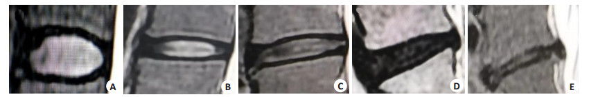
图 1 Pfirrmann分级的T2WI影像学表现
A~E分别表示椎间盘退变Ⅰ~Ⅴ级.
Figure 1. T2WI images of Pfirrmann grading.

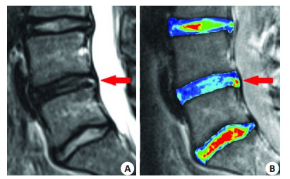
图 2 矢状位L3~S1的T2WI图和T2映射图
A: 矢状位L3~S1的T2WI图; B: 矢状位腰椎间盘T2映射(L3~S1)[29].
Figure 2. T2WI and T2 mapping images of sagittal L3-S1 images.
表 1 Pfirrmann分级
Table 1. Pfirrmann grading
等级 髓核信号 髓核结构 髓核与纤维环的分界 椎问盘的高度 Ⅰ 高 亮白、均一 清楚 正常 Ⅱ 高 不均、有水平带 清楚 正常 Ⅲ 中等 灰、不均 模糊 正常-轻度减低 Ⅳ 中低 灰-黑、不均 模糊 正常-中度减低 Ⅴ 低 不均、黑 消失 重度减低  下载: 导出CSV
表 2 改良Pfirrmann分级
Table 2. Improved Pfirrmann grading
分级 髓核及内层纤维信号 后方纤维内外层信号差别 椎间盘高度 1 均匀高信号, 与脑脊液相当 明显 正常 2 高信号(>骶骨前脂肪, <脑脊液)-髓核内低信号裂隙 明显 正常 3 高信号(<骶骨前脂肪) 不明显 正常 4 中等高信号(略>外层纤维环) 不明显 正常 5 低信号(=外层纤维环) 不明显 正常 6 低信号 不明显 椎间盘高度减小<30% 7 低信号 不明显 椎间盘高度减小30%~ 60% 8 低信号 不明显 椎间盘高度减小>60%
下载: 导出CSV
表 3 椎间盘退变组织学分级量表
Table 3. Histological grading scale for disc degeneration
椎间盘成分 1分 2分 3分 纤维环 纤维环正常, 纤维软骨板(向后呈u形, 向前略凸)纤维环无破裂, 环内任何部位无裂纹 纤维环破裂或呈裂纹样<30% 纤维环破裂或呈裂纹样>30% 纤维环与髓核的分界 正常 最小中断 中度、重度中断 髓核细胞结构 正常细胞, 基质凝胶状结构中有大液泡 细胞数量轻微减少, 空泡减少 细胞数量中度/重度减少(>50%), 无空泡 髓核基质 正常的凝胶状外观 细胞外基质轻度凝结 细胞外基质中度/重度凝结 髓核的细胞外基质 正常 细胞外基质轻度形成 中度/重度细胞外基质形成
下载: 导出CSV
表 4 髓核组织学退变评分量表
Table 4. Nucleus pulposus histological degeneration Scale
髓核成分 1分 2分 3分 髓核细胞 正常细胞形态, 在基质凝胶状结构中有大量的空泡 细胞数量轻度减少, 空泡轻度减少 细胞数量中度/重度减少(>50%), 无空泡 髓核基质 正常凝胶结构 细胞外基质轻度皱缩 细胞外基质中、重度皱缩
下载: 导出CSV
-
[1] Ji YY, Hong WF, Liu MY, et al. Intervertebral disc degeneration associated with vertebral marrow fat, assessed using quantitative magnetic resonance imaging[J]. Skeletal Radiol, 2020, 49(11): 1753-63. doi: 10.1007/s00256-020-03419-7 [2] Wenger HC, Cifu AS. Treatment of low back pain[J]. JAMA, 2017, 318(8): 743-4. doi: 10.1001/jama.2017.9386 [3] 张素芳. 3.0T MR T1 p及T2 mapping在颈椎间盘退行性变中的应用研究[D]. 泰安: 泰山医学院, 2018. [4] 王睿哲. CHI3L1在椎间盘退变中的作用及机制研究[D]. 上海: 中国人民解放军海军军医大学, 2020. [5] Abdollah V, Parent EC, Su A, et al. The effects of axial loading on the morphometric and T2 characteristics of lumbar discs in relation to disc degeneration[J]. Clin Biomech, 2021, 83: 105291. doi: 10.1016/j.clinbiomech.2021.105291 [6] Wang L, Han M, Wong J, et al. Evaluation of human cartilage endplate composition using MRI: spatial variation, association with adjacent disc degeneration, and in vivo repeatability[J]. J Orthop Res, 2021, 39(7): 1470-8. doi: 10.1002/jor.24787 [7] 曾菲菲, 査云飞, 邢栋, 等. 磁共振扩散峰度成像和T2*-mapping技术定量检测腰椎间盘退变的对比研究[J]. 放射学实践, 2018, 33 (10): 1087-92. https://www.cnki.com.cn/Article/CJFDTOTAL-FSXS201810034.htm [8] Wu XL, Liu C, Yang S, et al. Glycine-serine-threonine metabolic axis delays intervertebral disc degeneration through antioxidant effects: an imaging and metabonomics study[J]. Oxidative Med Cell Longev, 2021, 2021: 5579736. [9] 苏树燕, 刘源. 椎间盘退变的磁共振功能成像研究进展[J]. 汕头大学医学院学报, 2019, 32(1): 49-52. https://www.cnki.com.cn/Article/CJFDTOTAL-STDY201901015.htm [10] Verschueren J, Meuffels DE, Bron EE, et al. Possibility of quantitative T2-mapping MRI of cartilage near metal in high tibial osteotomy: a human cadaver study[J]. J Orthop Res, 2018, 36(4): 1206-12. [11] Raudner M, Schreiner MM, Hilbert T, et al. Clinical implementation of accelerated T2 mapping: quantitative magnetic resonance imaging as a biomarker for annular tear and lumbar disc herniation [J]. Eur Radiol, 2021, 31(6): 3590-9. doi: 10.1007/s00330-020-07538-6 [12] Sollmann N, Weidlich D, Klupp E, et al. T2 mapping of the distal sciatic nerve in healthy subjects and patients suffering from lumbar disc herniation with nerve compression[J]. Magn Reson Mater Phys Biol Med, 2020, 33(5): 713-24. doi: 10.1007/s10334-020-00832-w [13] Wang M, Tsang A, Tam V, et al. Multiparametric MR investigation of proteoglycan diffusivity, T 2 relaxation, and concentration in an ex vivo model of intervertebral disc degeneration[J]. J Magn Reson Imaging, 2020, 51(5): 1390-400. doi: 10.1002/jmri.26979 [14] Jiang YW, Yu L, Luo XJ, et al. Quantitative synthetic MRI for evaluation of the lumbar intervertebral disk degeneration in patients with chronic low back pain[J]. Eur J Radiol, 2020, 124: 108858. doi: 10.1016/j.ejrad.2020.108858 [15] 雷贞妮. 磁共振T2 Mapping成像对颈椎间盘退变的初步研究[D]. 广州: 南方医科大学, 2017. [16] 张新娟. 3.0T磁共振T1ρ与T2* mapping对腰椎间盘退行性变分级评价的对照研究[D]. 济南: 山东大学, 2015. [17] 朱记超, 张志强, 郝长胜, 等. 3.0 T磁共振定量T2及T2* mapping技术在颈椎间盘退变中的应用[J]. 临床放射学杂志, 2019, 38(4): 710-5. https://www.cnki.com.cn/Article/CJFDTOTAL-LCFS201904035.htm [18] Johannessen W, Auerbach JD, Wheaton AJ, et al. Assessment of human disc degeneration and proteoglycan content using T1ρ-weighted magnetic resonance imaging[J]. Spine, 2006, 31(11): 1253-7. doi: 10.1097/01.brs.0000217708.54880.51 [19] Clouet J, Fusellier M, Camus A, et al. Intervertebral disc regeneration: from cell therapy to the development of novel bioinspired endogenous repair strategies[J]. Adv Drug Deliv Rev, 2019, 146: 306-24. doi: 10.1016/j.addr.2018.04.017 [20] Antoniou J, Steffen T, Nelson F, et al. The human lumbar intervertebral disc: evidence for changes in the biosynthesis and denaturation of the extracellular matrix with growth, maturation, ageing, and degeneration[J]. J Clin Invest, 1996, 98(4): 996-1003. doi: 10.1172/JCI118884 [21] Yasuma T, Arai K, Yamauchi Y. The histology of lumbar intervertebral disc herniation[J]. Spine, 1993, 18(13): 1761-5. doi: 10.1097/00007632-199310000-00008 [22] 熊玉超, 曾旭文, 梁治平, 等. 磁共振T2-mapping及T2*-mapping对兔腰椎间盘退变的定量研究[J]. 放射学实践, 2021, 36(8): 1042-7. https://www.cnki.com.cn/Article/CJFDTOTAL-FSXS202108025.htm [23] Pfirrmann CWA, Metzdorf A, Zanetti M, et al. Magnetic resonance classification of lumbar intervertebral disc degeneration[J]. Spine, 2001, 26(17): 1873-8. doi: 10.1097/00007632-200109010-00011 [24] Griffith JF, Wang YXJ, Antonio GE, et al. Modified pfirrmann grading system for lumbar intervertebral disc degeneration[J]. Spine, 2007, 32(24): E708-12. doi: 10.1097/BRS.0b013e31815a59a0 [25] Imanishi T, Akeda K, Murata K, et al. Effect of diminished flow in rabbit lumbar arteries on intervertebral disc matrix changes using MRI T2-mapping and histology[J]. BMC Musculoskelet Disord, 2019, 20: 347. doi: 10.1186/s12891-019-2721-y [26] Masuda K, Aota Y, Muehleman C, et al. A novel rabbit model of mild, reproducible disc degeneration by an anulus needle puncture: correlation between the degree of disc injury and radiological and histological appearances of disc degeneration[J]. Spine, 2005, 30 (1): 5-14. doi: 10.1097/01.brs.0000148152.04401.20 [27] Nisolle JF, Bihin B, Kirschvink N, et al. Prevalence of age-related changes in ovine lumbar intervertebral discs during computed tomography and magnetic resonance imaging[J]. Comp Med, 2016, 66(4): 300-7. [28] 李金宝. MRI多种功能成像序列在椎间盘退变的诊断价值[J]. 黑龙江医学, 2020, 44(5): 659-61. doi: 10.3969/j.issn.1004-5775.2020.05.035 [29] Trattnig S, Stelzeneder D, Goed S, et al. Lumbar intervertebral disc abnormalities: comparison of quantitative T2 mapping with conventional MR at 3.0T[J]. Eur Radiol, 2010, 20(11): 2715-22. doi: 10.1007/s00330-010-1843-2 [30] Huang LT, Liu Y, Ding Y, et al. Quantitative evaluation of lumbar intervertebral disc degeneration by axial T2* mapping[J]. Medicine, 2017, 96(51): e9393. doi: 10.1097/MD.0000000000009393 [31] Chokan K, Murakami H, Endo H, et al. Evaluation of water retention in lumbar intervertebral disks before and after exercise stress with T2 mapping[J]. SPINE, 2016, 41(7): E430-6. doi: 10.1097/BRS.0000000000001283 [32] Schleich C, Müller-Lutz A, Eichner M, et al. Glycosaminoglycan chemical exchange saturation transfer of lumbar intervertebral discs in healthy volunteers[J]. SPINE, 2016, 41(2): 146-52. doi: 10.1097/BRS.0000000000001144 [33] Blumenkrantz G, Zuo J, Li XJ, et al. In vivo 3.0-tesla magnetic resonance T1ρ and T2 relaxation mapping in subjects with intervertebral disc degeneration and clinical symptoms[J]. Magn Reson Med, 2010, 63(5): 1193-200. doi: 10.1002/mrm.22362 [34] Ogon I, Takebayashi T, Takashima H, et al. Analysis of chronic low back pain with magnetic resonance imaging T2 mapping of lumbar intervertebral disc[J]. J Orthop Sci, 2015, 20(2): 295-301. doi: 10.1007/s00776-014-0686-0 [35] Cui YZ, Yang XH, Liu PF, et al. Preliminary study on diagnosis of lumbar disc degeneration with magnetic resonance T1p, T2 mapping and DWI quantitative detection technologies[J]. Eur Rev Med Pharmacol Sci, 2016, 20(16): 3344-50. [36] Wang YXJ, Zhao F, Griffith JF, et al. T1rho and T2 relaxation times for lumbar disc degeneration: an in vivo comparative study at 3.0-Tesla MRI[J]. Eur Radiol, 2013, 23(1): 228-34. doi: 10.1007/s00330-012-2591-2 [37] Yoo YH, Yoon CS, Eun NL, et al. Interobserver and test-retest reproducibility of T1ρ and T2 measurements of lumbar intervertebral discs by 3T magnetic resonance imaging[J]. Korean J Radiol, 2016, 17(6): 903. doi: 10.3348/kjr.2016.17.6.903 [38] Zhang W, Ma XH, Wang Y, et al. Assessment of apparent diffusion coefficient in lumbar intervertebral disc degeneration[J]. Eur Spine J, 2014, 23(9): 1830-6. doi: 10.1007/s00586-014-3285-z [39] Wang W, Hou J, Lv DY, et al. Multimodal quantitative magnetic resonance imaging for lumbar intervertebral disc degeneration[J]. Exp Ther Med, 2017, 14(3): 2078-84. doi: 10.3892/etm.2017.4786 [40] Paul CPL, Smit TH, de Graaf M, et al. Quantitative MRI in early intervertebral disc degeneration: T1rho correlates better than T2 and ADC with biomechanics, histology and matrix content[J]. PLoS One, 2018, 13(1): e0191442. doi: 10.1371/journal.pone.0191442 [41] Stadelmann MA, Maquer G, Voumard B, et al. Integrating MRI-based geometry, composition and fiber architecture in a finite element model of the human intervertebral disc[J]. J Mech Behav Biomed Mater, 2018, 85: 37-42. doi: 10.1016/j.jmbbm.2018.05.005 [42] Shinn RL, Pancotto TE, Stadler KL, et al. Magnetization transfer and diffusion tensor imaging in dogs with intervertebral disk herniation [J]. J Vet Intern Med, 2020, 34(6): 2536-44. doi: 10.1111/jvim.15899 [43] Vadapalli R, Mulukutla R, Vadapalli AS, et al. Quantitative predictive imaging biomarkers of lumbar intervertebral disc degeneration[J]. Asian Spine J, 2019, 13(4): 527-34. doi: 10.31616/asj.2018.0166 [44] Li L, Zhou ZG, Li J, et al. Diffusion kurtosis imaging provides quantitative assessment of the microstructure changes of disc degeneration: an in vivo experimental study[J]. Eur Spine J, 2019, 28 (5): 1005-13. doi: 10.1007/s00586-019-05924-3 [45] 曾菲菲, 查云飞. 椎间盘退变MR扩散成像的研究进展[J]. 中国医学影像学杂志, 2019, 27(2): 156-60. doi: 10.3969/j.issn.1005-5185.2019.02.018 [46] Li L, Zhou ZG, Xiong W, et al. Characterization of the microstructure of the intervertebral disc in patients with chronic low back pain by diffusion kurtosis imaging[J]. Eur Spine J, 2019, 28(11): 2517-25. doi: 10.1007/s00586-019-06095-x [47] 曾菲菲, 查云飞, 胡磊, 等. 磁共振扩散峰度成像对腰椎间盘退变的诊断价值[J]. 磁共振成像, 2018, 9(2): 113-6. https://www.cnki.com.cn/Article/CJFDTOTAL-CGZC201802008.htm [48] Wang CY, McArdle E, Fenty M, et al. Validation of sodium magnetic resonance imaging of intervertebral disc[J]. Spine, 2010, 35(5): 505-10. doi: 10.1097/BRS.0b013e3181b32d3b [49] Zhang C, Lin Y, Han ZH, et al. Feasibility of T2 mapping and magnetic transfer ratio for diagnosis of intervertebral disc degeneration at the cervicothoracic junction: a pilot study[J]. Biomed Res Int, 2019, 2019: 6396073. [50] Wang AM, Cao P, Yee A, et al. Detection of extracellular matrix degradation in intervertebral disc degeneration by diffusion magnetic resonance spectroscopy[J]. Magn Reson Med, 2015, 73 (5): 1703-12. doi: 10.1002/mrm.25289 [51] Schleich C, Miese F, Müller-Lutz A, et al. Value of delayed gadolinium-enhanced magnetic resonance imaging of cartilage for the pre-operative assessment of cervical intervertebral discs[J]. J Orthop Res, 2017, 35(8): 1824-30. doi: 10.1002/jor.23454 [52] Liu ZZ, Wen HQ, Zhu YQ, et al. Short-term effect of lumbar traction on intervertebral discs in patients with low back pain: correlation between the T2 value and ODI/VAS score[J]. CARTILAGE, 2021, 13(1_suppl): 414S-23S. doi: 10.1177/1947603521996793 [53] 冯国洋, 郭龙军, 王娟, 等. MRI参数对腰椎间盘突出症患者椎间盘退变程度的评估价值及与JOA、VAS评分相关性[J]. 影像科学与光化学, 2021, 39(2): 207-12. https://www.cnki.com.cn/Article/CJFDTOTAL-GKGH202102008.htm [54] 杨传红, 邱光, 杨骁驰, 等. 腰椎间盘水信号分数在椎间盘退变及突出的应用价值[J]. 临床放射学杂志, 2020, 39(11): 2326-9. https://www.cnki.com.cn/Article/CJFDTOTAL-LCFS202011041.htm [55] Wáng YXJ, Wáng JQ, Káplár Z. Increased low back pain prevalence in females than in males after menopause age: evidences based on synthetic literature review[J]. Quant Imaging Med Surg, 2016, 6(2): 199-206. doi: 10.21037/qims.2016.04.06 -

 点击查看大图
计量
- 文章访问数: 193
- HTML全文浏览量: 231
- PDF下载量: 16
- 被引次数: 0